. 2022 Jan 31;23(3):1680.
doi: 10.3390/ijms23031680.

Remodeling of Cell Wall Components in Root Nodules and Flower Abscission Zone under Drought in Yellow Lupine

Affiliations

Remodeling of Cell Wall Components in Root Nodules and Flower Abscission Zone under Drought in Yellow Lupine

Emilia Wilmowicz et al. Int J Mol Sci. .

Abstract

We recently showed that yellow lupine is highly sensitive to soil water deficits since this stressor disrupts nodule structure and functioning, and at the same time triggers flower separation through abscission zone (AZ) activation in the upper part of the plant. Both processes require specific transformations including cell wall remodeling. However, knowledge about the involvement of particular cell wall elements in nodulation and abscission in agronomically important, nitrogen-fixing crops, especially under stressful conditions, is still scarce. Here, we used immuno-fluorescence techniques to visualize dynamic changes in cell wall compounds taking place in the root nodules and flower AZ of Lupinus luteus following drought. The reaction of nodules and the flower AZ to drought includes the upregulation of extensins, galactans, arabinans, xylogalacturonan, and xyloglucans. Additionally, modifications in the localization of high- and low-methylated homogalacturonans and arabinogalactan proteins were detected in nodules. Collectively, we determined for the first time the drought-associated modification of cell wall components responsible for their remodeling in root nodules and the flower AZ of L. luteus. The involvement of these particular molecules and their possible interaction in response to stress is also deeply discussed herein.

Keywords: abscission zone; arabinan; cell wall; drought; extensins; galactans; root nodules; xyloglucans; yellow lupine; yielding.


Conflict of interest statement

The authors declare no conflict of interest.

Figures

Figure 1
Figure 1
Proline accumulates differently in various parts of Lupinus luteus L. root subjected to soil drought. Control plants were grown under optimal conditions (70% water holding capacity, WHC), whereas stressed plants were subjected to water deficit conditions for 2 weeks (25% WHC). Samples were collected from different parts of roots and nodules on the 48th day of cultivation. Data are presented as averages ± SE. ** p < 0.01, * p < 0.05 (root part from stressed plant versus non-stressed ones) (Student’s t-test).
Figure 2
Figure 2
The level and localization of hemoglobin in nodules of Lupinus luteus L. are strongly affected by drought. Lupines were cultivated under water deficit conditions (25% water holding capacity, WHC), while control plants were grown in the soil of optimal moisture (70% WHC). For analysis, nodules were excised on the 48th day of cultivation. Western blot analysis was performed with AHB2 antibody (A). M–molecular mass marker. A band reactive to this antibody (of ~17 kDa) was scanned, densitometry analysis was made, and results are presented on (B) (100% was set for the control). Data are presented as averages ± SE. ** p < 0.01 (n = 3), Coomassie-Blue-stained gel as reference protein standard (C). The localization pattern of hemoglobin (AHB2 antibody) in stressed (F,G) and control (D,E) nodules. Images (E,G) are magnifications of regions of the fixation zone and parenchyma cells (white squares) from control and stressed nodules, respectively. Green fluorescence indicates AHB2 presence, whereas blue color corresponds to nuclei (DAPI staining). Red arrows denote strong fluorescent labeling. Abbreviations: F: fixation zone; E: endodermis; PA: nodule parenchyma; PE: periderm; VB: vascular bundle. Bars = 100 µm (D,F), 50 µm (E,G).
Figure 3
Figure 3
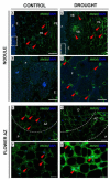
Drought stress changes the extensin (EXT) localization in nodules (AD) and flower abscission zone (AZ) (EH) of Lupinus luteus L. Immunolocalization of EXT (using JIM11 antibody) was performed in the nodules (C,D) and flower AZ (G,H) dissected from drought-treated plants (25% water holding capacity, WHC) and control lupines (70% WHC). Control nodules and AZ are presented on (A,B,E,F) respectively. For the analysis, tissue fragments were collected on the 48th day of plant cultivation. Images (B,D) are magnifications of parenchyma regions from control and stressed nodules, respectively. Images (F,H) show magnification of AZ region (white dotted lines) presented on (E,G). Green fluorescence indicates EXT detection, whereas DAPI blue labeling corresponds to nuclei. Red arrowheads indicate a strong signal. Abbreviations: F: fixation zone; E: endodermis cells; PA: nodule parenchyma; PE: periderm; AZ: abscission zone; P: proximal region of AZ, D: distal region of AZ. Bars = 50 µm (A,C), 25 µm (B,D), 40 µm (E,G), 20 µm (F,H).
Figure 4
Figure 4
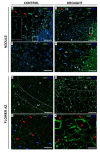
Extensin (EXT) distribution in nodules and flower abscission zone (AZ) cells of L. luteus L. is modified by soil drought stress. Immunolocalization of EXT (using JIM20 antibody) was performed in the nodules (C,D) and flower AZ (G,H) from drought-treated lupines (25% water holding capacity, WHC). Control nodules and AZs (70% WHC) are presented on (A,B,E,F) respectively. For each analysis fragments of nodules and AZs were collected on the 48th day of plant cultivation. Images B and D are magnified regions of the fixation zone from control and drought-treated nodules (squares on (A,C)). Images F and H show AZ region (white dotted lines) present in (E,G). EXT presence corresponds to green staining (red arrowheads), while blue fluorescence indicates nuclei (DAPI staining). Abbreviations: F: fixation zone; E: endodermis cells; PA: nodule parenchyma; PE: periderm; VB: vascular bundle; A: abscission zone; P: proximal region of AZ; D: distal region of AZ. Bars = 50 µm (A,C), 15 µm (B,D), 40 µm (E,G), 20 µm (F,H).
Figure 5
Figure 5
Drought affects pectin distribution and esterification degree in nodules of yellow lupine. Nodules were collected on the 48th day of cultivation from plants growing under soil drought (25% water holding capacity, WHC), and those grown in the soil of optimal moisture (70% WHC). The total pool of pectin, based on its absorbance, is presented on (A) (± SE, n = 3). Significant differences in the stressed plant in comparison to control are ** p < 0.01 (Student′s t-test). De-esterified pectin staining using ruthenium red in the control nodules (B) and drought-stressed ones (C). Black arrowheads are used to mark the strong pink staining reflecting the accumulation of de-esterified pectins (B,C). Abbreviations: F: fixation zone; E: endodermis cells; PA: nodule parenchyma. Bars = 50 µm (B,C).
Figure 6
Figure 6
Drought has an impact on pectin methylation in lupine nodules. Low-methylated (JIM5 antibody) and high-methylated (JIM7 antibody) pectin was localized in the nodules of yellow lupine grown under drought conditions (B,D) and optimal soil moisture (A,C). Nodules were excised from roots on the 48th day of cultivation. The insert was put in A to highlight the signal emitted by the periderm area, while the insert in B shows fluorescence in the parenchyma and periderm region. Green labeling indicates methylated pectin presence (marked by arrowheads), while blue fluorescence corresponds to nuclei (DAPI staining). Abbreviations: F: fixation zone; E: endodermis cells; PA: nodule parenchyma; PE: periderm cells; VB: vascular bundle. Bars = 50 µm (A,B), 100 µm (C,D). Dot blot assay using the same primary antibodies as for the microscopy experiment (E). Analysis was performed on control and drought-treated nodules. Dots were scanned and normalized; densitometry values were quantified using ImageJ software (F). A value of 100% was set for the controls. Data are presented as averages ± SE (n = 3). Significant differences in stressed-nodules versus control are * p < 0.05 for JIM5 and ** p < 0.01 for JIM7 (n = 3).
Figure 7
Figure 7
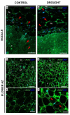
Galactans are accumulated in nodules and flower abscission zone (AZ) of yellow lupine following soil drought stress. Monoclonal antibody (LM5) was used to label (1-4)-β-D-galactans, RG–I side chain in the nodules (B) and AZ (D,F) of stressed plants (25% water holding capacity WHC), and also nodules (A) and AZ (C,E) of control lupines cultivated in optimal moisture (75% WHC). Insert is a magnified region of the fixation zone presented on (A). For immunofluorescence analyses, tissues were excised on the 48th day of cultivation. Green fluorescence indicates RG-I localization (red arrowheads), while a blue signal is emitted by nuclei (DAPI staining). AZ area is marked by white dotted lines on (C,D) and magnified on images (E,F). Abbreviations: F: fixation zone; E: endodermis; PA: nodule parenchyma; AZ: abscission zone; P: proximal region of AZ, D: distal region of AZ. Bars = 50 µm (A,B), 60 µm (C,D), 25 µm (E,F).
Figure 8
Figure 8
Drought affects the localization of arabinan in nodules and flower AZ of lupine. Monoclonal antibody LM6 served to detect (1-5)-α-L-arabinans RG-I side chains in the nodules (C,D) and flower AZs (G,H) of plants cultivated under drought conditions (25% water holding capacity, WHC). Nodules (A,B) and AZs (E,F) from lupines growing in optimal moisture (75% WHC) were the control. Material for analysis was collected from 48-day-old lupines. Green fluorescence indicates arabinan presence (red arrowheads), whereas DAPI blue signal is emitted by nuclei. Images (B,D) are magnifications of fixation zone areas from (A,C) (white squares). White dotted lines on (E,G) correspond to the AZ area, which is magnified precisely on (F,H), respectively. Additionally, a small square on E shows an enlarged region of vascular tissue from the pedicel. Abbreviations: F: fixation zone; E: endodermis; PA: nodule parenchyma; PE: periderm; VB: vascular bundle; AZ: abscission zone; P: proximal region of AZ, D: distal region of AZ. Bars = 50 µm (A,C), 15 µm (B,D), 100 µm (E,G), 20 µm (F,H).
Figure 9
Figure 9
Soil drought stress changes xylogalacturonan (XGA) localization in the nodules and flower abscission zone (AZ) of yellow lupine. Monoclonal antibody (LM8) was used to label XGA in the nodules (C,D) and AZ (G,H) of stressed plants (25% water holding capacity, WHC). Reactions were made also for control nodules (A,B) and AZ (E,F) from plants cultivated under optimal moisture (75% WHC). Material for analysis was harvested on the 48th day of lupine cultivation. Green fluorescence corresponds to XGA localization, while blue labeling is visible in the presence of nuclei after DAPI staining. (B,D) are magnifications of regions of fixation zones. Left insert on (A) shows the parenchyma region. Enlarged regions of AZ (marked by white dotted lines on (E,G) are presented on (F,H). Red arrowheads indicate a strong signal emitted from different compartments. Abbreviations: F: fixation zone; E: endodermis; PA: nodule parenchyma; PE: periderm; VB: vascular bundle; P: proximal region of AZ, D: distal region of AZ. Bars = 50 µm (A,C), 25 µm (B,D), 80 µm (E,G), 40 µm (F,H).
Figure 10
Figure 10
Nodule- and flower-AZ-specific distribution of xyloglucan (XyG) in yellow lupine subjected to drought. LM24 antibody was used to detect XyG in nodules (B) and AZ (D,F) collected on the 48th day of lupine cultivation under drought stress (25% water holding capacity, WHC). Control sections were prepared from nodules (A) and AZ (C,E) of plants growing in the soil of optimal moisture (75% WHC). Green and DAPI-blue fluorescence indicate XyG and nucleic acid staining, respectively. AZ region is limited by white dotted lines (C,D). Enlarged regions of AZ are on images (E,F). Red arrowheads indicate the areas characterized by strong fluorescence. Abbreviations: F: fixation zone; E: endodermis; PA: nodule parenchyma; PE: periderm; VB: vascular bundle; AZ: abscission zone; P: proximal region of AZ, D: distal region of AZ. Bars = 30 µm (A,B), 100 µm (C,D), 25 µm (E,F).
Figure 11
Figure 11
Pectin antigens are differentially present in the drought-treated nodules (A) and flower abscission zone (AZ) (C) of lupine. Immuno-dot-blot by using LM5, LM6, LM8, and LM24 antibodies was applied to detect galactans, arabinans, xylogalacturonans, and xyloglucans, respectively. The same primary antibodies as for the microscopy experiment were used. Tissues were collected on the 48th day of lupine cultivation under drought stress (25% water holding capacity, WHC) or plants growing in the soil of optimal moisture (75% WHC, control). The same amount of proteins (2.5 µg) was loaded for each dot. Dots were scanned and normalized densitometry values were quantified using ImageJ software for nodules (B) and AZ (D) tissues. A value of 100% was set for the controls. Data are presented as averages ± SE (n = 3). Significant differences in stressed tissues versus control for each antibody are * p < 0.05 and ** p < 0.01 (n = 3).
Figure 12
Figure 12
Water deficit stress affects arabinogalactan protein (AGP) localization in yellow lupine nodules. Immunofluorescence localization of different AGPs epitopes was made using JIM13 (A,B) and JIM8 (C,D) antibodies. Reactions were made for the control nodules (A,C) and those excised from plants growing under drought conditions (B,D). Tissues were collected from 48-day-old lupines. AGP presence corresponds to green fluorescence, while blue labeling indicates nuclei stained with DAPI. Abbreviations: F: fixation zone; E: endodermis cells; PA: nodule parenchyma; VB: vascular bundle. Bars = 75 µm.

Similar articles

Cited by

References

    1. Fernández-Pascual M., Pueyo J.J., Felipe M., Golvano M.P., Lucas M.M. Singular features of the Bradyrhizobium-Lupinus symbiosis. Soil Dyn. Plant. 2007;1:1–16.
    1. Kunert K.J., Vorster B.J., Fenta B.A., Kibido T., Dionisio G., Foyer C.H. Drought stress responses in soybean roots and nodules. Front. Plant Sci. 2016;7:1015. doi: 10.3389/fpls.2016.01015. - DOI - PMC - PubMed
    1. Frankowski K., Wilmowicz E., Kućko A., Zienkiewicz A., Zienkiewicz K., Kopcewicz J. Molecular cloning of the BLADE-ON-PETIOLE gene and expression analyses during nodule development in Lupinus luteus. J. Plant Physiol. 2015;179:35–39. doi: 10.1016/j.jplph.2015.01.019. - DOI - PubMed
    1. Wilmowicz E., Kućko A., Golińska P., Burchardt S., Przywieczerski T., Świdziński M., Brzozowska P., Kapuścińska D. Abscisic acid and ethylene in the control of nodule-specific response on drought in yellow lupine. Environ. Exp. Bot. 2020;169:103900. doi: 10.1016/j.envexpbot.2019.103900. - DOI
    1. Wilmowicz E., Kućko A., Burchardt S., Przywieczerski T. Molecular and hormonal aspects of drought-triggered flower shedding in yellow lupine. Int. J. Mol. Sci. 2019;20:3731. doi: 10.3390/ijms20153731. - DOI - PMC - PubMed

MeSH terms

LinkOut - more resources