Review
. 2022 Feb 2;23(3):1731.
doi: 10.3390/ijms23031731.

The Therapeutic Prospects of Targeting IL-1R1 for the Modulation of Neuroinflammation in Central Nervous System Disorders

Affiliations
Review

The Therapeutic Prospects of Targeting IL-1R1 for the Modulation of Neuroinflammation in Central Nervous System Disorders

João P Luís et al. Int J Mol Sci. .

Abstract

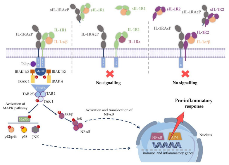
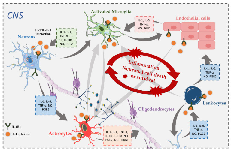
The interleukin-1 receptor type 1 (IL-1R1) holds pivotal roles in the immune system, as it is positioned at the "epicenter" of the inflammatory signaling networks. Increased levels of the cytokine IL-1 are a recognized feature of the immune response in the central nervous system (CNS) during injury and disease, i.e., neuroinflammation. Despite IL-1/IL-1R1 signaling within the CNS having been the subject of several studies, the roles of IL-1R1 in the CNS cellular milieu still cause controversy. Without much doubt, however, the persistent activation of the IL-1/IL-1R1 signaling pathway is intimately linked with the pathogenesis of a plethora of CNS disease states, ranging from Alzheimer's disease (AD), Parkinson's disease (PD), amyotrophic lateral sclerosis (ALS) and multiple sclerosis (MS), all the way to schizophrenia and prion diseases. Importantly, a growing body of evidence is showing that blocking IL-1R1 signaling via pharmacological or genetic means in different experimental models of said CNS diseases leads to reduced neuroinflammation and delayed disease progression. The aim of this paper is to review the recent progress in the study of the biological roles of IL-1R1, as well as to highlight key aspects that render IL-1R1 a promising target for the development of novel disease-modifying treatments for multiple CNS indications.

Keywords: CNS diseases; interleukin-1; interleukin-1 receptor type 1; neuroinflammation; therapeutic target.


Conflict of interest statement

The authors declare no conflict of interest. This review was conducted in the absence of any commercial or financial relationships that could be construed as a potential conflict of interest.

Figures

Figure 2
Figure 2
Schematic representation of IL-1 signaling. Upon binding of IL-1α/β to the extracellular domain of membrane-bound receptor IL-1R1, IL-1RAcP is recruited and signaling is initiated by the interaction of the intracellular TIR domains of the two polypeptide chains. When IL-1Ra binds IL-1R1, the IL-1RAcP is not recruited, thereby blocking signaling. Similarly, when IL-1β binds IL-1R2, no signaling occurs as IL-1R2 lacks a cytoplasmic TIR domain. IL-1 signaling may also be inhibited by soluble forms of the receptor, sIL-1R1 and sIL-1R2, lacking the transmembrane and intracellular regions of the native form, binding both IL-1α, IL-1β and sIL-1RAcP. Adapted from [39]. Figure abbreviations: IL-1, interleukin-1; IL-1R1, interleukin-1 receptor type 1; IL-1Ra, interleukin 1 receptor antagonist; IL-1RAcP, interleukin-1 receptor accessory protein; IL-1R2, interleukin-1 receptor type 2; TIR domain, the toll-interleukin-1 receptor homology domain; IRAK, interleukin-1 receptor-associated kinases; TRAF6, tumor necrosis factor receptor associated factor 6; TAK1, transforming growth factor-β-activated kinase 1; TAB, TAK1-binding proteins; MAPK, mitogen-activated protein kinases; p42/p44 MAPK, p42/p44 mitogen-activated protein kinases; p38 MAPK, p38 mitogen-activated protein kinases; JNK., c-Jun N-terminal kinase; IKKβ, I kappa B kinase β; NF-κB, nuclear factor-kappa B; AP-1, activator protein 1.
Figure 7
Figure 7
Comparison of human, rat and mouse interleukin-1 receptor type 1 (IL-1R1) protein sequences and structure. Upper panel: (A) Structural alignment of predicted rat (rIL-1R1, colored in green) and mouse (mIL-1R1, colored in blue) structures (full-length) retrieved from the AlphaFold Protein Structure Database [201] with the crystallographic structure of the extracellular domain of human IL-1R1 (hIL-1R1, colored in yellow) (PDB entry 4GAF). The protein structures were superimposed using the MatchMaker tool of the UCSF Chimera 1.15 software program [202]. The D1-D2 surface representation enclosed by the black circle is magnified, along with the conserved and non-conserved residues for the largest pocket (PO_0) predicted by DogSiteScorer. Lower panel: Protein sequence alignment for residues (one-letter code) comprising three “druggable” binding pockets—PO_0_0 (B), PO_0_1 (C) and PO_0_6 (D) predicted in the crystal structure of hIL-1R1 by DoGSiteScorer [200]. The sequence alignment was produced using the Clustal Omega Server [192]. Conservation across orthologs is color-coded as green, for full conservation across the three orthologs (a whole green table line represents 100% conservation); yellow represents amino acid differences with similar side-chain properties; red represents amino acid differences with distinct side-chain properties.
Figure 8
Figure 8
Main strategies targeting IL-1 signaling. Therapeutic approaches targeting IL-1R1 include a recombinant form of IL-1Ra (anakinra), a chimeric IL-1Ra-IL-1β protein (isunakinra), a fully human monoclonal antibody (MEDI-896) and peptides AF10847 and rytvela. Rilonacept stands as an engineered dimeric fusion protein consisting of the ECDs of the human IL-1R1 and IL-1RAcP linked to the Fc portion of human immunoglobulin G1 (IgG1), acting as a decoy receptor, binding IL-1α, IL-1β and IL-1Ra. The monoclonal antibodies (mAb) antagonizing IL-1β are canakinumab, gevokizumab and LYS2189102, whilst bermekimab works by blocking IL-1α. The small molecules inzomelid and glyburide directly inhibit NLRP3 inflammasome activation, while belnacasan specifically inhibits caspase-1 and consequently the maturation of pro-IL-1β into the respective active form. The figure was prepared using BioRender. Adapted from [243]. Figure abbreviations: IL-1, interleukin-1; IL-1R1, interleukin-1 receptor type 1; IL-1Ra, interleukin 1 receptor antagonist; IL-1RAcp, interleukin-1 receptor accessory protein; TIR domain, the toll-interleukin-1 receptor homology domain; NLRP3, NLR family pyrin-domain-containing 3.
Figure 1
Figure 1
An illustration of the IL-1β/IL-1R1/IL-1RAcP ternary complex. Binding of the IL-1 cytokine (colored in pink) to the membrane-bound IL-1R1-ECD (colored in green) recruits transmembrane IL-1RAcP (colored in lime), initiating intracellular signaling via the TIR domains (colored in blue). Both IL-1R1 and IL-1RAcP can participate in the negative regulation of IL-1 signaling when cleaved to their soluble ECD forms. The lipid bilayer (cell membrane) is represented in yellow and transmembrane domains are colored in red. The image was generated using imported VMD visualization states [33] uploaded in the Blender software [34]. Figure abbreviations: IL-1, interleukin-1; IL-1R1, interleukin-1 receptor type 1; IL-1R1-ECD, extracellular domain of IL-1R1; IL-1RAcp, interleukin-1 receptor accessory protein; TIR domain, the toll-interleukin-1 receptor homology domain.
Figure 3
Figure 3
CNS cellular communication of IL-1 signaling in neuronal injury. In the normal brain, the expression of IL-1 is low, although acute CNS injury causes a sharp increase in IL-1 production. Different CNS cell types express IL-1R1, thereby enabling them to respond to IL-1β in an autocrine manner, as well as in a paracrine manner, after neuronal injury. Upon IL-1/IL-1R1 interactions, these different cells can produce a wide range of pro-inflammatory and immune-regulatory mediators that contribute to neuronal death or survival through different signal transduction pathways. The figure was prepared using Servier Medical Art. Figure abbreviations: CNS, central nervous system; IL-1, interleukin-1; IL-6, interleukin-6; IL-10, interleukin-10; IL-1Ra, interleukin-1 receptor antagonist; IL-1R1, interleukin-1 receptor type 1; TNF-α, tumor necrosis factor-α; NO, nitric oxide; PGE2, prostaglandin E2; NGF, nerve growth factor; BDNF, brain-derived neurotrophic factor.
Figure 4
Figure 4
(A) Molecular structure the IL-1R1 ectodomain. The coordinates were retrieved from the PDB (PDB entry 4GAF). The Ig-like domains are labeled as D1 (orange), D2 (green) and D3 (blue), and the 6 amino acid flexible linker located between D2 and D3 is represented in yellow. (B) Alignment of IL-1R1, IL-1R2, IL-18Rα, ST2 and IL-36R ectodomains. The β-strands were assigned using the DSSP algorithm [191], and are shown for the IL-1R1 sequence (a1-g1 located in D1, a2-g2 located in D2 and a3-g3 located in D3, represented as blue arrows). Brown circles show the conserved cysteine residues, and the connectivity between the disulfide bonds in the IL-1R1 structure is represented by brown dashed lines. IL-1R1 shares 25.5%, 18.4%, 19.6% and 29.3% sequence identity with IL-1R2, IL-18Rα, ST2 and IL-36R, respectively. The alignments were generated using the Clustal Omega Server [192]. Adapted from [193]. Figure abbreviations: IL-1R1, interleukin-1 receptor type 1; IL-1R2, interleukin-1 receptor type 2; IL-18Rα, interleukin-18 receptor-α; ST2, defined as interleukin-33 receptor; IL-36R, interleukin-36 receptor.
Figure 5
Figure 5
Structures of the binary IL-1R1-ECD/IL-1β (PDB entry 1ITB) and IL-1R1-ECD/IL-1Ra (PDB entry 1IRA) complexes. In both panels, IL-1R1-ECD is depicted in the surface representation (gray), while IL-1β (magenta) and IL-1Ra (lime) are shown in cartoon representations. The two binding regions are shown as spheres and colored in blue (site A) and red (site B), respectively. Left panel: IL-1β interacts with the two sites on the receptor surface (IL-1R1-ECD residues involved are presented in blue and red). Right panel: IL-1Ra interacts mostly with site A (IL-1R1-ECD residues involved are presented in blue and red). Figure abbreviations: IL-1β, interleukin-1β; IL-1R1-ECD, extracellular domain of IL-1R1; IL-1Ra, interleukin 1 receptor antagonist.
Figure 6
Figure 6
Binding pocket prediction on the IL-1R1 ectodomain surface (PDB entry 4GAF), using the DoGSiteScorer tool. (A) Ten binding pockets are shown: PO_0 (red); PO_1 (blue); PO_2 (purple); PO_3 (mauve); PO_4 (dark grey); PO_5 (yellow); PO_6 (green); PO_7 (cyan); PO_8 (violet); PO_9 (brown). (B) The sub-pockets comprising PO_0 are illustrated in detail, with respective volume and druggability scores. Seven sub-pockets are shown: PO_0_0 (red); PO_0_1 (yellow); PO_0_2 (lime); PO_0_3 (blue); PO_0_4 (green); PO_0_5 (purple); PO_0_6 (cyan).

Similar articles

Cited by

References

    1. Fantuzzi G. Body Messages. Harvard University Press; Cambridge, MA, USA: 2016. 3. Evolution of message discovery: The Interleukin-1 family; pp. 29–73.
    1. Dinarello C.A. Overview of the IL-1 family in innate inflammation and acquired immunity. Immunol. Rev. 2018;281:8–27. doi: 10.1111/imr.12621. - DOI - PMC - PubMed
    1. Sims J.E., Smith D.E. The IL-1 family: Regulators of immunity. Nat. Rev. Immunol. 2010;10:89–102. doi: 10.1038/nri2691. - DOI - PubMed
    1. Mantovani A., Dinarello C.A., Molgora M., Garlanda C. Interleukin-1 and related cytokines in the regulation of inflammation and immunity. Immunity. 2019;50:778–795. doi: 10.1016/j.immuni.2019.03.012. - DOI - PMC - PubMed
    1. Elzinga B.M., Twomey C., Powell J.C., Harte F., McCarthy J.V. Interleukin-1 receptor type 1 is a substrate for γ-secretase- dependent regulated intramembrane proteolysis. J. Biol. Chem. 2009;284:1394–1409. doi: 10.1074/jbc.M803108200. - DOI - PubMed

MeSH terms

Substances

LinkOut - more resources