. 2022 Feb 3;23(3):1740.
doi: 10.3390/ijms23031740.

Conversion of Human Fibroblasts into Induced Neural Stem Cells by Small Molecules

Affiliations

Conversion of Human Fibroblasts into Induced Neural Stem Cells by Small Molecules

Donghui Liu et al. Int J Mol Sci. .

Abstract

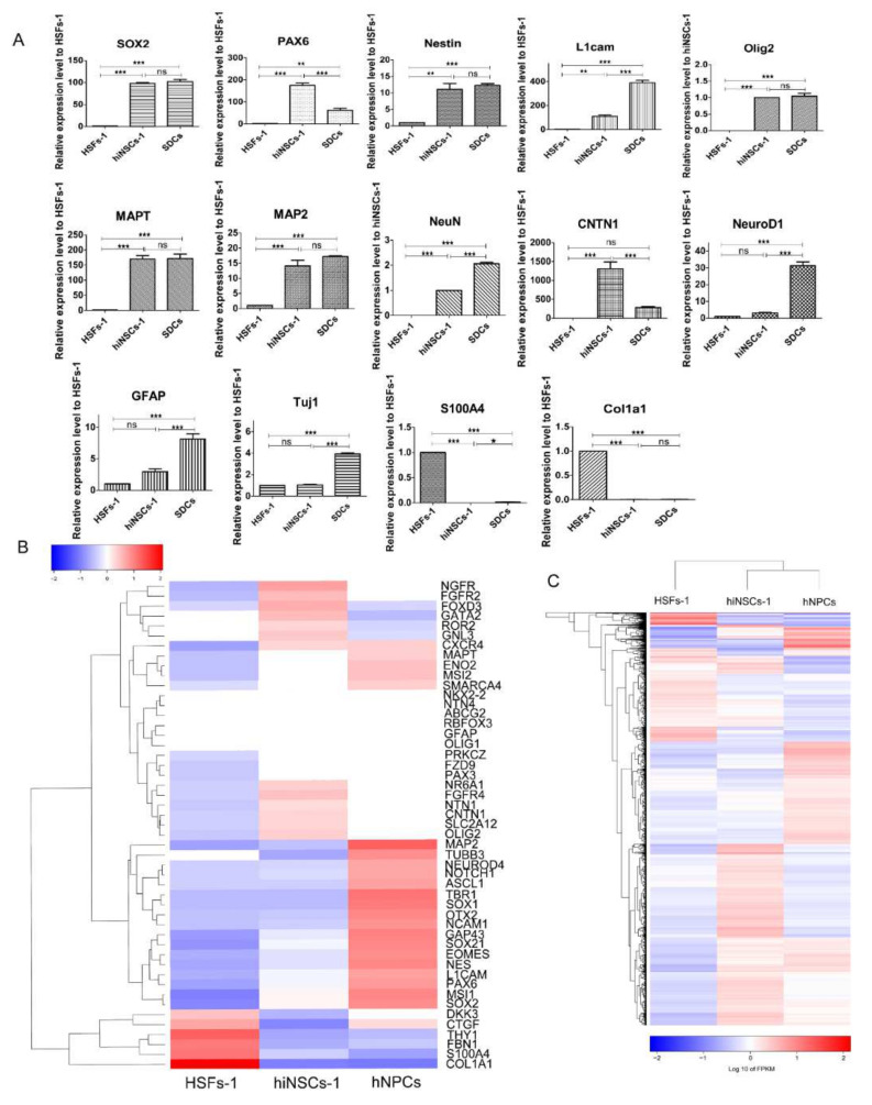
Induced neural stem cells (iNSCs) reprogrammed from somatic cells hold great potentials for drug discovery, disease modelling and the treatment of neurological diseases. Although studies have shown that human somatic cells can be converted into iNSCs by introducing transcription factors, these iNSCs are unlikely to be used for clinical application due to the safety concern of using exogenous genes and viral transduction vectors. Here, we report the successful conversion of human fibroblasts into iNSCs using a cocktail of small molecules. Furthermore, our results demonstrate that these human iNSCs (hiNSCs) have similar gene expression profiles to bona fide NSCs, can proliferate, and are capable of differentiating into glial cells and functional neurons. This study collectively describes a novel approach based on small molecules to produce hiNSCs from human fibroblasts, which may be useful for both research and therapeutic purposes.

Keywords: human fibroblasts; induced neuron stem cells; small molecules.


Conflict of interest statement

The authors declare no conflict of interest.

Figures

Figure 1
Figure 1
Generation of human iNSCs from fibroblasts by small molecules. (A) Scheme of induction procedure. See Methods and Supplemental Experimental Procedures for detailed protocol. (B) Phase contrast images of cells during induction time, scale bars are 200 μm. (C) Morphology of hiNSCs-1 in monolayer culture and suspension culture, scale bars are 200 μm. (D) hiNSCs-1 stained for neural stem cell makers, SOX2, Nestin, PAX6, and Ncam1, scale bars are 40 μm. (E) The percentage of Sox2+ and Pax6+ cells in total cells at the time of quantification (means ± SEM, cell counting was from 3 triplicate samples). (F) Representative immunoblots for SOX2, Nestin (upper band), and Ncam1 in different cells, B-actin (beta-actin) was used as a loading control.
Figure 2
Figure 2
hiNSCs-1 express multiple up-regulated NSCs makers and down-regulated fibroblast-specific genes. (A) qRT-PCR results of the expression of some neural-lineage makers and fibroblast-specific genes in HSFs-1, hiNSCs-1 and spontaneous differentiated cells (SDCs). * p < 0.05, ** p < 0.01, *** p < 0.001, ns = not significant. (B) Heatmap of the expression of neural lineage makers and fibroblast-specific genes in HSFs-1, hiNSCs-1, and hNPCs using RNA-sequencing. (C) Heatmap and hierarchical clustering of genes with significance in HSFs-1, hiNSCs-1, and hNPCs using RNA-sequencing.
Figure 3
Figure 3
Differentiation of hiNSCs-1 in vitro. hiNSCs-1 differentiated into neurons (marked by Tuj1, Map2, and NeuN, AC), astrocytes (marked by GFAP, D), and Oligodendrocytes (marked by O4 and Olig2, E) after 2–3 weeks culture in NDM, ADM, and ODM, respectively. Scale bars are 40 μm.
Figure 4
Figure 4
Electrophysiological properties of hiNSCs-1 cells. A, B, and C. Examples of membrane currents recorded in hiNSCs-1 cells in response to 50 ms (A,B) or 300 ms (C) voltage steps ranging from −60 mV to 60 mV, applied in 10 mV increments from a holding potential of −75 mV. (D) Average current-voltage plots of the Na+ (solid symbols) and K+ (clear symbols) currents constructed using recordings similar to those shown in panels (AC) (n = 22). (E) Scatter plots of the absolute peak amplitudes of Na+ and K+ currents shown in panel (D). (F) Membrane currents recorded in response to 100 ms voltage ramps ranging from −120 to 120 mV before and after replacement of 145 mM NaCl in the bath solution with 100 mM BaCl2. (G) Action potentials recorded in a typical hiNSCs-1 cell in response to depolarizing current injections in the current clamp mode G. Action potentials recorded in a typical hiNSCs-1 cell in response to depolarizing current injections in the current clamp mode. Data were from 4 cell preparations.
Figure 5
Figure 5
Generation of hiNSCs from another fibroblast cell line. (A) Scheme of induction procedure of hiNSCs-2. (B) Phase contrast images of cells during induction time, scale bars are 200 μm. (C) hiNSCs-1 stained for neural stem cell makers, SOX2, Nestin, PAX6, and Ncam1, scale bars are 40 μm. (D) The percentage of Sox2+ and Pax6+ cells in total cells at the time of quantification (means ± SEM, cell counting was from 3 triplicate samples) (E) Representative immunoblots for SOX2, Nestin (upper band), and Ncam1 in different cells, B-actin (beta-actin) was used as a loading control. (F) Differentiation of hiNSCs-2 in vitro, differentiated cells were immuno-positive for Tuj1, GFAP and O4 after 2–3 weeks culture in NDM, ADM, and ODM, respectively. (G) Electrophysiological properties of hiNSCs-2 derived neurons. i. Representative membrane currents recorded in hiNSCs-2 cell in response to the same voltage protocol as in Figure 4A. ii. Average current–voltage plots of the Na+ (solid symbols) and K+ (clear symbols) currents constructed using recordings similar to those shown in panels A (n = 9). iii. Scatter plots of the absolute peak amplitudes of Na+ and K+ currents shown in panel ii. Data were from 2 cell preparations.
Figure 5
Figure 5
Generation of hiNSCs from another fibroblast cell line. (A) Scheme of induction procedure of hiNSCs-2. (B) Phase contrast images of cells during induction time, scale bars are 200 μm. (C) hiNSCs-1 stained for neural stem cell makers, SOX2, Nestin, PAX6, and Ncam1, scale bars are 40 μm. (D) The percentage of Sox2+ and Pax6+ cells in total cells at the time of quantification (means ± SEM, cell counting was from 3 triplicate samples) (E) Representative immunoblots for SOX2, Nestin (upper band), and Ncam1 in different cells, B-actin (beta-actin) was used as a loading control. (F) Differentiation of hiNSCs-2 in vitro, differentiated cells were immuno-positive for Tuj1, GFAP and O4 after 2–3 weeks culture in NDM, ADM, and ODM, respectively. (G) Electrophysiological properties of hiNSCs-2 derived neurons. i. Representative membrane currents recorded in hiNSCs-2 cell in response to the same voltage protocol as in Figure 4A. ii. Average current–voltage plots of the Na+ (solid symbols) and K+ (clear symbols) currents constructed using recordings similar to those shown in panels A (n = 9). iii. Scatter plots of the absolute peak amplitudes of Na+ and K+ currents shown in panel ii. Data were from 2 cell preparations.

Similar articles

Cited by

References

    1. McGinley L.M., Kashlan O.N., Bruno E.S., Chen K.S., Hayes J.M., Kashlan S.R., Raykin J., Johe K., Murphy G.G., Feldman E.L. Human neural stem cell transplantation improves cognition in a murine model of alzheimer’s disease. Sci. Rep. 2018;8:14776. doi: 10.1038/s41598-018-33017-6. - DOI - PMC - PubMed
    1. Yasuhara T., Kameda M., Sasaki T., Tajiri N., Date I. Cell therapy for parkinson’s disease. Cell Transpl. 2017;26:1551–1559. doi: 10.1177/0963689717735411. - DOI - PMC - PubMed
    1. Kokaia Z., Darsalia V. Human neural stem cells for ischemic stroke treatment. Results Probl. Cell Differ. 2018;66:249–263. - PubMed
    1. Takahashi K., Yamanaka S. Induction of pluripotent stem cells from mouse embryonic and adult fibroblast cultures by defined factors. Cell. 2006;126:663–676. doi: 10.1016/j.cell.2006.07.024. - DOI - PubMed
    1. Li W., Wei W., Zhu S., Zhu J., Shi Y., Lin T., Hao E., Hayek A., Deng H., Ding S. Generation of rat and human induced pluripotent stem cells by combining genetic reprogramming and chemical inhibitors. Cell Stem Cell. 2009;4:16–19. doi: 10.1016/j.stem.2008.11.014. - DOI - PubMed

MeSH terms

LinkOut - more resources