. 2022 Feb 3;23(3):1744.
doi: 10.3390/ijms23031744.

Immature Vascular Smooth Muscle Cells in Healthy Murine Arteries and Atherosclerotic Plaques: Localization and Activity

Affiliations

Immature Vascular Smooth Muscle Cells in Healthy Murine Arteries and Atherosclerotic Plaques: Localization and Activity

Alexander Balatskiy et al. Int J Mol Sci. .

Abstract

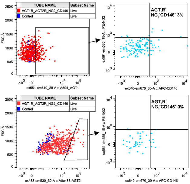
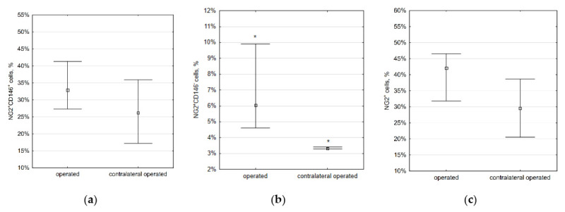
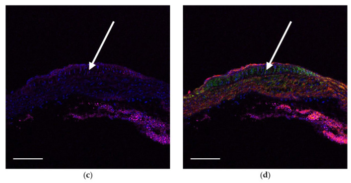
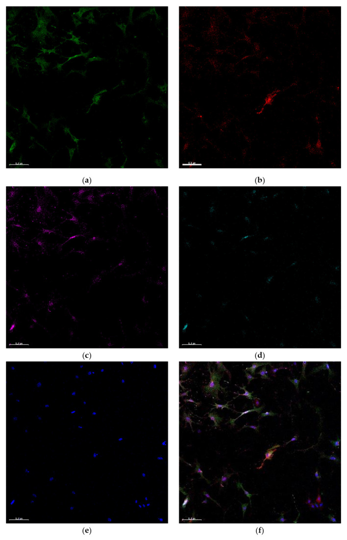
The local development of atherosclerotic lesions may, at least partly, be associated with the specific cellular composition of atherosclerosis-prone regions. Previously, it was demonstrated that a small population of immature vascular smooth muscle cells (VSMCs) expressing both CD146 and neuron-glial antigen 2 is postnatally sustained in atherosclerosis-prone sites. We supposed that these cells may be involved in atherogenesis and can continuously respond to angiotensin II, which is an atherogenic factor. Using immunohistochemistry, flow cytometry, wound migration assay xCELLigence system, and calcium imaging, we studied the functional activities of immature VSMCs in vitro and in vivo. According to our data, these cells do not express nestin, CD105, and the leptin receptor. They are localized in atherosclerosis-prone regions, and their number increases with age, from 5.7% to 23%. Immature VSMCs do not migrate to low shear stress areas and atherosclerotic lesions. They also do not have any unique response to angiotensin II. Thus, despite the localization of immature VSMCs and the presence of the link between their number and age, our study did not support the hypothesis that immature VSMCs are directly involved in the formation of atherosclerotic lesions. Additional lineage tracing studies can clarify the fate of these cells during atherogenesis.

Keywords: CD146; NG2; angiotensin II; angiotensin II receptor type 2; atherosclerosis; immature smooth muscle cells; pericytes.


Conflict of interest statement

The authors declare no conflict of interest.

Figures

Figure A1
Figure A1
The aortic arch and the innominate artery in an ApoE knockout mouse fed with chow diet: (a) NG2, (b) CD146, (c) lipids, and (d) merged. Nuclei are stained with DAPI (blue). Scale bars 100 µm.
Figure A2
Figure A2
Co-expressing of NG2, CD146, AT1R, and AT2R in VSMCs. The subpopulation of AT1R positive cells contains immature NG2+CD146+ VSMCs, but immature VSMCs are absent in AT2R-positive subpopulation.
Figure A3
Figure A3
The gating strategy for calculating the percentage of NG2+CD146+ cells and for cell sorting.
Figure 1
Figure 1
Representative images showing the bifurcation of the aorta and the innominate artery. No pericytic markers were found in atherosclerosis-prone sites (bifurcations of large arteries), except for NG2, CD146, and CD140b (which is a common marker for cells of mesenchymal origin): (a,b) staining with antibodies to CD140b (red) and to leptin receptor (green, arrow); (c) staining with antibodies to CD105 (red) and to NG2 (green); (d) GFP (green) and staining with antibodies to GFP (magenta) in nestin-GFP mice. In all images, nuclei are blue. Scale bars (a) 200 µm, (b,d) 50 µm, and (c) 20 µm.
Figure 2
Figure 2
Representative images showing the bifurcations of the aortic arch and its major branches: (a) 10-week-old mice, (b) 14-week-old mice, and (c) 19-week-old mice. In young animals, a small number of NG2+CD146+ are located in bifurcations and other areas with low shear stress. In aged animals, the number of these cells is larger throughout all major arteries. The percentage of double-positive NG2+CD146+ cells in the vascular wall is shown on (d). Staining with antibodies to CD146 (red) and to NG2 (green). Nuclei are stained with DAPI (blue). Scale bars 50 µm.
Figure 3
Figure 3
The percentage of different VSMCs changes with age: (a) NG2+CD146+ cells, * p = 0.036; (b) NG2+CD146 cells, * p = 0.012; (c) all NG2+ cells. This means that NG2+CD146 cells can de-differentiate into immature NG2+CD146+ cells with age. Squares show medians; whiskers show upper and lower quartiles.
Figure 4
Figure 4
The percentage of wound closure by different subpopulations indicating the migration rate. Immature NG2+CD146+ VSMCs migrate better than other vascular cells, but the difference with mature NG2+CD146 cells is not significant. The difference between columns marked with the same symbols is significant (p < 0.05).
Figure 5
Figure 5
The proliferation rate of different vascular cells: (a) delta cell index changing over time; (b) delta cell index doubling time. Immature NG2+CD146+ VSMCs migrate better than other vascular cells, but the difference with mature NG2+CD146 cells is not significant. The difference between columns marked with the same symbols is significant (p < 0.05). Squares show medians; whiskers show upper and lower quartiles.
Figure 6
Figure 6
The percentage of different VSMCs cells in common carotid arteries—three months after partial carotid ligation. Immature VSMCs do not migrate into low shear stress areas and do not proliferate there: (a) NG2+CD146+ cells: operated, 55.9% (43.7%, 69.2%); contralateral operated, 50.7% (35.5%, 61.0%); sham operated, 69.5% (56.9%, 69.9%); (b) NG2+CD146 cells: operated, 4.6% (1.5%, 7.3%); contralateral operated, 4.0% (3.3%, 5.0%); sham operated, 2.2% (1.5%, 2.5%); (c) all NG2+ cells: operated, 59.7% (47.9%, 73.4%); contralateral operated, 54.0% (39.5%, 67.8%); sham operated, 70.1% (67.4%, 72.1%). Squares show medians; whiskers show upper and lower quartiles. Data are given as median (interquartile range).
Figure 7
Figure 7
The percentage of NG2+ cells in common carotid arteries, two weeks after partial carotid ligation. The number of mature NG2+CD146 (but not immature NG2+CD146+) VSMCs slightly increases in low shear stress areas: (a) NG2+CD146+ cells: operated, 32.9% (27.3%, 41.3%); contralateral operated, 26.2% (17.2%, 35.9%); (b) NG2+CD146 cells: operated, 6.1% (4.6%, 9.9%); contralateral operated, 3.3% (3.3%, 3.4%); (c) all NG2+ cells: operated, 42.0% (31.8%, 46.5%); contralateral operated, 29.5% (20.5%, 38.7%). Squares show medians; whiskers show upper and lower quartiles. Data are given as median (interquartile range).
Figure 8
Figure 8
Fatty streak (arrowed) in the lesser curvature of the aortic arch in an 18-week-old ApoE knockout mouse fed with a Western-type diet for two weeks. Immature NG2+CD146+ VSMCs (green and red) are located at the streak’s borders, but inside the streak, only mature NG2+CD146 (green only) VSMCs are present: (a) NG2 (green), (b) CD146 (red), (c) lipids (magenta), and (d) merged. Nuclei are stained with DAPI (blue). Scale bars 100 µm.
Figure 8
Figure 8
Fatty streak (arrowed) in the lesser curvature of the aortic arch in an 18-week-old ApoE knockout mouse fed with a Western-type diet for two weeks. Immature NG2+CD146+ VSMCs (green and red) are located at the streak’s borders, but inside the streak, only mature NG2+CD146 (green only) VSMCs are present: (a) NG2 (green), (b) CD146 (red), (c) lipids (magenta), and (d) merged. Nuclei are stained with DAPI (blue). Scale bars 100 µm.
Figure 9
Figure 9
Atherosclerotic plaque in the aortic root in a 20-week-old ApoE knockout mouse fed with a Western-type diet for four weeks. Immature NG2+CD146+ VSMCs (green and red) are located at plaque’s borders, but inside the plaque, only mature NG2+CD146 VSMCs (green only) are present: (a) NG2 (green), (b) CD146 (red), (c) lipids (magenta), and (d) merged. Nuclei are stained with DAPI (blue). Scale bars 100 µm.
Figure 9
Figure 9
Atherosclerotic plaque in the aortic root in a 20-week-old ApoE knockout mouse fed with a Western-type diet for four weeks. Immature NG2+CD146+ VSMCs (green and red) are located at plaque’s borders, but inside the plaque, only mature NG2+CD146 VSMCs (green only) are present: (a) NG2 (green), (b) CD146 (red), (c) lipids (magenta), and (d) merged. Nuclei are stained with DAPI (blue). Scale bars 100 µm.
Figure 10
Figure 10
(a) The bifurcation of the aortic arch and the innominate artery. In such sites, immature NG2+CD146+ cells express only AT1R but not AT2R. Square indicates the area shown in (bf): VSMCs are stained with antibodies to (b) NG2 (green), (c) CD146 (red), (d) AT1R (magenta), and (e) AT2R (cyan); (f) merged image. On (e), AT2R staining is not observed. Visible fluorescence is the autofluorescence of collagen fibers induced by a 488 nm laser. Nuclei are stained with DAPI (blue). Scalebars (a) 50 µm, (bf) 10 µm.
Figure 10
Figure 10
(a) The bifurcation of the aortic arch and the innominate artery. In such sites, immature NG2+CD146+ cells express only AT1R but not AT2R. Square indicates the area shown in (bf): VSMCs are stained with antibodies to (b) NG2 (green), (c) CD146 (red), (d) AT1R (magenta), and (e) AT2R (cyan); (f) merged image. On (e), AT2R staining is not observed. Visible fluorescence is the autofluorescence of collagen fibers induced by a 488 nm laser. Nuclei are stained with DAPI (blue). Scalebars (a) 50 µm, (bf) 10 µm.
Figure 11
Figure 11
Cultured VSMCs express AT2R only in nuclei, and this expression is not limited to NG2+CD146+ cells: (a) NG2 (green), (b) CD146 (red), (c) AT1R (magenta), (d) AT2R (cyan), (e) nuclei (DAPI, blue), and (f) merged. Scalebars 100 µm.
Figure 12
Figure 12
The response of sorted VSMCs to the applications of angiotensin II. Both immature and mature VSMCs have no uniform or population-specific response pattern: (a) NG2+CD146+ cells and (b) NG2+CD146 cells.
Figure 13
Figure 13
The showing all experiments and findings.

Similar articles

References

    1. Steffensen L.B., Mortensen M.B., Kjolby M., Hagensen M.K., Oxvig C., Bentzon J.F. Disturbed Laminar Blood Flow Vastly Augments Lipoprotein Retention in the Artery Wall: A Key Mechanism Distinguishing Susceptible from Resistant Sites. Arterioscler. Thromb. Vasc. Biol. 2015;35:1928–1935. doi: 10.1161/ATVBAHA.115.305874. - DOI - PubMed
    1. Andrés V., Dorado B. Methods in Mouse Atherosclerosis. Volume 1339 Springer; New York, NY, USA: 2015.
    1. Langhaas T. Beiträge zur normalen und pathologischen Anatomie der Arterien. Arch. Pathol. Anat. Physiol. Klin. Med. 1866;36:187–226. doi: 10.1007/BF01927642. - DOI
    1. Orekhov A.N., Karpova I.I., Tertov V.V., Rudchenko S.A., Andreeva E.R., Krushinsky A.V., Smirnov V.N. Cellular composition of atherosclerotic and uninvolved human aortic subendothelial intima. Light-microscopic study of dissociated aortic cells. Am. J. Pathol. 1984;115:17–24. - PMC - PubMed
    1. Andreeva E.R., Pugach I.M., Gordon D., Orekhov A.N. Continuous subendothelial network formed by pericyte-like cells in human vascular bed. Tissue Cell. 1998;30:127–135. doi: 10.1016/S0040-8166(98)80014-1. - DOI - PubMed

Substances

LinkOut - more resources