. 2022 Feb 3;23(3):1747.
doi: 10.3390/ijms23031747.

Gene Transfer of Skeletal Muscle-Type Myosin Light Chain Kinase via Adeno-Associated Virus 6 Improves Muscle Functions in an Amyotrophic Lateral Sclerosis Mouse Model

Affiliations

Gene Transfer of Skeletal Muscle-Type Myosin Light Chain Kinase via Adeno-Associated Virus 6 Improves Muscle Functions in an Amyotrophic Lateral Sclerosis Mouse Model

Ryohei Oya et al. Int J Mol Sci. .

Abstract

Amyotrophic lateral sclerosis (ALS) is a neurodegenerative disease that shows progressive muscle weakness. A few treatments exist including symptomatic therapies, which can prolong survival or reduce a symptom; however, no fundamental therapies have been found. As a therapeutic strategy, enhancing muscle force is important for patients' quality of life. In this study, we focused on skeletal muscle-specific myosin regulatory light chain kinase (skMLCK), which potentially enhances muscle contraction, as overexpression of skMLCK was thought to improve muscle function. The adeno-associated virus serotype 6 encoding skMLCK (AAV6/skMLCK) and eGFP (control) was produced and injected intramuscularly into the lower limbs of SOD1G37R mice, which are a familial ALS model. AAV6/skMLCK showed the successful expression of skMLCK in the muscle tissues. Although the control did not affect the muscle force in both of the WT and SOD1G37R mice, AAV6/skMLCK enhanced the twitch force of SOD1G37R mice and the tetanic force of WT and SOD1G37R mice. These results indicate that overexpression of skMLCK can enhance the tetanic force of healthy muscle as well as rescue weakened muscle function. In conclusion, the gene transfer of skMLCK has the potential to be a new therapy for ALS as well as for other neuromuscular diseases.

Keywords: adeno-associated virus 6; amyotrophic lateral sclerosis; gene therapy; muscle function; myosin light chain kinase.


Conflict of interest statement

The authors have no conflict of interest to declare.

Figures

Figure 1
Figure 1
The observed expression of target protein in 293T cells by AAV vectors. (A) The scheme of the produced viral vectors. The left panel shows the structure of the skMLCK vector, which contains the eGFP, T2A peptide, FLAG tag, and skMLCK gene downstream of the cytomegalovirus (CMV) promoter. The right panel shows the structure of the control vector, which contains only the eGFP gene downstream of the CMV promoter. (B) Immunofluorescence staining against actin (phalloidin), eGFP, and FLAG in 293T cells infected by the skMLCK vector (0, 5 × 10⁸, 5 × 10⁹, and 5 × 10¹⁰ vg/mL); scale bar: 50 μm. (C) Immunofluorescence staining against actin (phalloidin) and eGFP in 293T cells infected by the control vector (0, 5 × 10⁹ and 5 × 10¹⁰ vg/mL); scale bar: 50 μm. (D) Immunoblot analysis of the skMLCK proteins using anti-FLAG antibodies in 293T cells infected by the skMLCK vector (0, 5 × 10⁸, 5 × 10⁹, and 5 × 10¹⁰ vg/mL). α-tubulin was used as a loading control. (E) Immunoblot analysis of the eGFP proteins using anti-eGFP antibodies in 293T cells infected by the control vector (0, 5 × 10⁹ and 5 × 10¹⁰ vg/mL). α-tubulin was used as a loading control.
Figure 2
Figure 2
The gene transfer to wild-type mice using the control vector. (A) The scheme of the injection of the control vector. The control vector (1 × 10¹¹ vg/mouse) was injected into the right lower limb, and saline was injected into the left lower limb at the age of 8 weeks. A series of analysis was performed after 6 weeks. (B) Immunoblot analysis of the eGFP proteins in the extensor digitorum longus (EDL) (upper panel) and tibialis anterior (TA) muscles (lower panel). R (right side) is the AAV-injected side, and L (left side) is the sham side. α-tubulin was used as a loading control. (C) Immunofluorescence staining against actin (phalloidin) and eGFP in the TA muscle; scale bar: 50 μm. (D) Hematoxylin and eosin (HE) stain of the TA muscle; scale bar: 50 μm. (E) The twitch force of the isolated EDL muscle of the sham and the AAV sides. The data shown in the same color were from the same mouse. (F) The normalized twitch force calculated from E. No significant difference was observed between the sham (62.1 ± 4.38 mN/mm2) and the AAV sides (57.6 ± 3.40 mN/mm2). (G) The tetanic force of isolated EDL muscle of the sham and the AAV sides. The data shown in the same color were from the same mouse. (H) The normalized tetanic force calculated from G. No significant difference was observed between the sham (205.6 ± 31.7 mN/mm2) and the AAV sides (204.9 ± 14.7 mN/mm2).
Figure 3
Figure 3
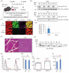
The gene transfer to wild-type mice using the skMLCK vector. (A) The scheme of the injection of the skMLCK vector. The control vector (1 × 10¹¹ vg/mouse) was injected into the right lower limb, and saline was injected into the left lower limb at the age of 8 weeks. A series of analysis was performed after 6 weeks. (B) Immunoblot analysis of the FLAG peptide in the extensor digitorum longus (EDL) (upper panel) and tibialis anterior (TA) muscles (lower panel). R (right side) is the AAV-injected side, and L (left side) is the sham side. α-tubulin was used as a loading control. (C) Immunofluorescence staining against actin (phalloidin), eGFP, and FLAG in the TA muscle; scale bar: 50 μm. (D) Gene expression of human (Hs) MYLK2 gene in the TA muscle. Gene expression is demonstrated as the ratio of HsMYLK2 to mouse (Mm) MYLK2. (E) Hematoxylin and eosin (HE) stain of the TA muscle; scale bar: 50 μm. (F) The Phos-tag PAGE of EDL muscle. The upper band showed the phosphorylated MYLPF protein (pMYLPF, white arrow) and the lower band showed the non-phosphorylated MYLPF protein (black arrow). R (right side) is the AAV-injected side, and L (left side) is the sham side. (G) The twitch force of the isolated EDL muscle of the sham and the AAV sides. The data shown in the same color were from the same mouse. (H) The normalized twitch force calculated from G. No significant difference was observed between the sham (57.4 ± 7.09 mN/mm2) and the AAV sides (56.3 ± 5.65 mN/mm2). (I) The normalized tetanic force of the isolated EDL muscle of the sham and the AAV sides. The data shown in the same color were from the same mouse. (J) The normalized tetanic force calculated from I. The normalized tetanic force of the AAV sides (260.6 ± 32.5 mN/mm2) was significantly greater than that of the sham sides (202.1 ± 24.2 mN/mm2, p = 0.028).
Figure 4
Figure 4
The gene transfer to SOD1 mice using the control vector. (A) The scheme of the injection of the control vector. The control vector (1 × 10¹¹ vg/mouse) was injected into the right lower limb, and saline was injected into the left lower limb at the age of 8 weeks. A series of analysis was performed after 6 weeks. (B) Immunoblot analysis of the eGFP proteins in the extensor digitorum longus (EDL) (upper panel) and tibialis anterior (TA) muscles (lower panel). R (right side) is the AAV-injected side, and L (left side) is the sham side. α-tubulin was used as a loading control. (C) Immunofluorescence staining against actin (phalloidin) and eGFP in the TA muscle; scale bar: 50 μm. (D) Hematoxylin and eosin (HE) stain of the TA muscle; scale bar: 50 μm. (E) The twitch force of the isolated EDL muscle of the sham and the AAV sides. The data shown in the same color were from the same mouse. (F) The normalized twitch force calculated from E. No significant difference was observed between the sham (36.0 ± 3.01 mN/mm2) and AAV sides (35.8 ± 3.25 mN/mm2). (G) The tetanic force of the isolated EDL muscle of the sham and the AAV sides. The data shown in the same color were from the same mouse. (H) The normalized tetanic force calculated from G. No significant difference was observed between the sham (67.8 ± 1.59 mN/mm2) and the AAV sides (77.3 ± 11.3 mN/mm2).
Figure 5
Figure 5
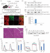
The gene transfer to SOD1 mice using the skMLCK vector. (A) The scheme of the injection of the skMLCK vector. The control vector (1 × 10¹¹ vg/mouse) was injected into the right lower limb, and saline was injected into the left lower limb at the age of 8 weeks. A series of analysis was performed after 6 weeks. (B) Immunoblot analysis of the FLAG peptide in the extensor digitorum longus (EDL) (upper panel) and tibialis anterior (TA) muscles (lower panel). R (right side) is the AAV-injected side, and L (left side) is the sham side. α-tubulin was used as a loading control. (C) Immunofluorescence staining against actin (phalloidin), eGFP, and FLAG in the TA muscle; scale bar: 50 μm. (D) Gene expression of the human (Hs) MYLK2 gene in the TA muscle. Gene expression was demonstrated as the ratio of HsMYLK2 to mouse (Mm) MYLK2. (E) Hematoxylin and eosin (HE) stain of the TA muscle; scale bar: 50 μm. (F) The Phos-tag PAGE of EDL muscle. The upper band showed the phosphorylated MYLPF protein (pMYLPF, white arrow) and the lower band showed the non-phosphorylated MYLPF protein (black arrow). R (right side) is the AAV-injected side, and L (left side) is the sham side. (G) The twitch force of isolated EDL muscle of sham and AAV sides. The data shown in the same color were from the same mouse. (H) The normalized twitch force calculated from G. The normalized twitch force of the AAV sides (45.1 ± 4.02 mN/mm2) was significantly greater than that of the sham sides (36.4 ± 1.22 mN/mm2, p < 0.01). (I) The tetanic force of the isolated EDL muscle of the sham and the AAV sides. The data shown in the same color were from the same mouse. (J) The normalized tetanic force calculated from I. The normalized tetanic force of the AAV sides (114.6 ± 16.0 mN/mm2) was significantly greater than that of the sham sides (61.2 ± 4.44 mN/mm2, p < 0.01).

Similar articles

Cited by

References

    1. Van Es M.A., Hardiman O., Chio A., Al-Chalabi A., Pasterkamp R.J., Veldink J.H., van den Berg L.H. Amyotrophic lateral sclerosis. Lancet. 2017;390:2084–2098. doi: 10.1016/S0140-6736(17)31287-4. - DOI - PubMed
    1. Robberecht W., Philips T. The changing scene of amyotrophic lateral sclerosis. Nat. Rev. Neurosci. 2013;14:248–264. doi: 10.1038/nrn3430. - DOI - PubMed
    1. Hardiman O., Al-Chalabi A., Chio A., Corr E.M., Logroscino G., Robberecht W., Shaw P.J., Simmons Z., van den Berg L.H. Amyotrophic lateral sclerosis. Nat. Rev. Dis. Primers. 2017;3:17071. doi: 10.1038/nrdp.2017.71. - DOI - PubMed
    1. Julien J.P., Kriz J. Transgenic mouse models of amyotrophic lateral sclerosis. Biochim. Biophys. Acta. 2006;1762:1013–1024. doi: 10.1016/j.bbadis.2006.03.006. - DOI - PubMed
    1. Al-Chalabi A., Hardiman O. The epidemiology of ALS: A conspiracy of genes, environment and time. Nat. Rev. Neurol. 2013;9:617–628. doi: 10.1038/nrneurol.2013.203. - DOI - PubMed

Substances

Supplementary concepts