. 2022 Feb 4;23(3):1769.
doi: 10.3390/ijms23031769.

Structural Analysis of Human Serum Albumin in Complex with the Fibrate Drug Gemfibrozil

Affiliations

Structural Analysis of Human Serum Albumin in Complex with the Fibrate Drug Gemfibrozil

Stefano Liberi et al. Int J Mol Sci. .

Abstract

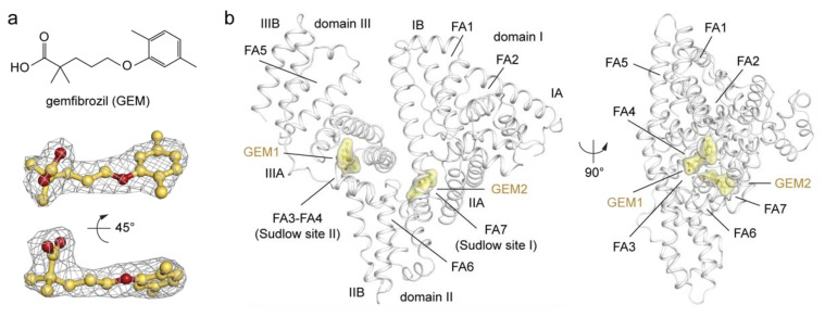
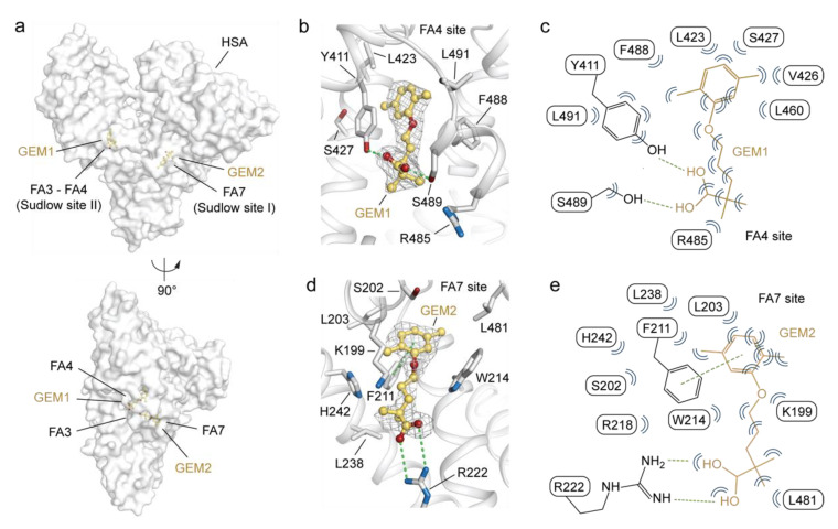
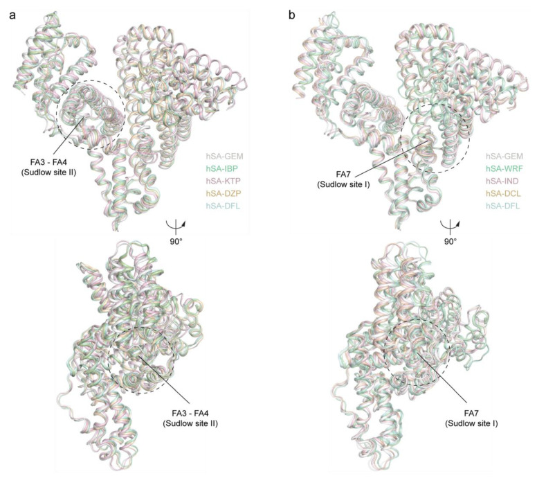
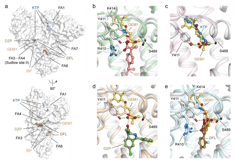
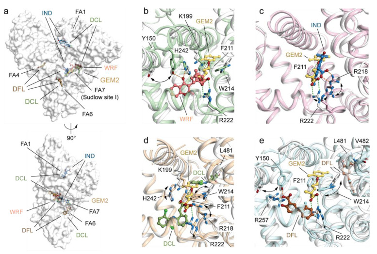
Gemfibrozil (GEM) is an orally administered lipid-regulating fibrate derivative drug sold under the brand name Lopid®, among others. Since its approval in the early 80s, GEM has been largely applied to treat hypertriglyceridemia and other disorders of lipid metabolism. Though generally well tolerated, GEM can alter the distribution and the free, active concentration of some co-administered drugs, leading to adverse effects. Most of them appear to be related to the ability of GEM to bind with high affinity human serum albumin (HSA), the major drug-carrier protein in blood plasma. Here, we report the crystal structure of HSA in complex with GEM. Two binding sites have been identified, namely Sudlow's binding sites I (FA7) and II (FA3-FA4). A comparison of the crystal structure of HSA in complex with GEM with those of other previously described HSA-drug complexes enabled us to appreciate the analogies and differences in their respective binding modes. The elucidation of the molecular interaction between GEM and HSA might offer the basis for the development of novel GEM derivatives that can be safely and synergistically co-administered with other drugs, enabling augmented therapeutic efficacies.

Keywords: Lopid; Sudlow’s site; fibrate; fibric acid; gemfibrozil; hypercholesterolemia; hyperlipidaemia; hypertriglyceridemia; hypolipidemic drug; serum albumin.


Conflict of interest statement

The authors declare no conflict of interest. The funders had no role in the design of the study; in the collection, analyses, or interpretation of data; in the writing of the manuscript; or in the decision to publish the results.

Figures

Figure 1
Figure 1
Structure of HSA in complex with GEM. (a) Chemical structure (top) and composite omit maps depicting the (FoFc) electron density (bottom) of GEM1 ligand contoured at 2.5σ and shown in two orientations (90° rotation); (b) crystal structure of the HSA-GEM complex (white) shown in two orientations (90° rotation, PDB identification code: 7QFE). The structure of HSA is organised in homologues domains (I, II, and III), subdomains (A and B), fatty acids binding sites (FA), and Sudlow’s binding sites. The α-helices of HSA are represented by cartoon loops (white). Bound GEM1 and GEM2 ligands are shown in a ball-and-stick representation with a semi-transparent van der Waals surface (yellow) and coloured by atom type (GEM: carbon = yellow orange, oxygen = firebrick). The electron density of GEM ligands is shown as a grey mesh. The three-dimensional structure models were generated and rendered using Pymol [29].
Figure 2
Figure 2
Details on the binding mode of GEM ligands to HSA. (a) Molecular surface representation of the overall HSA-GEM complex (white) shown in two orientations (90° rotation); (b) detailed view of GEM1 bound to FA4 in sub-domain IIIA; (c) schematic representation of molecular interactions between HSA residues and GEM1; (d) detailed view of GEM2 bound to FA7 in sub-domain IIA; (e) schematic representation of molecular interactions between HSA residues and GEM2. The α-helices of HSA are depicted as cartoon loops (white), and the selected labelled amino acid side chains are represented as sticks and coloured by atom type (carbon = white, oxygen = firebrick, and nitrogen = sky blue). Bound GEM molecules are depicted as ball-and-stick models coloured by atom type (carbon = yellow orange and oxygen = firebrick). The composite omit maps representing the (FoFc) electron density of GEM ligands are contoured at 2.5σ and shown as a grey mesh. Hydrogen bonds, salt bridges, and π–π intermolecular interactions are shown as green dashed lines. Bent blue lines indicate residues of HSA in close contact with GEM ligands (distances shorter than 4.0 Å that are not hydrogen bonds). The three-dimensional structure models were generated and rendered using Pymol [29].
Figure 3
Figure 3
Structural comparison of different HSA-drug crystal structure complexes. (a) Superimposed HSA-GEM complex (white; PDB identification code: 7QFE) aligned with HSA-ibuprofen (pale green; PDB identification code: 2BXG), HSA-ketoprofen (light pink; PDB identification code: 7JWN), HSA-diazepam (wheat; PDB identification code: 2BXF), and HSA-diflunisal (pale cyan; PDB identification code: 2BXE) complexes shown in two orientations (90° rotation); (b) superimposed HSA-GEM complex (white; PDB identification code: 7QFE) aligned with HSA-warfarin (pale green; PDB identification code: 2BXD), HSA-indomethacin (light pink; PDB identification code: 2BXM), HSA-diclofenac (wheat; PDB identification code: 4Z69), and HSA-diflunisal (pale cyan; PDB identification code: 2BXE) complexes shown in two orientations (90° rotation). The α-helices of HSA are represented by cartoon loops and coloured. The three-dimensional structure models were generated and rendered using Pymol [29].
Figure 4
Figure 4
Structural comparison of the ligand binding mode of GEM1 with that of other known drugs binding HSA Sudlow’s site II (FA3-FA4) in sub-domain IIIA. (a) Molecular surface representation of the overall HSA-GEM complex (white; PDB identification code: 7QFE) aligned with HSA-ibuprofen (IBP; PDB identification code: 2BXG), HSA-ketoprofen (KTP; PDB identification code: 7JWN), HSA-diazepam (DZP; PDB identification code: 2BXF), and HSA-diflunisal (DFL; PDB identification code: 2BXE) complexes. The superimposed complexes are shown in two orientations (90° rotation). Detailed view of the superimposed GEM1 (yellow orange) to (b) IBP (salmon), (c) KTP (sky blue), (d) DZP (smudge), and (e) DFL (brown) molecules bound to Sudlow’s site II (FA4). The α-helices of HSA in complex with GEM1, IBP, KTP, DZP, and DFL are represented by cartoon loops and coloured in white, pale green, light pink, wheat, and pale cyan, respectively. The selected labelled amino acid side chains are represented as sticks and coloured by atom type (carbon = white for the HSA-GEM1 complex, pale green for the HSA-IBP complex, light pink for the HSA-KTP complex, wheat for the HSA-DZP complex, pale cyan for the HSA-DFL complex, firebrick for oxygen, and sky blue for nitrogen). Bound ligands are shown in a ball-and-stick representation and coloured by atom type (GEM1: carbon = yellow orange and oxygen = firebrick; IBP: carbon = salmon and oxygen = firebrick; KTP: carbon = sky blue, oxygen = firebrick, and nitrogen = sky blue; DZP: carbon = smudge, oxygen = firebrick, nitrogen = sky blue, and chlorine = light green; and DFL: carbon = brown, oxygen = firebrick, and fluorine = pale cyan). For visualisation, only the side chains of amino acids of HSA forming inter-molecular polar interactions below 4.0 Å are shown. Inter-molecular interactions are represented as dashed lines. Those established by GEM1 are coloured in light green, while those formed by IBP, KTP, DZP, and DFL are coloured in orange. The three-dimensional structure models were generated and rendered using Pymol [29].
Figure 5
Figure 5
Structural comparison of the ligand binding mode of GEM2 with that of other known drugs binding HSA Sudlow’s site I (FA7) in sub-domain IIA. (a) Molecular surface representation of the overall HSA-GEM complex (white; PDB identification code: 7QFE) aligned with HSA-warfarin (WRF; PDB identification code: 2BXD), HSA-indomethacin (IND; PDB identification code: 2BXM), HSA-diclofenac (DCL; PDB identification code: 4Z69), and HSA-diflunisal (DFL; PDB identification code: 2BXE) complexes. The superimposed complexes are shown in two orientations (90° rotation). Detailed view of the superimposed GEM2 (yellow orange) to (b) WRF (salmon), (c) IND (sky blue), (d) DCL (smudge), (e) DFL (brown) molecules bound to Sudlow’s site II (FA4). The α-helices of HSA in complex with GEM2, WRF, IND, DCL, and DFL are represented by cartoon loops and coloured in white, pale green, light pink, wheat, and pale cyan, respectively. The selected labelled amino acid side chains are represented as sticks and coloured by atom type (carbon = white for the HSA-GEM2 complex, pale green for the HSA-WRF complex, light pink for the HSA-IND complex, wheat for the HSA-DCL complex, pale cyan for the HSA-DFL complex, firebrick for oxygen, and sky blue for nitrogen). Bound ligands are shown in a ball-and-stick representation and coloured by atom type (GEM2: carbon = yellow orange and oxygen = firebrick; WRF: carbon = salmon and oxygen = firebrick; IND: carbon = sky blue, oxygen = firebrick, and nitrogen = sky blue; DCL: carbon = smudge, oxygen = firebrick, nitrogen = sky blue, and chlorine = light green; and DFL: carbon = brown, oxygen = firebrick, and fluorine = pale cyan). For visualisation, only the side chains of amino acids of HSA forming inter-molecular polar interactions below 4.0 Å or presenting a notable difference are shown. Inter-molecular interactions are represented as dashed lines. Those established by GEM2 are coloured in light green while those formed by WRF, IND, DCL, and DFL are coloured in orange. The three-dimensional structure models were generated and rendered using Pymol [29].

Similar articles

Cited by

References

    1. Todd P.A., Ward A. Gemfibrozil. Drugs. 1988;36:314–339. doi: 10.2165/00003495-198836030-00004. - DOI - PubMed
    1. Hermens W.A.J.J., Lohman J.J.H.M. Gemfibrozil (Lopid®) Pharm. Weekbl. 1990;125:984–986.
    1. Kundu S.C., Roxy S., Batabyal S.K. Gemfibrozil in dyslipidaemia. J. Assoc. Physicians India. 1990;38:156–159. - PubMed
    1. Gaw A., Packard C.J., Shepherd J. In: Fibrates-Principles and Treatment of Lipoprotein Disorders. Schettler G., Habenicht A.J.R., editors. Springer; Berlin/Heidelberg, Germany: 1994. pp. 325–348.
    1. Duval C., Müller M., Kersten S. PPARα and dyslipidemia. Biochim. Biophys. Acta Mol. Cell Biol. Lipids. 2007;1771:961–971. doi: 10.1016/j.bbalip.2007.05.003. - DOI - PubMed