. 2022 Feb 4;23(3):1778.
doi: 10.3390/ijms23031778.

Neuronal Phenotype of col4a1 and col25a1: An Intriguing Hypothesis in Vertebrates Brain Aging

Affiliations

Neuronal Phenotype of col4a1 and col25a1: An Intriguing Hypothesis in Vertebrates Brain Aging

Adele Leggieri et al. Int J Mol Sci. .

Abstract

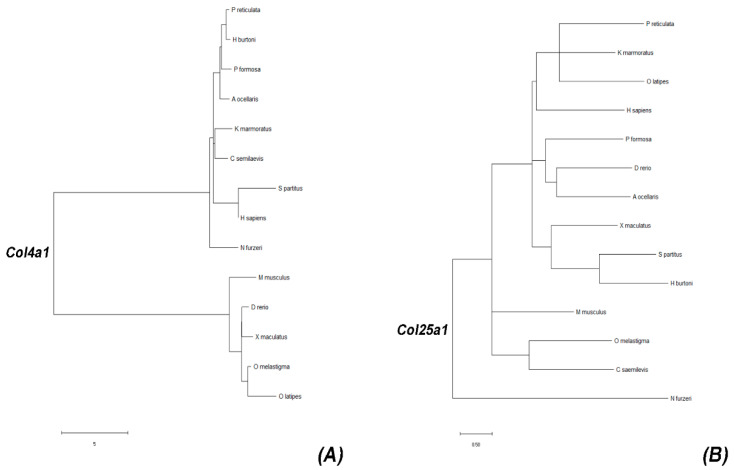
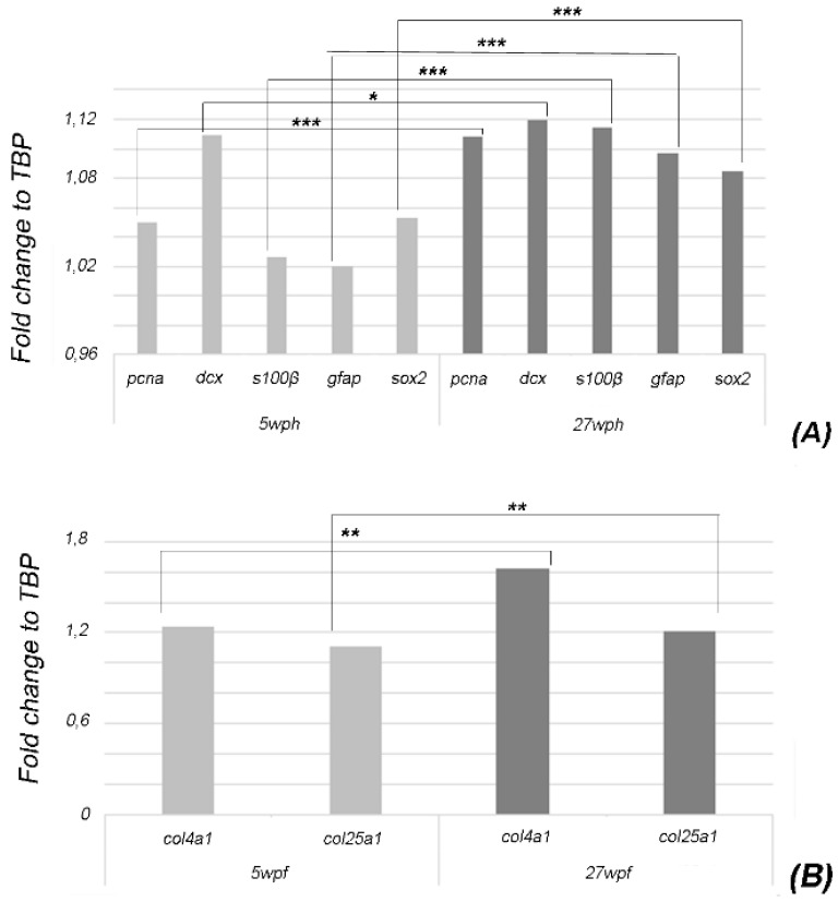
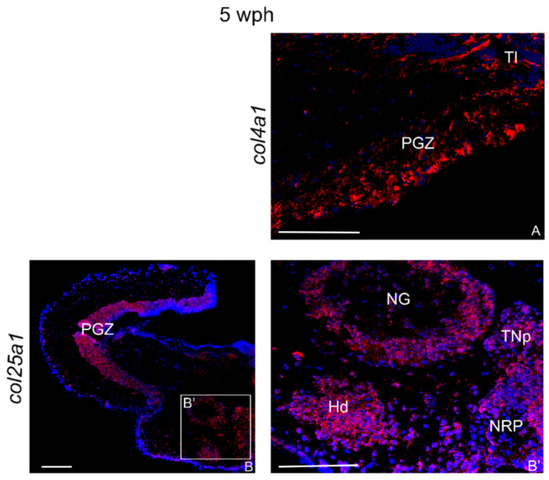
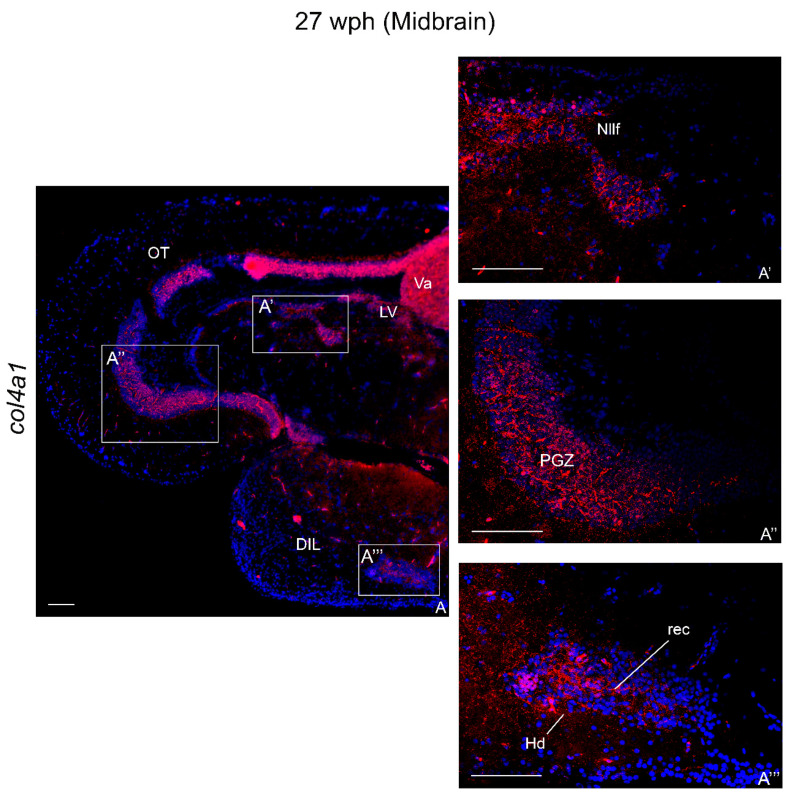
Collagens are the most abundant proteins in vertebrates and constitute the major components of the extracellular matrix. Collagens play an important and multifaceted role in the development and functioning of the nervous system and undergo structural remodeling and quantitative modifications during aging. Here, we investigated the age-dependent regulation of col4a1 and col25a1 in the brain of the short-lived vertebrate Nothobranchius furzeri, a powerful model organism for aging research due to its natural fast-aging process and further characterized typical hallmarks of brain aging in this species. We showed that col4a1 and col25a1 are relatively well conserved during vertebrate evolution, and their expression significantly increases in the brain of N. furzeri upon aging. Noteworthy, we report that both col4a1 and col25a1 are expressed in cells with a neuronal phenotype, unlike what has already been documented in mammalian brain, in which only col25a1 is considered a neuronal marker, whereas col4a1 seems to be expressed only in endothelial cells. Overall, our findings encourage further investigation on the role of col4a1 and col25a1 in the biology of the vertebrate brain as well as the onset of aging and neurodegenerative diseases.

Keywords: Nothobranchius furzeri; aging markers; central nervous system; collagens; fish.


Conflict of interest statement

The authors declare no conflict of interest.

Figures

Figure 1
Figure 1
Gene tree representing Col4a1 and Col25a1 evolutionary history. (A) Branching pattern of col4a1 of N. furzeri revealing a common ancestor with human COL4A1 in comparison to mice and zebrafish. (B) Branching pattern of col25a1 of N. furzeri displaying the phylogenetic distance from the other selected species.
Figure 2
Figure 2
qPCR to evaluate the brain expression of pcna, dcx, sox2, s100β, gfap, col4a1, and col25a1 mRNAs in young (5wph) and old (27wph) animals. Expression levels were analyzed by the ΔΔCt method and normalized to the housekeeping gene TATA-box binding protein (TBP). The analysis was performed using the relative delta curve threshold (ΔΔCT) method. Graphic was built on fold change values to TBP. (A) Bar plot shows upregulation of gfap, pcna, s100β, and sox2. (B). Bar plot shows upregulation of col4a1 and col25a1 “*” indicates p ≤ 0.5, “**” indicates p ≤ 0.05, “***” indicates p ≤ 0.0001.
Figure 3
Figure 3
col4a1 and col25a1 mRNA distribution in transverse sections of the brains of young N. furzeri. (A) Optic tectum with intense staining in the periventricular gray zone and near the layer of the longitudinal tori. (B) Overview of diencephalon/midbrain with strong labeling in the optic tectum. (B’) Higher magnification of the inlet in (B) showing intense labeling of neurons in glomerular nucleus, nucleus of posterior recess, posterior tuberal nuclei, and dorsal hypothalamus. Abbreviations: Hd, dorsal hypothalamus; NG, glomerular nucleus; NRP, nucleus of the posterior recess; PGZ, periventricular gray zone; TNp, posterior tuberal nucleus. Scale bars: (B) 100 µm; (A,B’) 200 µm.
Figure 4
Figure 4
col4a1 mRNA distribution in transverse sections of forebrain of old N. furzeri. (A) Overview of the rostral telencephalon including olfactory bulbs. (A’) Higher magnifications of positive cells in the internal cellular layer of olfactory bulbs and in cell group 1 of the medial zone of dorsal telencephalon. (A’’) Higher magnifications of positive cells in cell groups 1–2 of medial zone and dorsal zone of dorsal telencephalon. (B) Expression in a few cells in cell group 4 of the medial zone of dorsal telencephalon, ventro-ventral zone of ventral telelencephalon, and in the anterior pre-optic area. (C) Expression in cells of habenular nucleus, thalamic nuclei, magnocellular, and posterior preoptic nuclei. (D) Numerous and intense expression in cells of habenular nucleus. Abbreviations: A, anterior thalamic nucleus; Dd, dorsal region of dorsal telencephalon; Dld, dorso-lateral region of dorsal telencephalon; Dlv, ventro-lateral region of dorsal telencephalon; Dm1-4, layers of the medial region of dorsal telencephalon; ECL, external cellular layer; Ha, habenular nucleus; ICL, internal cellular layer; PM, magnocellular pre-optic nucleus; PPa, anterior pre-optic nucleus; PPp, parvocellular portion of pre-optic nucleus; Vd, dorsal region of ventral telencephalon; VM, ventro-medial thalamic nucleus; Vs, supracommissural region of ventral telencephalon; Vv, ventral region of ventral telencephalon. Scale bars: (A) 100 µm; (A’,A’’,B,C,D) 200 µm.
Figure 5
Figure 5
col4a1 mRNA distribution in transverse section of midbrain of old N. furzeri. (A) Overview of the midbrain. (A’) Strong expression in the nucleus of lateral longitudinal fascicle. (A’’) Strong expression in the periventricular gray zone of the optic tectum. (A’’’) Higher magnification of positive cells in the dorsal and inferior lobe of hypothalamus. Abbreviations: DIL, diffuse inferior lobe of hypothalamus; Hd, dorsal hypothalamus; Nllf, nucleus of lateral longitudinal fascicle; LV, nucleus of lateral valvula; OT, optic tectum; PGZ, periventricular gray zone; Tl, longitudinal tori; rec, hypothalamic recess; Va, valvula of cerebellum. Scale bars: (A) 50 µm; (A’,A’’,A’’’) 200 µm.
Figure 6
Figure 6
col25a1 mRNA distribution in transverse section of old N. furzeri forebrain. (A) Overview of labeling in the telencephalon and olfactory bulbs. (BD) Higher magnification of positive cells in the cell groups 1 of medial, dorsal and central zones of dorsal telencephalon. (E) Intense expression in the cells of diencephalic periventricular pre-tectal and thalamic nuclei. (F) Labeling in cells of the ventro-lateral thalamic nucleus. (G) Labeling in sparse cells of the diffuse inferior lobe of the hypothalamus. Abbreviations: A, anterior thalamic nucleus; Dc, central zone of dorsal telencephalon; Dd, dorsal zone of dorsal telencephalon; Dld, dorso-lateral zone of dorsal telencephalon; Dlv, ventro-lateral zone of dorsal telencephalon; Dm1-2, layers of the medial zone of dorsal telencephalon; ECL, external cellular layer; I, inferior thalamic nucleus; ICL, internal cellular layer; PM, magnocellular pre-optic nucleus; PPd, dorsal periventricular pre-tectal nucleus; PPv, ventral periventricular pre-tectal nucleus; VM, ventro-medial thalamic nucleus; VL, ventro-lateral thalamic nucleus; Vs, supracommissural zone of ventral telencephalon; Vv, ventral zone of ventral telencephalon. Scale bars: (A) 100 µm; (B) 200 µm; (CG) 300 µm.
Figure 7
Figure 7
col25a1 mRNA distribution in transverse section of caudal diencephalon and mesencephalon of old N. furzeri. (A) Diffuse positive cells in the glomerular nucleus, nucleus of posterior recess, and posterior tuberal nucleus. (B,C) Labeling in the longitudinal tori and optic tectum. (D) Diffuse and intense labeling over the different layers of optic tectum. (E) Diffuse signal probe in the optic tectum, semicircular tori, nucleus of lateral longitudinal fascicle, nucleus of lateral valvula. Abbreviations: CZ, central zone; DWZ, deep white zone; EG, granular eminentiae; Nllf, nucleus of lateral longitudinal fascicle; LV, nucleus of lateral valvula; NG, glomerular nucleus; NRP, nucleus of posterior recess; PGZ, periventricular gray zone; SWGZ, superficial white gray zone; TNp, posterior tuberal nucleus; TS, semicircular tori. Scale bars: (E) 100 µm; (AD) 200 µm.
Figure 8
Figure 8
Co-localization of col4a1 and col25a1 with anti-S100β in transverse sections of old N. furzeri brain. (A) col4a1/s100β faint co-localization along the diencephalic ventricle. (B) Faint col25a1/s100β co-localization in in the parvocellular portion of pre-optic nucleus. (C) col25a1/s100β co-localization along the diencephalic ventricle and in a few sparse glial cells. Abbreviations: A, anterior thalamic nucleus; Cmin, minor commissure; Ha, habenular nucleus; ON, optic nerve; PM, magnocellular pre-optic nucleus; PPd, dorsal periventricular pre-tectal nucleus; PPv, ventral periventricular pre-tectal nucleus; VM, ventro-medial thalamic nucleus. Scale bars: (A,B) 200 µm; (C) 100 µm.
Figure 9
Figure 9
Co-localization of col4a1 and col25a1 with anti-DCX, anti-HuC/HuD, and anti-MAP2 in transverse sections of old N. furzeri brain. (A) Faint col4a1/DCX co-staining along the telencephalic ventricle. (B) Faint col25a1/DCX co-staining along the telencephalic ventricle and in a few neurons of dorsal telencephalon. (C,D) Weak col4a1/HuC/HuD co-localization in the optic tectum, the body of cerebellum and granular eminentiae. (EG) Co-localization of col25a1/HuC/HuD in very few neurons of magnocellular pre-optic nucleus, in the body of the cerebellum and in the semicircular tori. (H) Weak col4a1/MAP2 co-localization in a few neurons of the optic tectum. (I) Faint col25a1/MAP2 co-localization in few neurons of the optic tectum. Abbreviations: A, anterior thalamic nucleus; CCe, body of cerebellum; CZ, central zone of the optic tectum; Dm4, central zone of dorsal telencephalon, layer 4; DWGZ, deep white and gray zone of the optic tectum; EG, granular eminentiae; gc, central griseum; I, inferior thalamic nucleus; PGZ, periventricular gray zone; PM, magnocellular pre-optic nucleus; PPd, dorsal periventricular pre-tectal nucleus; PPv, ventral periventricular pre-tectal nucleus; TS, layer of semicircular tori; Vd, dorsal zone of the ventral telencephalon; VM, ventro-medial thalamic nucleus; Vs, supracommissural zone of ventral telencephalon. Scale bars: (AG) 100 µm; (H,I) 200 µm.

Similar articles

Cited by

References

    1. Hou Y., Dan X., Babbar M., Wei Y., Hasselbalch S.G., Croteau D.L., Bohr V.A. Ageing as a risk factor for neurodegenerative disease. Nat. Rev. Neurol. 2019;15:565–581. doi: 10.1038/s41582-019-0244-7. - DOI - PubMed
    1. Mayne K., White J.A., McMurran C.E., Rivera F.J., de la Fuente A.G. Aging and Neurodegenerative Disease: Is the Adaptive Immune System a Friend or Foe? Front. Aging Neurosci. 2020;12 doi: 10.3389/fnagi.2020.572090. - DOI - PMC - PubMed
    1. Tony Wyss C. Ageing, neurodegeneration and brain rejuvenation. Nature. 2016;10:180–186. doi: 10.1038/nature20411. - DOI - PMC - PubMed
    1. Podtelezhnikov A.A., Tanis K.Q., Nebozhyn M., Ray W.J., Stone D.J., Loboda A.P. Molecular insights into the pathogenesis of Alzheimer’s disease and its relationship to normal aging. PLoS ONE. 2011;13:6. doi: 10.1371/journal.pone.0029610. - DOI - PMC - PubMed
    1. Wakabayashi T. Transmembrane Collagens in Neuromuscular Development and Disorders. Front. Mol. Neurosci. 2021;13:1–8. doi: 10.3389/fnmol.2020.635375. - DOI - PMC - PubMed