. 2022 Feb 4;23(3):1787.
doi: 10.3390/ijms23031787.

Amniotic LPS-Induced Apoptosis in the Fetal Brain Is Suppressed by Vaginal LPS Preconditioning but Is Promoted by Continuous Ischemic Reperfusion

Affiliations

Amniotic LPS-Induced Apoptosis in the Fetal Brain Is Suppressed by Vaginal LPS Preconditioning but Is Promoted by Continuous Ischemic Reperfusion

Yupeng Dong et al. Int J Mol Sci. .

Abstract

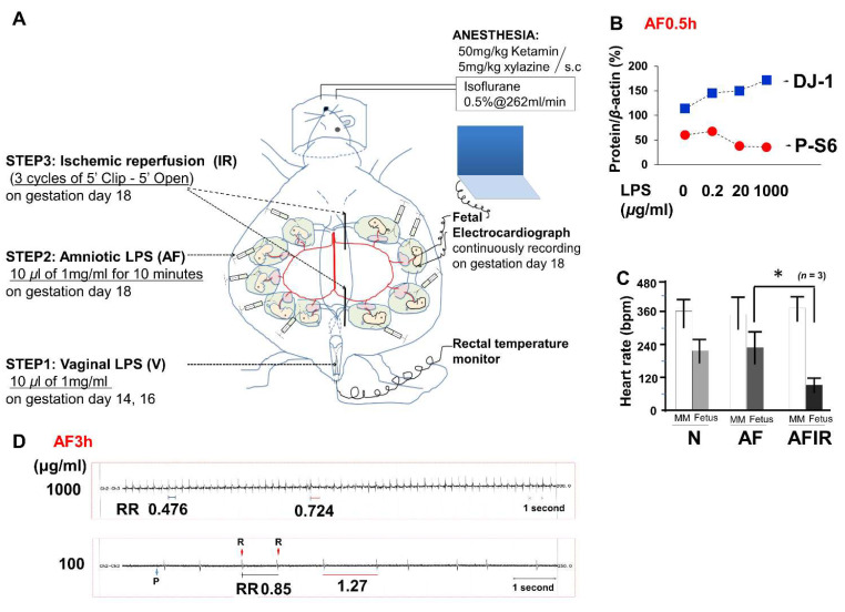
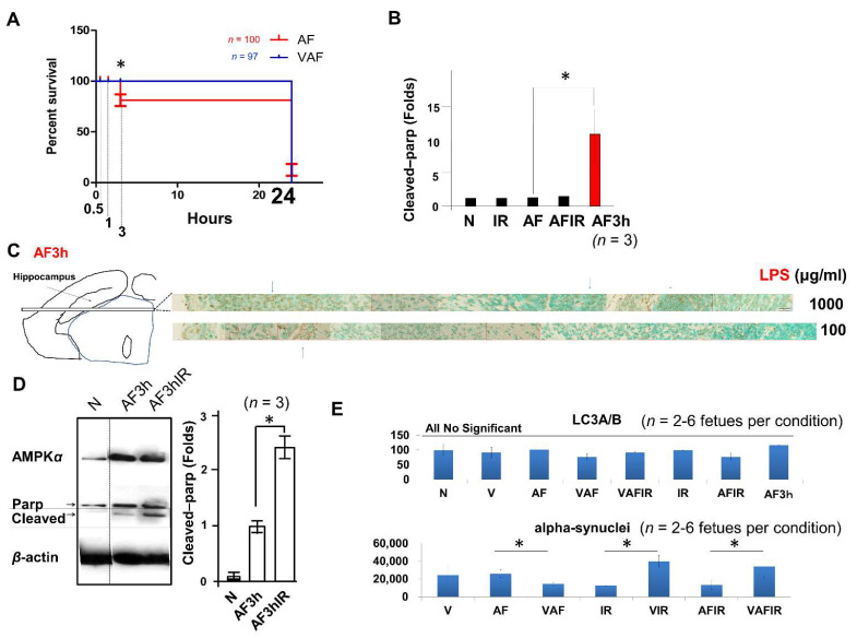
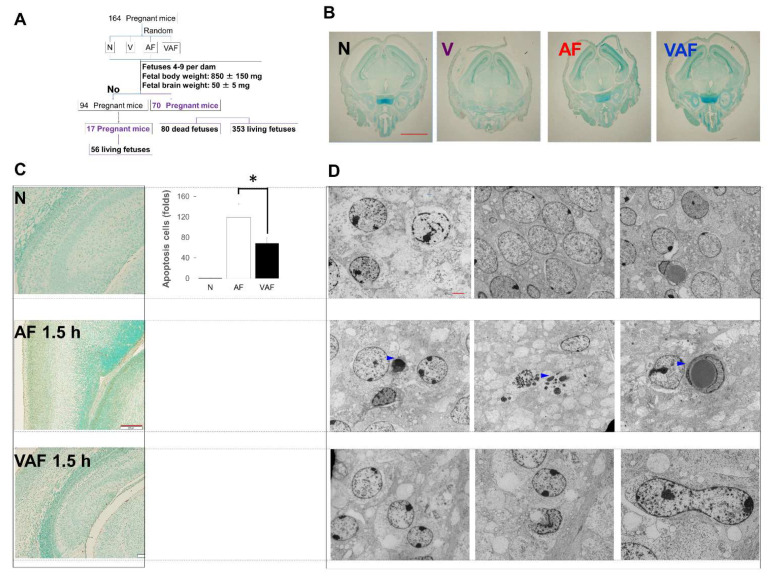
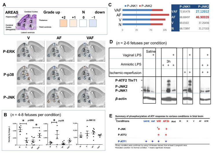
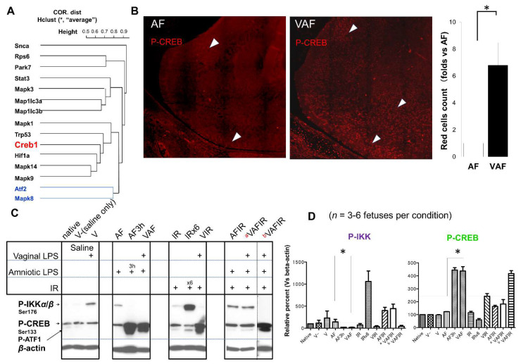
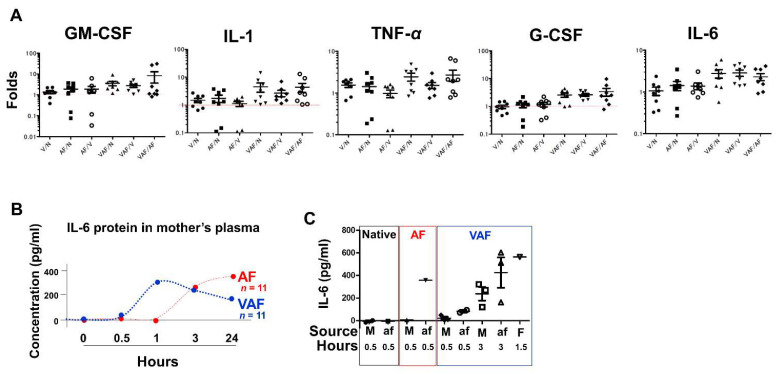
Chorioamnionitis (CAM) is an increasingly common disease affecting pregnant women which derives from bacterial vaginosis. In different clinical cases, it has been shown that CAM can cause multiple risk factors for fetal brain damage, such as infection, and intra-uterine asphyxia. However, the molecular mechanism remains unknown. In this study, we established a novel CAM mouse model by exposing pregnant mice to a combination of three risk factors: vaginal lipopolysaccharides (LPS), amniotic LPS, and ischemic reperfusion. We found amniotic LPS caused Parkinson's disease-like fetal brain damage, in a dose and time-dependent manner. Moreover, the mechanism of this fetal brain damage is apoptosis induced by amniotic LPS but it was inhibited by being pretreated with a vaginal LPS challenge before amniotic LPS injection. In contrast, amniotic LPS with continuous ischemic reperfusion caused a higher level of apoptotic cell death than amniotic LPS alone. In particular, a potential neuroprotective biomarker phosphorylation (p)-CREB (ser133) appeared in only vaginal LPS preconditioned before amniotic LPS, whereas ischemic reperfusion triggered IKK phosphorylation after amniotic LPS. Despite the need for many future investigations, this study also discussed a developed understanding of the molecular mechanism of how these phenotypes occurred.

Keywords: bacterial vaginosis; chorioamnionitis; fetal brain damage; mouse model; vaginal lipopolysaccharide.


Conflict of interest statement

The authors declare no conflict of interest.

Figures

Figure 1
Figure 1
Fetal electrocardiography detects ischemic reperfusion. (A) A novel mouse model to expose fetuses to vaginal lipopolysaccharide (LPS) preconditioning, amniotic LPS, and ischemic reperfusion from gestational days (GDs) 14–18. On GD18, fetal electrocardiography (FECG) was used as a real-time monitor. (B) Western blot assays of amniotic fluid (AF) LPS 0.5 h fetal brain tissue lysate (1% NP40) to detect Parkinson-disease 7 (PARK7, also known as DJ)-1 under indicated LPS concentration. (C) Heart rate detected by FECG of indicated conditions including ischemic reperfusion after exposure to lethal amniotic LPS (AFIR). * p < 0.05. (D) FECG of fetuses (AF condition) exposed to amniotic LPS at both 1 mg/mL and 0.1 mg/mL concentrations.
Figure 2
Figure 2
Amniotic LPS causes fetal death. (A) Dead fetuses were counted in the AF (red) and VAF (blue) groups from the endpoint 3 h. Almost all fetuses died by liquefactive necrosis of brains after 24-h exposure to amniotic LPS, even after vaginal LPS preconditioning. (B) An increased cleaved Parp protein (10 folds) supports the apoptosis-like cell death in the AF. (C) Significant cell death occurred in indicated concentrations (AF). (D) Protein level of cleaved Parp in the indicated conditions (E) Protein level of LC3A/B and alpha-synuclein (SNCA) in the fetal brain at different condition by western blot. * p < 0.05.
Figure 3
Figure 3
Time-course investigation of cell death in the fetal brain. (A) Quantitative and qualitative analyzes of fetal mouse brains, details in materials and methods. (B) No detectable cell death in the whole fetal brains of N, V, AF 0.5 h and VAF 0.5 h (×1.25 microscopy). Scale bar means 2 mm. (C) 1.5 h of AF and VAF cell death measure (brown doses are positive apoptosis cells). Scale bar means 200 μm. * p < 0.05. (D) Transmission electron microscopy images of cell death in the Native, Apoptosis [blue triangles in AF (1.5 h), Scale bar means 2 μm.
Figure 4
Figure 4
The phosphorylation of mitogen-activated protein kinase pathways and transcription factors. (A) Three fetuses derived from three pregnant mice per group were used for immunochemical staining. Each fetal brain was separated into six fields (1–6). The grades (0–4) of phosphorylated protein in the paraffin-embedded sections areas, which we indicated using different colors. (B) Western blot data of phosphorylated ERK1/2, P38, and JNK1/2. * p < 0.05. (C) The average percentage of phosphorylation of JNK1 and JNK2, normal condition (N) is 100%. (D) Phosphorylated ATF2 and JNK in the whole fetal brain under indicated conditions. (E) Summary of western blot data: strong phosphorylation (++) of both JNK and ATF conditions were indicated.
Figure 5
Figure 5
The phosphorylation of CREB was activated in VAF group but not in AF group. (A) hclust average of correlation distance of indicated proteins. (B) Immunochemistry images of phosphorylated CREB (ser133) in fetal brains derived from the amniotic LPS and VAF groups. * means p < 0.05. (C,D) Phosphorylated CREB and IKK were measured by western blotting. The bands under P-CREB are phosphorylation of ATF1. β-actin was used as a loading control. Samples from the VAFIR condition showed two distinct molecular phenotypes described as a VAFIR and b VAFIR respectively. * p < 0.05.
Figure 6
Figure 6
Cytokine expression measured by ELISA. (A) The relative folds of cytokine gene expression by comparing with the indicated conditions, V: vaginal; N: normal/native; AF: amniotic LPS; VAF: vaginal LPS plus amniotic LPS. Hprt is a housekeeping gene used as a control. (B) IL-6 protein in the maternal plasma (M) was measured at each endpoint time (0–24 h) after amniotic LPS exposure. Either saline injection into the amniotic fluid, or the vaginal LPS group, were used as control samples to compare with the AF (red) or VAF (blue) groups, respectively (for detailed information refer to raw data). (C) IL-6 concentration in M plasma and amniotic fluid (af) at the indicated time. Fetal plasma (F) represents a mixture derived from 12 fetuses from 3 dams.

Similar articles

Cited by

References

    1. Faro S. Bacterial Vaginitis. Clin. Obstet. Gynecol. 1991;34:582–586. doi: 10.1097/00003081-199134030-00014. - DOI - PubMed
    1. Gibbs R.S., Romero R., Hillier S.L., Eschenbach D.A., Sweet R.L. A Review of Premature Birth and Subclinical Infection. Am. J. Obstet. Gynecol. 1992;166:1515–1528. doi: 10.1016/0002-9378(92)91628-N. - DOI - PubMed
    1. Kimberlin D.F., Andrews W.W. Bacterial Vaginosis: Association with Adverse Pregnancy Outcome. Semin. Perinatol. 1998;22:242–250. doi: 10.1016/S0146-0005(98)80012-8. - DOI - PubMed
    1. Peebles K., Velloza J., Balkus J.E., McClelland R.S., Barnabas R.V. High Global Burden and Costs of Bacterial Vaginosis: A Systematic Review and Meta-Analysis. Sex Transm. Dis. 2019;46:304–311. doi: 10.1097/OLQ.0000000000000972. - DOI - PubMed
    1. Shatrov J.G., Birch S.C., Lam L.T., Quinlivan J.A., McIntyre S., Mendz G.L. Chorioamnionitis and Cerebral Palsy: A Meta-analysis. Obstet. Gynecol. 2010;116:387–392. doi: 10.1097/AOG.0b013e3181e90046. - DOI - PubMed

Substances

LinkOut - more resources