

Dual RNA Sequencing of Mycobacterium tuberculosis-Infected Human Splenic Macrophages Reveals a Strain-Dependent Host-Pathogen Response to Infection
- PMID: 35163725
- PMCID: PMC8836425
- DOI: 10.3390/ijms23031803
Dual RNA Sequencing of Mycobacterium tuberculosis-Infected Human Splenic Macrophages Reveals a Strain-Dependent Host-Pathogen Response to Infection
Abstract
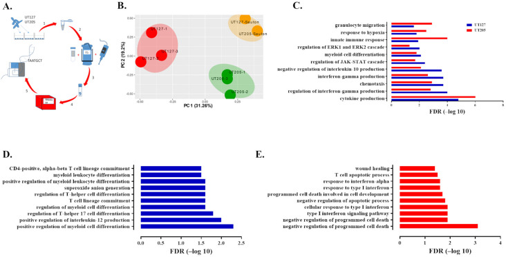
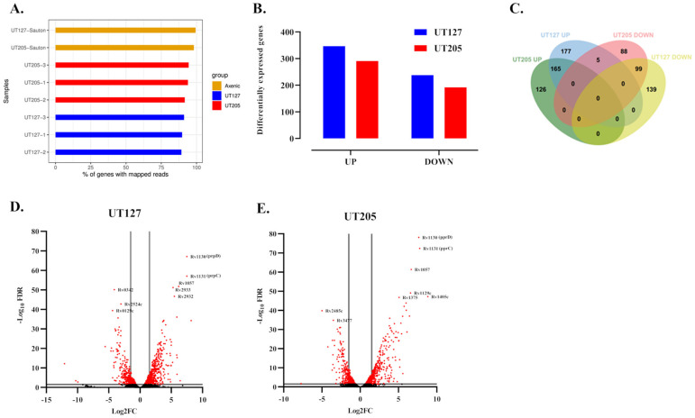
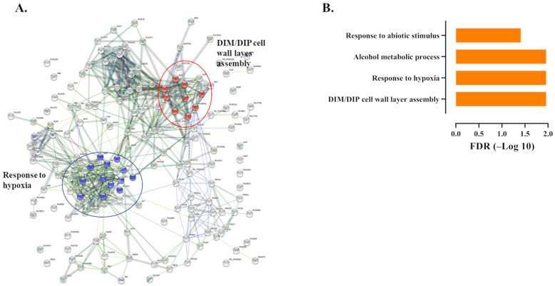
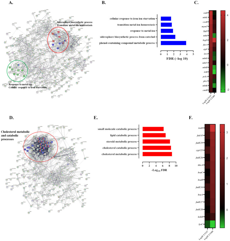
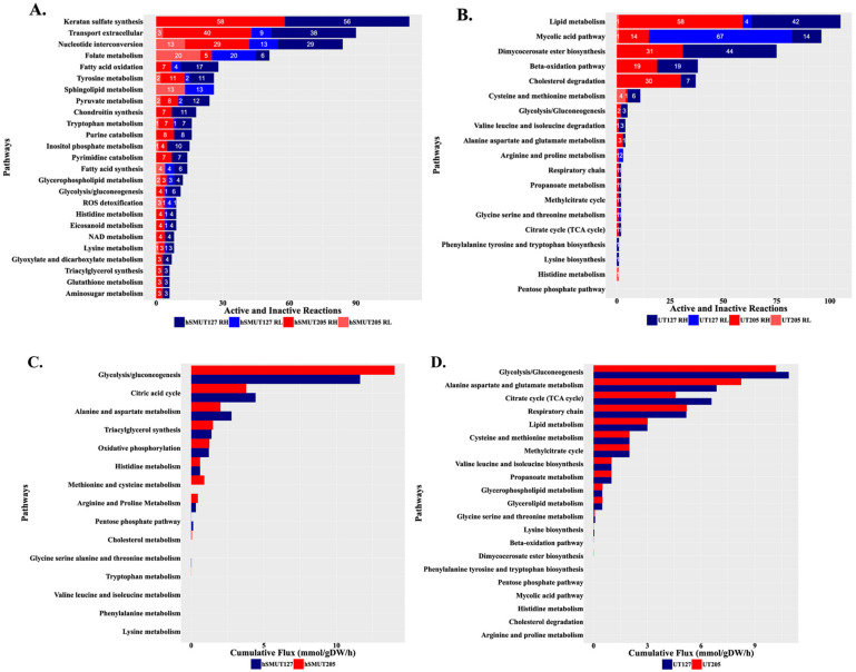
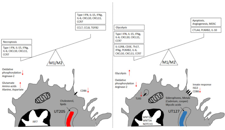
Tuberculosis (TB) is caused by Mycobacterium tuberculosis (Mtb), leading to pulmonary and extrapulmonary TB, whereby Mtb is disseminated to many other organs and tissues. Dissemination occurs early during the disease, and bacteria can be found first in the lymph nodes adjacent to the lungs and then later in the extrapulmonary organs, including the spleen. The early global gene expression response of human tissue macrophages and intracellular clinical isolates of Mtb has been poorly studied. Using dual RNA-seq, we have explored the mRNA profiles of two closely related clinical strains of the Latin American and Mediterranean (LAM) family of Mtb in infected human splenic macrophages (hSMs). This work shows that these pathogens mediate a distinct host response despite their genetic similarity. Using a genome-scale host-pathogen metabolic reconstruction to analyze the data further, we highlight that the infecting Mtb strain also determines the metabolic response of both the host and pathogen. Thus, macrophage ontogeny and the genetic-derived program of Mtb direct the host-pathogen interaction.
Keywords: Mycobacterium tuberculosis; clinical strains; dual RNA-seq; human; metabolic reconstruction; splenic macrophages.
Conflict of interest statement
The authors declare no conflict of interest.
Figures
Similar articles
-
Dual RNA-Seq of Mtb-Infected Macrophages In Vivo Reveals Ontologically Distinct Host-Pathogen Interactions.Cell Rep. 2020 Jan 14;30(2):335-350.e4. doi: 10.1016/j.celrep.2019.12.033. Cell Rep. 2020. PMID: 31940480 Free PMC article.
-
Integrative Analysis of Human Macrophage Inflammatory Response Related to Mycobacterium tuberculosis Virulence.Front Immunol. 2021 Jun 28;12:668060. doi: 10.3389/fimmu.2021.668060. eCollection 2021. Front Immunol. 2021. PMID: 34276658 Free PMC article.
-
Human Alveolar and Splenic Macrophage Populations Display a Distinct Transcriptomic Response to Infection With Mycobacterium tuberculosis.Front Immunol. 2020 Apr 21;11:630. doi: 10.3389/fimmu.2020.00630. eCollection 2020. Front Immunol. 2020. PMID: 32373118 Free PMC article.
-
Mycobacterium tuberculosis-macrophage interaction: Molecular updates.Front Cell Infect Microbiol. 2023 Mar 3;13:1062963. doi: 10.3389/fcimb.2023.1062963. eCollection 2023. Front Cell Infect Microbiol. 2023. PMID: 36936766 Free PMC article. Review.
-
Mycobacterium tuberculosis Infection-Driven Foamy Macrophages and Their Implications in Tuberculosis Control as Targets for Host-Directed Therapy.Front Immunol. 2020 May 12;11:910. doi: 10.3389/fimmu.2020.00910. eCollection 2020. Front Immunol. 2020. PMID: 32477367 Free PMC article. Review.
Cited by
-
The functional response of human monocyte-derived macrophages to serum amyloid A and Mycobacterium tuberculosis infection.Front Immunol. 2023 Sep 15;14:1238132. doi: 10.3389/fimmu.2023.1238132. eCollection 2023. Front Immunol. 2023. PMID: 37781389 Free PMC article.
-
Dual RNA-seq reveals a type 6 secretion system-dependent blockage of TNF-α signaling and BicA as a Burkholderia pseudomallei virulence factor important during gastrointestinal infection.Gut Microbes. 2022 Jan-Dec;14(1):2111950. doi: 10.1080/19490976.2022.2111950. Gut Microbes. 2022. PMID: 35984745 Free PMC article.
-
Developing New Tools to Fight Human Pathogens: A Journey through the Advances in RNA Technologies.Microorganisms. 2022 Nov 21;10(11):2303. doi: 10.3390/microorganisms10112303. Microorganisms. 2022. PMID: 36422373 Free PMC article. Review.
-
Whole Blood vs Serum-Derived Exosomes for Host and Pathogen-Specific Tuberculosis Biomarker Identification: RNA-Seq-Based Machine-Learning Approach.Biochem Genet. 2024 Dec 23. doi: 10.1007/s10528-024-11002-1. Online ahead of print. Biochem Genet. 2024. PMID: 39715973
-
An approach to analyze spatiotemporal patterns of gene expression at single-cell resolution in Candida albicans-infected mouse tongues.mSphere. 2024 Sep 25;9(9):e0028224. doi: 10.1128/msphere.00282-24. Epub 2024 Aug 22. mSphere. 2024. PMID: 39171917 Free PMC article.
References
-
- WHO Global Tuberculosis Report 2018. 2018. [(accessed on 1 November 2021)]. Available online: https://www.who.int/tb/publications/global_report/en/
MeSH terms
Grants and funding
LinkOut - more resources
Full Text Sources
Medical
