. 2022 Feb 4;23(3):1807.
doi: 10.3390/ijms23031807.

Up and Down γ-Synuclein Transcription in Dopamine Neurons Translates into Changes in Dopamine Neurotransmission and Behavioral Performance in Mice

Affiliations

Up and Down γ-Synuclein Transcription in Dopamine Neurons Translates into Changes in Dopamine Neurotransmission and Behavioral Performance in Mice

Rubén Pavia-Collado et al. Int J Mol Sci. .

Abstract

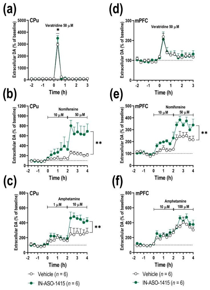
The synuclein family consists of α-, β-, and γ-Synuclein (α-Syn, β-Syn, and γ-Syn) expressed in the neurons and concentrated in synaptic terminals. While α-Syn is at the center of interest due to its implication in the pathogenesis of Parkinson's disease (PD) and other synucleinopathies, limited information exists on the other members. The current study aimed at investigating the biological role of γ-Syn controlling the midbrain dopamine (DA) function. We generated two different mouse models with: (i) γ-Syn overexpression induced by an adeno-associated viral vector and (ii) γ-Syn knockdown induced by a ligand-conjugated antisense oligonucleotide, in order to modify the endogenous γ-Syn transcription levels in midbrain DA neurons. The progressive overexpression of γ-Syn decreased DA neurotransmission in the nigrostriatal and mesocortical pathways. In parallel, mice evoked motor deficits in the rotarod and impaired cognitive performance as assessed by novel object recognition, passive avoidance, and Morris water maze tests. Conversely, acute γ-Syn knockdown selectively in DA neurons facilitated forebrain DA neurotransmission. Importantly, modifications in γ-Syn expression did not induce the loss of DA neurons or changes in α-Syn expression. Collectively, our data strongly suggest that DA release/re-uptake processes in the nigrostriatal and mesocortical pathways are partially dependent on substantia nigra pars compacta /ventral tegmental area (SNc/VTA) γ-Syn transcription levels, and are linked to modulation of DA transporter function, similar to α-Syn.

Keywords: AAV vector; antisense oligonucleotide; cognitive dysfunction; dopamine; motor deficits; γ-synuclein.


Conflict of interest statement

A.B. is an inventor of the issued patents WO2011131693, WO-2014064257-A1, WO2014064258-A1 for ligand-conjugated siRNA and ASO molecules and the targeting approach for monoamine systems related to this work. The rest of the authors declare no competing interest.

Figures

Figure 1
Figure 1
Overexpression of γ-Syn mRNA in SNc/VTA DA neurons of mice. Mice received unilaterally 1 μL of AAV10 vector containing a cytomegalovirus promoter to drive the expression of γ-Syn or vehicle into SNc/VTA and were euthanized at 1, 4, 8 and 16 weeks (W) post-injection. An empty vector (AAV-EV) with noncoding stuffer DNA was also used as a control group (see Supplementary Materials Figures S1 and S3). (a) Coronal brain sections showing γ-Syn mRNA levels in the SNc/VTA assessed by in situ hybridization. Scale bar: 500 μm. (b) Progressive increases of γ-Syn mRNA expression in the ipsilateral SNc/VTA of AAV10-injected mice compared to vehicle- and AAV-EV-injected mice (n = 5 mice/group). (c) Photomicrographs showing TH-positive cells expressing γ-Syn mRNA (33P-oligonucleotide silver grains) in the SNc/VTA of vehicle- and AAV10-injected mice at 4 W post-injection. Scale bar: 20 μm. Frame within the AAV10 4W image shows contralateral and ipsilateral SNc enlargements from AAV10-injected mice. Scale bar: 5 μm. Note the higher density of intracellular γ-Syn mRNA in TH-positive neurons on the ipsilateral versus contralateral side. (d) Dipping analyses revealed a significant increase in TH-positive cells expressing γ-Syn mRNA in the ipsilateral SNc/VTA at different antero-posterior coordinates from bregma (−2.46 to −3.64 mm). No differences in the number of TH-positive cells were found in the SNc/VTA of vehicle- and AAV10-injected mice. (e,f) Images of Western blot (e) and relative quantification (f) of murine γ-Syn and α-Syn proteins in ipsilateral SNc/VTA lysates from mice injected with vehicle or AAV10 at 4 W (n = 8 mice/group). β-actin was used as house-keeping control. Values are presented as mean ± SEM. ** p < 0.01, *** p < 0.001, **** p < 0.0001 compared to vehicle- injected mice. Abbreviations: contra, contralateral; ipsi, ipsilateral.
Figure 2
Figure 2
Local injection of AAV10 construct into ipsilateral SNc/VTA also induces increased γ-Syn expression in non-TH cells. Mice received unilaterally 1 μL of AAV10 vector containing a cytomegalovirus promoter to drive the expression of γ-Syn or vehicle into SNc/VTA and were euthanized at 4 weeks (W) post-injection (n = 5 mice/group, same mice used in Figure 1). (a) Photomicrographs showing GAD67-positive cells expressing γ-Syn mRNA (33P-oligonucleotide silver grains) in the SNc of vehicle- and AAV10-injected mice at 4 weeks (W) later. Scale bar: 20 μm. The frames inserted in the images show contralateral and ipsilateral SNc enlargements from vehicle- and AAV10-injected mice. Scale bar: 5 μm. Note the higher density of intracellular γ-Syn mRNA in GAD67-positive cells on the ipsilateral versus the contralateral side of AAV10-injected mice. (b) Dipping analyses revealed a significant increase in the number of GAD67-positive cells expressing γ-Syn mRNA in the ipsilateral SNc/VTA at different antero-posterior coordinates from bregma (−2.46 to −3.64 mm). Likewise, increases in intracellular γ-Syn mRNA density were found in GAD67-positive cells in the ipsilateral side of AAV10-injected mice. No differences in the number of GAD67-positive cells were detected in the SNc/VTA of vehicle- and AAV10-injected mice. Values are presented as mean ± SEM. *** p < 0.001, **** p < 0.0001 compared to vehicle- injected mice. Abbreviations: contra, contralateral; ipsi, ipsilateral. Black circles represent the number of mice used for each group.
Figure 3
Figure 3
Overexpression of γ-Syn in the SNc/VTA alters forebrain DA neurotransmission. Mice received unilaterally 1 μL of AAV10 vector containing a cytomegalovirus promoter to drive the expression of γ-Syn or vehicle into SNc/VTA and were examined at 4 or 16 weeks (W) post-injection (n = 6 mice/group). At the end of the microdialysis procedures, the mice were euthanized and γ-Syn expression was confirmed. (a,e) Local veratridine infusion (depolarizing agent, 50 μM) by reverse dialysis increased extracellular DA release in CPu both at 4 and 16 W later. This effect was more pronounced in vehicle-injected than in AAV10-injected mice. (b,f) Nomifensine (DAT/NET inhibitor, 10 and 50 μM) increased more significantly extracellular DA levels in CPu of control group than in AAV10-injected mice. (c,g) Unlike CPu, local infusion of veratridine into mPFC augmented DA release, but this effect was similar in both phenotypes. (d,h) Local infusion of nomifensine in mPFC produced a greater effect on extracellular DA levels in vehicle-treated mice than in mice injected with AAV10 at both 4 and 16 W post-injection. Values are presented as mean ± SEM. *** p < 0.001, compared to vehicle-injected mice. Abbreviations: CPu, caudate-putamen; mPFC, medial prefrontal cortex.
Figure 4
Figure 4
Overexpression of γ-Syn in the SNc/VTA impairs motor and cognitive behaviors. Mice received unilaterally 1 μL of AAV10 vector containing a cytomegalovirus promoter to drive the expression of γ-Syn or vehicle into SNc/VTA and were examined at 4, 8, or 16 weeks (W) post-injection (n = 8–12 mice/group). All mice were examined in the different behavioral paradigms. (a) Mice overexpressing γ-Syn showed alterations in the motor coordination in the rotarod, evaluated by a shorter latency to fall at 16 W post-AAV10 injection compared to vehicle-injected mice. (b,c) No differences were observed in the behavioral performance between the different groups in the cylinder test (b) as well as in the open field test (c). (d) Mice overexpressing γ-Syn failed to discriminate between familiar and novel objects (F and N, respectively) compared to vehicle-treated mice at 16 W post-injection. (e) Similarly, mice overexpressing γ-Syn also showed a cognitive deterioration with a shorter latency to cross in the passive avoidance test at 16 W post-injection compared to vehicle-injected mice. (f) In the Morris water maze, mice with increased γ-Syn expression in the SNc/VTA evoked greater latencies to reach the platform than vehicle-injected mice starting at 8 W after injection. However, no differences in time elapsed in the target quadrant were detected between the different groups of mice. Values are presented as mean ± SEM. ** p < 0.01, **** p < 0.0001, compared to vehicle- injected mice.
Figure 5
Figure 5
Single acute administration of IN-ASO-1415 into SNc/VTA downregulates γ-Syn expression in DA neurons. Mice received unilaterally 1 μL of IN-ASO-1415 (total dose of 60 μg) or vehicle into SNc/VTA and were euthanized at 24 or 72 h post-infusion. An IN-conjugated nonsense ASO sequence (IN-ASO-1227) was also used as a control group. (a) Coronal brain sections showing γ-Syn mRNA levels in the SNc/VTA assessed by in situ hybridization. Yellow arrowheads shown γ-Syn mRNA expression in SNc/VTA. Scale bar: 500 μm. (b) Significant reductions of γ-Syn mRNA expression in the ipsilateral SNc/VTA of IN-ASO-1415-injected mice compared to vehicle- and IN-ASO-1227-injected mice (n = 4–5 mice/group). (c) Photomicrographs showing TH-positive cells expressing γ-Syn mRNA (33P-oligonucleotide silver grains) in the ipsilateral SNc from mice injected with vehicle or IN-ASO sequences. Scale bar: 20 μm. Note the lower density of intracellular γ-Syn mRNA in SNc TH-positive neurons in mice that received IN-ASO-1415 than in those that received vehicle. (d) Dipping analyses revealed significant reductions in TH-positive cells expressing γ-Syn mRNA in the ipsilateral SNc/VTA at different antero-posterior coordinates from bregma (−2.46 to −3.64 mm). Likewise, decreases of intracellular γ-Syn mRNA density were found in TH-positive cells in the ipsilateral side of IN-ASO-1415-injected mice. No differences in the number of TH-positive cells were detected in the SNc/VTA of vehicle-, IN-ASO-1227-, and IN-ASO-1415-injected mice. (e,f) Images of Western blot (e) and relative quantification (f) of murine γ-Syn and α-Syn proteins in ipsilateral SNc/VTA lysates from mice injected with vehicle or IN-ASO-1415 at 24 and 72 h post-injection (n = 6–9 mice/group). β-actin was used as house-keeping control. Values are presented as mean ± SEM. ** p < 0.01, *** p < 0.001, **** p < 0.0001 compared to vehicle-injected mice. Abbreviations: contra, contralateral; ipsi, ipsilateral. (See Supplementary Materials Figure S3).
Figure 6
Figure 6
Neurochemical effects on forebrain DA neurotransmission of IN-ASO-1415-induced γ- Syn knockdown. Mice received unilaterally 1 μL of IN-ASO-1415 (total dose of 60 μg) or vehicle into SNc/VTA. Microdialysis experiments were conducted 3 days after administration (n = 6 mice/group). (a,d) Local veratridine infusion (depolarizing agent, 50 mM) by reverse dialysis increased extracellular DA release in CPu (a) and mPFC (d). This effect was significantly higher in the CPu of IN-ASO-1415-treated mice, but not in mPFC, compared to vehicle-treated mice. (b,e) Nomifensine (DAT/NET inhibitor, 10 and 50 μM) markedly increased extracellular DA levels in CPu (b) and mPFC (e) of γ-Syn knockdown mice than in control groups. (c,f) Similarly, local infusion of amphetamine (DA releaser and DAT inhibitor, 1–10–100 μM) induced a higher DA release in CPu (c), but not in mPFC (f) of IN-ASO-1415-treated mice versus control mice. Values are presented as mean ± SEM. * p < 0.05, ** p < 0.01, compared to vehicle- injected mice. Abbreviations: CPu, caudate-putamen; mPFC, medial prefrontal cortex.

Similar articles

Cited by

References

    1. George J.M. The synucleins. Genome Biol. 2002;3:REVIEWS3002. - PMC - PubMed
    1. Li J.Y., Henning Jensen P., Dahlström A. Differential localization of alpha-, beta- and gamma-synucleins in the rat CNS. Neuroscience. 2002;113:463–478. doi: 10.1016/S0306-4522(02)00143-4. - DOI - PubMed
    1. Winham C.L., Le T., Jellison E.R., Silver A.C., Levesque A.A., Koob A.O. γ-Synuclein induces human cortical astrocyte proliferation and subsequent BDNF expression and release. Neuroscience. 2019;410:41–54. doi: 10.1016/j.neuroscience.2019.04.057. - DOI - PubMed
    1. Abeliovich A., Schmitz Y., Fariñas I., Choi-Lundberg D., Ho W.H., Castillo P.E., Shinsky N., Verdugo J.M., Armanini M., Ryan A., et al. Mice lacking alpha-synuclein display functional deficits in the nigrostriatal dopamine system. Neuron. 2000;25:239–252. doi: 10.1016/S0896-6273(00)80886-7. - DOI - PubMed
    1. Chandra S., Chen X., Rizo J., Jahn R., Südhof T.C. A broken alpha helix in folded alpha-synuclein. J. Biol. Chem. 2003;278:15313–15318. doi: 10.1074/jbc.M213128200. - DOI - PubMed