Review
. 2022 Feb 5;23(3):1811.
doi: 10.3390/ijms23031811.

The Role of Oncostatin M and Its Receptor Complexes in Cardiomyocyte Protection, Regeneration, and Failure

Affiliations
Review

The Role of Oncostatin M and Its Receptor Complexes in Cardiomyocyte Protection, Regeneration, and Failure

Thomas Kubin et al. Int J Mol Sci. .

Abstract

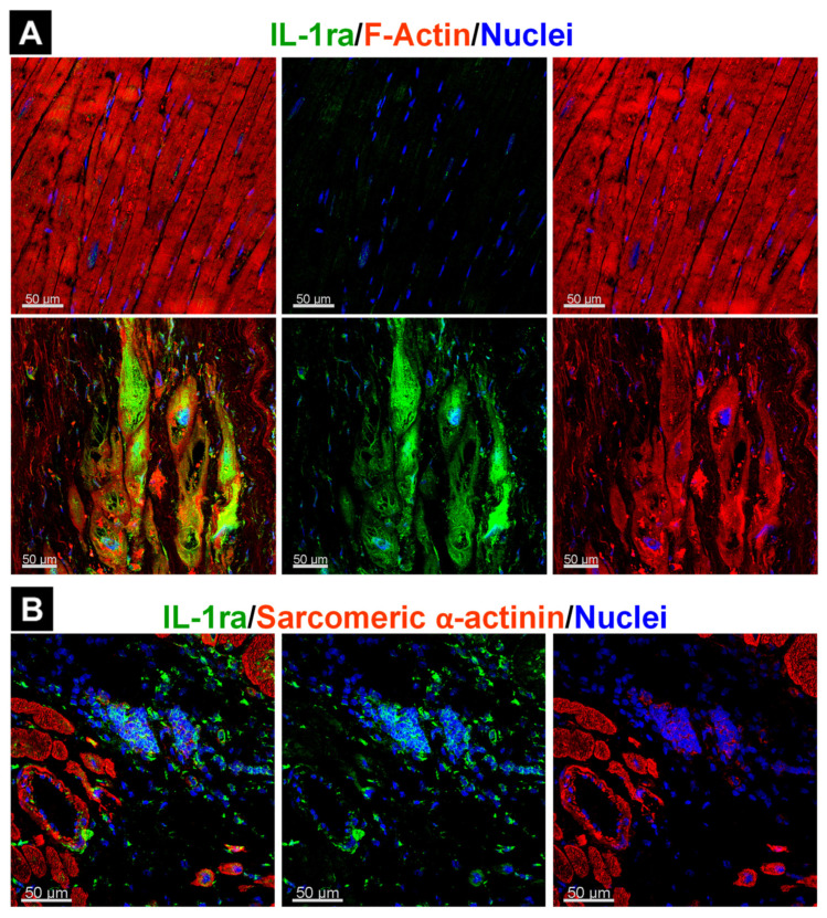
Oncostatin M (OSM), a member of the interleukin-6 family, functions as a major mediator of cardiomyocyte remodeling under pathological conditions. Its involvement in a variety of human cardiac diseases such as aortic stenosis, myocardial infarction, myocarditis, cardiac sarcoidosis, and various cardiomyopathies make the OSM receptor (OSMR) signaling cascades a promising therapeutic target. However, the development of pharmacological treatment strategies is highly challenging for many reasons. In mouse models of heart disease, OSM elicits opposing effects via activation of the type II receptor complex (OSMR/gp130). Short-term activation of OSMR/gp130 protects the heart after acute injury, whereas chronic activation promotes the development of heart failure. Furthermore, OSM has the ability to integrate signals from unrelated receptors that enhance fetal remodeling (dedifferentiation) of adult cardiomyocytes. Because OSM strongly stimulates the production and secretion of extracellular proteins, it is likely to exert systemic effects, which in turn, could influence cardiac remodeling. Compared with the mouse, the complexity of OSM signaling is even greater in humans because this cytokine also activates the type I leukemia inhibitory factor receptor complex (LIFR/gp130). In this article, we provide an overview of OSM-induced cardiomyocyte remodeling and discuss the consequences of OSMR/gp130 and LIFR/gp130 activation under acute and chronic conditions.

Keywords: cardiomyocyte; dedifferentiation; gp130; heart failure; inflammation; interleukin-6; leukemia inhibitory factor receptor; myocardial infarction; oncostatin M receptor; remodeling.


Conflict of interest statement

The authors have no conflict of interest to disclose.

Figures

Figure 1
Figure 1
Simplified scheme illustrating the members of the interleukin-6 family of cytokines and their receptor complexes involved in adult cardiomyocyte remodeling and the differential formation of receptor complexes by OSM in the human, rat, and mouse. IL-6, IL-11, CT-1, LIF, and OSM elicit receptor complex activation in cardiomyocytes. Amongst them, OSM showed the strongest morphological effect on cultured cardiomyocytes, whereas the other members exerted a strong and comparable effect, except for IL-6, which was less effective because the weakly expressed IL-6 receptor probably needs trans-signaling events. The common co-receptor gp130 may explain the similar morphological responses of cardiomyocytes exposed to the IL-6 family. IL-6 and IL-11 activate two homodimeric gp130 receptor complexes containing the IL-6 receptor and the IL-11 receptor, respectively. In contrast, CT-1, LIF, and OSM do not require an additional non-signaling receptor to form functional complexes because they signal through a complex consisting of gp130 and the LIFR. OSM is unique in terms of receptor binding in that it can bind to both the type I (LIFR/gp130) receptor complex and type II (OSMR/gp130) receptor complex in humans and rats, whereas it binds only to the type I complex in mice.
Figure 2
Figure 2
Schematic representation of morphological effects of activated type I and type II receptor complexes on cultured adult rat cardiomyocytes and IL-6 secretion. In all our screens, we utilized 20 ng/mL of albumin (Con), rat leukemia inhibitory factor (LIF, activates the rat LIFR/gp130 complex), mouse oncostatin M (mOSM, activates the rat OSMR/gp130 receptor complex), rat oncostatin M (rOSM, activates the rat LIFR/gp130 and the rat OSMR/gp130 complex), cardiotrophin-1 (CT1), transforming growth factor-β (TGFβ), tumor necrosis factor-α (TNFα), interleukin-1α (IL-1α), and fibroblast growth factor-2 (FGF2). (A) IL-6 ELISA showing concentrations of IL-6 in the cardiomyocyte culture supernatants 36 h after stimulation with albumin (n = 8), OSM (n = 8), LIF (n = 6), CT-1 (n = 6), TGFß (n = 4), TNFα (n = 4), and IL-1α (n = 2). Data represent the mean ± SEM. Statistical analysis was performed through an unpaired t test with Welch’s correction showing significances between Con and OSM **** p < 0.0001 and between Con and IL-1α ** p < 0.002. (B) Bright-field micrograph of a freshly isolated rat cardiomyocyte showing a complex three-dimensional structure with typical cross-striation. (C) Intercalated discs (stained with connexin-43 in red) are organized laterally (yellow arrows) and at the cell ends (white arrows). White ovals indicate nuclei and the green color marks sarcomeres (stained with sarcomeric α-actinin) in a freshly isolated cardiomyocyte. (D) Scheme summarizing the main morphological effects of rOSM, mOSM, LIF, and FGF2 after 5–7 days in culture. Cellular elongation and formation of multiple extensions were the most obvious changes after oncostatin M treatment. While elongation after rOSM stimulation was dominant, a certain amount of spreading might have become visible with increased culture time when cellular contacts were reestablished. It is important to note that serum (fetal calf serum, FCS) was only utilized for the initial plating of cardiomyocytes and then the cultures were kept without serum or any further growth enhancer/stimulant in these studies. For comparison, FGF-2-stimulated cultures show an increase in surface area but comparatively little elongation or formation of extensions.
Figure 3
Figure 3
The architecture of intercalated discs is changed in the diseased myocardium. (A) Connexin 43, which marks intercalated discs in the normal heart, is downregulated/rearranged in cardiomyocytes of the border and remote zone in the infarcted mouse myocardium. (B) P-ERM (a marker of activated Ezrin/Radixin/Moesin proteins) characterizes the formation of cardiomyocyte extensions (yellow arrows) in the infarcted mouse myocardium. (C) Similarly, connexin 43 is downregulated in patients with ischemic (ICM) and dilated (DCM) cardiomyopathy, indicating remodeling of intercalated discs. CON represents human control tissue.
Figure 4
Figure 4
Infiltration of OSM-releasing cells is associated with fetal remodeling, increased expression of the OSMR, and secreted proteins in the human myocardium. MyoC indicates myocarditis, CS is cardiac sarcoidosis, ICM means ischemic cardiomyopathy, and CON represents human control tissue. Actn1 is non-muscle α-actinin-1 (Actn1). (A) Increases in OSM-positive infiltrates (mainly macrophages) are consistent with an increased expression of the OSMR in cardiomyocytes. (B) Increased dedifferentiation characterizes fetal remodeling (image is modified from [13]). Note the strong and thin cardiomyocyte elongation. (C) Both single FGF23-positive and clusters of positive cardiomyocytes can be identified. OSM is currently the only known cytokine that induces FGF23 expression in cardiomyocytes.
Figure 5
Figure 5
Localization of the interleukin-1 receptor antagonist (IL-1ra) is influenced by the underlying disease. (A) IL-1ra in cardiomyocytes of a patient with ischemic cardiomyopathy. (B) IL-1ra in the granuloma of a patient with cardiac sarcoidosis.
Figure 6
Figure 6
Hypothetical overall model of OSM-driven protection, regeneration, and failure of the heart. (A) The development of hypertrophy is initiated by activation of the OSMR in patients with aortic stenosis. When activation of the OSMR cascades decreases, the amount of hypertrophic signals increases (FGFs, IGFs). (B) After a cardiac injury such as acute myocardial infarction, OSM-releasing infiltrates reduce damage/infarct expansion and extension by inducing fetal remodeling of cardiomyocytes. Macrophage infiltration is controlled by various chemokine families (Reg3, IL-7, MCPs). (C) Cardiomyocytes form extensions and restore cell-cell contacts. (D) Infiltration and inflammatory processes are downregulated by anti-inflammatory molecules (IL-1ra, TGF-β) and hypertrophic pathways are activated (FGFs, IGFs). (E) Fetal remodeling is downregulated and myocytes undergo hypertrophic remodeling. Surviving cardiomyocytes adapt to the increased workload by enlarging and accumulating sarcomeres. (F) If infiltration and inflammatory processes persist, chronically activated OSM receptors cause degeneration of cardiomyocytes. (G) Dying cells and elongation of surviving cardiomyocytes lead to dilatation of the myocardium.

Similar articles

Cited by

References

    1. Pang P.S., Komajda M., Gheorghiade M. The current and future management of acute heart failure syndromes. Eur. Heart J. 2010;31:784–793. doi: 10.1093/eurheartj/ehq040. - DOI - PubMed
    1. Roger V.L. Epidemiology of heart failure. Circ. Res. 2013;113:646–659. doi: 10.1161/CIRCRESAHA.113.300268. - DOI - PMC - PubMed
    1. Hou Y., Adrian-Segarra J.M., Richter M., Kubin N., Shin J., Werner I., Walther T., Schönburg M., Pöling J., Warnecke H., et al. Animal Models and “Omics” Technologies for Identification of Novel Biomarkers and Drug Targets to Prevent Heart Failure. BioMed Res. Int. 2015;2015:212910. doi: 10.1155/2015/212910. - DOI - PMC - PubMed
    1. Mann D.L. Innate immunity and the failing heart: The cytokine hypothesis revisited. Circ. Res. 2015;116:1254–1268. doi: 10.1161/CIRCRESAHA.116.302317. - DOI - PMC - PubMed
    1. Seta Y., Shan K., Bozkurt B., Oral H., Mann D. Basic mechanisms in heart failure: The cytokine hypothesis. J. Card. Fail. 1996;2:243–249. doi: 10.1016/S1071-9164(96)80047-9. - DOI - PubMed