Review
. 2022 Feb 5;23(3):1818.
doi: 10.3390/ijms23031818.

Human Papillomaviruses as Infectious Agents in Gynecological Cancers. Oncogenic Properties of Viral Proteins

Affiliations
Review

Human Papillomaviruses as Infectious Agents in Gynecological Cancers. Oncogenic Properties of Viral Proteins

Daria A Haręża et al. Int J Mol Sci. .

Abstract

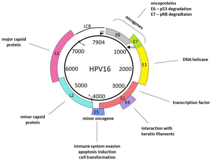
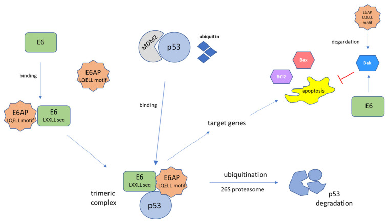
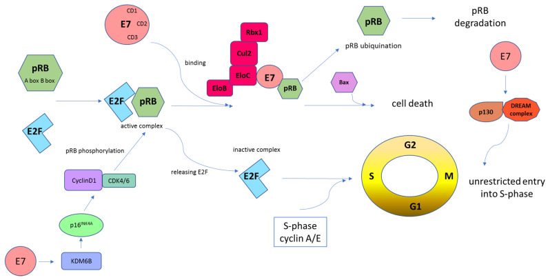
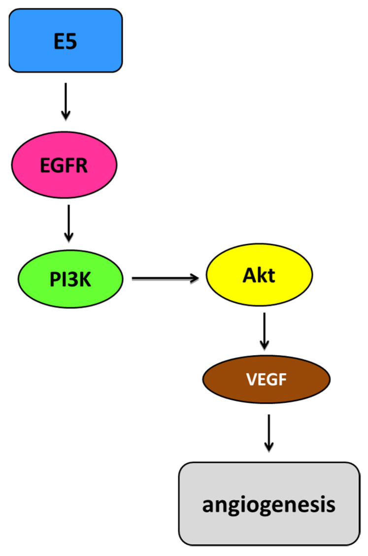
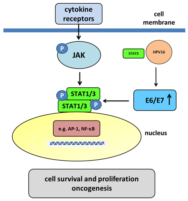
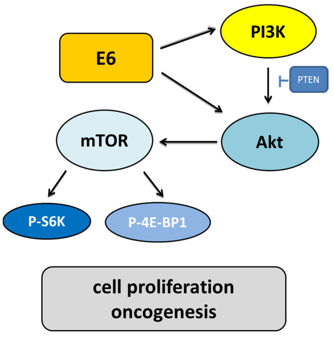
Human papillomaviruses (HPVs), which belong to the Papillomaviridae family, constitute a group of small nonenveloped double-stranded DNA viruses. HPV has a small genome that only encodes a few proteins, and it is also responsible for 5% of all human cancers, including cervical, vaginal, vulvar, penile, anal, and oropharyngeal cancers. HPV types may be classified as high- and low-risk genotypes (HR-HPVs and LR-HPVs, respectively) according to their oncogenic potential. HR-HPV 16 and 18 are the most common types worldwide and are the primary types that are responsible for most HPV-related cancers. The activity of the viral E6 and E7 oncoproteins, which interfere with critical cell cycle points such as suppressive tumor protein p53 (p53) and retinoblastoma protein (pRB), is the major contributor to HPV-induced neoplastic initiation and progression of carcinogenesis. In addition, the E5 protein might also play a significant role in tumorigenesis. The role of HPV in the pathogenesis of gynecological cancers is still not fully understood, which indicates a wide spectrum of potential research areas. This review focuses on HPV biology, the distribution of HPVs in gynecological cancers, the properties of viral oncoproteins, and the molecular mechanisms of carcinogenesis.

Keywords: gynecological cancers; human papillomavirus; oncoprotein.


Conflict of interest statement

The authors declare no conflict of interest.

Figures

Figure 1
Figure 1
Schematic representation of the HPV16 genome. The viral genome consists of the L1 and L2 genes, encoding major capsid protein L1, minor capsid protein L2, and the long regulatory region (LCR). LCR is the least conserved genome region. It contains a p97 promoter and numerous sequences that function as enhancers and silencers of viral transcription. The remaining HPV genome sequences comprise early genes: E1, E2, E4, E5, E6, and E7. E1 participates in viral DNA replication. E2 functions in transcriptional control, tethering of viral episomes, and, similarly to E1, participates in viral DNA replication. E4 interact with the cytoskeleton, while E5 participates in genome amplification. E6 and E7 interact with tumor suppressor proteins. Furthermore, both E5 and E6, as well as E7, also have many different functions, which are described in the “Oncoproteins” section. In the viral genome, we also distinguish late polyadenylation sites, AL; early polyadenylation sites, AE; and a late promoter, P670 [15].
Figure 2
Figure 2
Schematic structure of the E6 oncoprotein (based on Boulet G. et al., 2007) [71]. The E6 protein contains four CXXC motifs (blue). The functions of these motifs are associated with cellular proteins, transcriptional activation, transformation, and immortalization. The E6 carboxy-terminal domain contains a PDZ (PSD95/DLG/ZO-1-)-binding motif (red) engaged in the interaction with PDZ domain-containing proteins such as discs, large homolog 1 (DLG1), DLG4, scribble planar cell polarity protein (SCRIB), membrane-associated guanylate kinase (MAGI1), and tyrosine–protein phosphatase non-receptor type 13 (PTPN13) [10,71,72,73].
Figure 3
Figure 3
Schematic structure of the E7 oncoprotein (based on Boulet G. et al., 2007) [71]. The E7 oncoprotein contains three conserved regions (CR1/2/3). The NH2-terminal CR1 domain (green) is necessary for cellular transformation and pRB degradation but does not directly contribute to pRB binding. This domain interacts with host proteins such as E3 ubiquitin-protein ligase UBR4/p600 and p300/CBP-associated factor (PCAF), also known as K (lysine) acetyltransferase 2B [75]. The CR2 domain (pink) contains the pRB-binding core sequence LXCXE and a phosphorylation site for casein kinase II (CKII). The COOH-terminal CR3 domain (blue) is conserved and encodes a zinc finger domain containing two copies of the CXXC motif. This region is implicated in the association of pRB and other host cellular proteins. It is also critical for zinc-dependent dimerization and for mediating E7 interactions with cellular proteins crucial for cell cycle regulation and apoptosis (p21 and pRB) [10,71,75,76].
Figure 4
Figure 4
Schematic representation of E6-mediated tumor suppressor p53 protein degradation. The E6 protein, through a conserved binding motif containing the sequence LXXLL, binds to the LXXLL motif (LQELL) on the cellular E3 ubiquitin ligase E6AP. E6AP, E6, and p53 bind to each other and form a trimeric complex. This is followed by ubiquitin-dependent proteasomal degradation of the p53 protein. The polyubiquitinated p53 is then degraded by the 26S proteasome complex. The result of p53 degradation is the elimination of the trophic sentinel response to viral DNA synthesis and an increase in telomerase activity, leading to uncontrolled cell proliferation [94]. The function of E6 is the proteolytic inactivation of certain proapoptotic factors, such as p53, Bak, or Bax, through the ubiquitin–proteasome pathway. E6 can interact with Bak, Bax, and BCl2 directly, leading to the degradation of Bak in vivo. Moreover, E6 may block the Bak-mediated intrinsic mode of apoptosis through p53–E6AP interaction. Bak is also a target of the E6AP, while E6 stimulates the ubiquitin-mediated degradation of Bak through its interaction with Bak and E6AP [97]. MDM2—mouse double minute 2 homolog, a transcriptional target of p53.
Figure 5
Figure 5
Schematic representation of E7-mediated retinoblastoma tumor suppressor protein (pRB) inhibition. Cells must pass the G1 restriction point, which is under the control of pRB, to progress from the G1 to the S cell cycle phase. E2F transcription factors are bound and repressed by pRB via the A and B boxes. Then, this complex binds the HR-HPV E7 protein. E7, through the CD2 and CD3 regions, binds to the B box of pRB via its LXCXE motif. Following these interactions, the pRB–E2F complex is disturbed, which leads to abnormal cell progression into the S-phase of the cell cycle. HPV E7-mediated pRB degradation can be mediated by the cullin 2 (Cul2) ubiquitin ligase complex. This interaction occurs via the E7 CR1 domain and the C-terminal sequences and drives cell cycle progression by degradation of pRB and upregulation of CDK2 and cyclins A/E. In addition, the cyclin D1/CDK4/6 complex phosphorylates pRB, which promotes E2F release. Subsequently, cyclin A/E facilitates pRB phosphorylation, allowing S-phase entry. This whole mechanism leads to unrestricted entry into the S-phase and unrestrained cell proliferation [55,76]. The E7 oncoprotein causes the transcriptional induction of KDM6B and, as a result, the p16INK4A expression. Induced expression of p16INK4A results in a G1 cell cycle arrest by inhibiting phosphorylation of pRB by CDK4/6 kinases [101]. Moreover, E7 targets p130 specifically in the DREAM complex to remove the barrier to entry into the S-phase [103,104].
Figure 6
Figure 6
Simplified representation of the EGFR pathway mediated by E5. HPV E5 is involved in the activation of and increase in the epidermal growth factor receptor (EGFR) pathway depending on the ligand. Activated EGFR homodimers autophosphorylate, leading to increased activation of EGFR-related pathways such as the phosphoinositide 3-kinase (PI3K)/protein kinase B (Akt) pathway. Akt can affect upregulation of the vascular endothelial growth factor (VEGF), which consequently increases angiogenesis [113].
Figure 7
Figure 7
The JAK/STAT pathway mediated by the E6 and E7 oncoproteins. Membrane cytokine receptors have cytoplasmic tails in which inactive JAKs associate constitutively. The cytokine interaction with their receptors induces dimerization of these receptors. Interaction between the cytokine and its receptor results in the juxtaposition of JAKs, leading to their autophosphorylation. The activated JAKs then phosphorylate the receptor’s cytoplasmic tails on tyrosine residues, creating sites that allow the binding of other signaling molecules, such as STAT proteins. Cytoplasmic STATs bind to phosphorylated receptors, becoming substrates for the JAKs, which phosphorylate STATs on highly conserved tyrosine residues. After their phosphorylation, the STATs form homodimers or heterodimers that are capable of translocating to the nucleus and activating gene transcription. The E6/E7 oncoproteins decrease the translocation of STAT1 to the nucleus. A decrease in STAT1 is necessary for the amplification of the viral genome in the early stages of infection, meaning that STAT1 plays a protective role in the early phase of HPV infection. In the nucleus, transcription factors such as AP-1 and NF-κB, as well as STAT3 may play a regulatory role in HPV infection. HPV-infected cells produce large amounts of IL-6 for autocrine signaling and for increasing STAT3 activation. Some studies have suggested that STAT3 could bind to HPV16 upstream of the URR, driving the expression of E6/E7. Activated STAT3 results in an increase in the E6 and E7 oncoproteins. The oncoproteins promote a decrease in pRB and p53, which are the proteins that are responsible for the inhibition and arrest of the cell cycle and the promotion of apoptosis. HPV16 oncogenes downregulate the expression of IFN-responsive genes and upregulate proliferation-associated and NF-κB-responsive genes in cervical keratinocytes [121,130].
Figure 8
Figure 8
The role of the PI3K/Akt/mTOR signaling pathway induced by HPV infection. E6 activates PI3K through the receptor protein tyrosine kinase or direct interaction with PI3K. Throughout phosphorylation, PI3K activates Akt. Activated Akt influences cell growth through the mammalian targets of rapamycin (mTOR) and angiogenesis. Due to the upregulation of the ribosomal protein S6 kinase (S6K) and the blocking of the eukaryotic initiation factor 4E-binding protein (4E-BP), mTOR may increase cell proliferation. E6 also blocks tuberous sclerosis complex 1/2 (TSC1/2) to increase the mammalian target of rapamycin complex 1 (mTORC1) activity to increase cell growth and block the proapoptotic Bad and Bax proteins.

References

    1. de Martel C., Georges D., Bray F., Ferlay J., Clifford G.M. Global burden of cancer attributable to infections in 2018: A worldwide incidence analysis. Lancet Glob. Health. 2020;8:e180–e190. doi: 10.1016/S2214-109X(19)30488-7. - DOI - PubMed
    1. Bosch F.X., Broker T.R., Forman D., Moscicki A.B., Gillison M.L., Doorbar J., Stern P.L., Stanley M., Arbyn M., Poljak M., et al. ICO Monograph ‘Comprehensive Control of HPV Infections and Related Diseases’. Comprehensive control of human papillomavirus infections and related diseases. Vaccine. 2013;31:I1–I31. doi: 10.1016/j.vaccine.2013.07.026. - DOI - PMC - PubMed
    1. Walboomers J.M., Jacobs M.V., Manos M.M., Bosch F.X., Kummer J.A., Shah K.V., Snijders P.J., Peto J., Meijer C.J., Muñoz N. Human papillomavirus is a necessary cause of invasive cervical cancer worldwide. J. Pathol. 1999;189:12–19. doi: 10.1002/(SICI)1096-9896(199909)189:1<12::AID-PATH431>3.0.CO;2-F. - DOI - PubMed
    1. de Sanjose S., Quint W.G., Alemany L., Geraets D.T., Klaustermeier J.E., Lloveras B., Tous S., Felix A., Bravo L.E., Shin H.R., et al. Human papillomavirus genotype attribution in invasive cervical cancer: A retrospective cross-sectional worldwide study. Lancet Oncol. 2010;11:1048–1056. doi: 10.1016/S1470-2045(10)70230-8. - DOI - PubMed
    1. Bilyk O.O., Pande N.T., Pejovic T., Buchynska L.G. The frequency of Human Papillomavirus types 16, 18 in upper genital tract of women at high risk of developing ovarian cancer. Exp. Oncol. 2014;36:121–124. - PubMed

Substances