

Innate Lymphoid Cells and Intestinal Inflammatory Disorders
- PMID: 35163778
- PMCID: PMC8836863
- DOI: 10.3390/ijms23031856
Innate Lymphoid Cells and Intestinal Inflammatory Disorders
Abstract
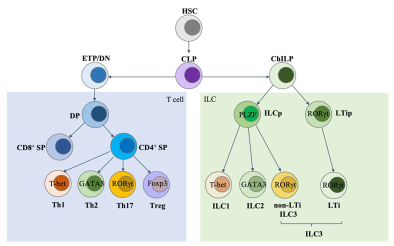
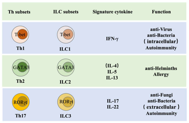
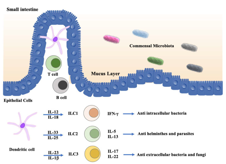
Innate lymphoid cells (ILCs) are a population of lymphoid cells that do not express T cell or B cell antigen-specific receptors. They are largely tissue-resident and enriched at mucosal sites to play a protective role against pathogens. ILCs mimic the functions of CD4 T helper (Th) subsets. Type 1 innate lymphoid cells (ILC1s) are defined by the expression of signature cytokine IFN-γ and the master transcription factor T-bet, involving in the type 1 immune response; ILC2s are characterized by the expression of signature cytokine IL-5/IL-13 and the master transcription factor GATA3, participating in the type 2 immune response; ILC3s are RORγt-expressing cells and are capable of producing IL-22 and IL-17 to maintain intestinal homeostasis. The discovery and investigation of ILCs over the past decades extends our knowledge beyond classical adaptive and innate immunology. In this review, we will focus on the roles of ILCs in intestinal inflammation and related disorders.
Keywords: ILC1; ILC2; ILC3; intestinal inflammation.
Conflict of interest statement
The authors declare no conflict of interest.
Figures
Similar articles
-
A T-bet gradient controls the fate and function of CCR6-RORγt+ innate lymphoid cells.Nature. 2013 Feb 14;494(7436):261-5. doi: 10.1038/nature11813. Epub 2013 Jan 16. Nature. 2013. PMID: 23334414
-
Heterogeneity and diversity of group 3 innate lymphoid cells: new cells on the block.Int Immunol. 2016 Jan;28(1):29-34. doi: 10.1093/intimm/dxv054. Epub 2015 Oct 13. Int Immunol. 2016. PMID: 26462712 Review.
-
Group 3 innate lymphoid cells (ILC3s): Origin, differentiation, and plasticity in humans and mice.Eur J Immunol. 2015 Aug;45(8):2171-82. doi: 10.1002/eji.201545598. Epub 2015 Jun 18. Eur J Immunol. 2015. PMID: 26031799 Review.
-
Subsets of ILC3-ILC1-like cells generate a diversity spectrum of innate lymphoid cells in human mucosal tissues.Nat Immunol. 2019 Aug;20(8):980-991. doi: 10.1038/s41590-019-0425-y. Epub 2019 Jun 17. Nat Immunol. 2019. PMID: 31209406 Free PMC article.
-
Enrichment of IL-17A+ IFN-γ+ and IL-22+ IFN-γ+ T cell subsets is associated with reduction of NKp44+ ILC3s in the terminal ileum of Crohn's disease patients.Clin Exp Immunol. 2017 Oct;190(1):143-153. doi: 10.1111/cei.12996. Epub 2017 Jul 7. Clin Exp Immunol. 2017. PMID: 28586085 Free PMC article.
Cited by
-
Therapeutic potential of mesenchymal stem cell-derived extracellular vesicles: A focus on inflammatory bowel disease.Clin Transl Med. 2024 Nov;14(11):e70075. doi: 10.1002/ctm2.70075. Clin Transl Med. 2024. PMID: 39488745 Free PMC article. Review.
-
Overview of the development, characterization, and function of human types 1, 2, and 3 innate lymphoid cells.Einstein (Sao Paulo). 2024 Nov 29;22:eRW1042. doi: 10.31744/einstein_journal/2024RW1042. eCollection 2024. Einstein (Sao Paulo). 2024. PMID: 39630753 Free PMC article. Review.
-
Reduced type 3 innate lymphoid cells related to worsening kidney function in renal dysfunction.Exp Biol Med (Maywood). 2023 Feb;248(3):242-252. doi: 10.1177/15353702221147561. Epub 2023 Jan 20. Exp Biol Med (Maywood). 2023. PMID: 36670544 Free PMC article.
-
The Genetic Background of Ankylosing Spondylitis Reveals a Distinct Overlap with Autoimmune Diseases: A Systematic Review.J Clin Med. 2025 May 23;14(11):3677. doi: 10.3390/jcm14113677. J Clin Med. 2025. PMID: 40507438 Free PMC article. Review.
-
Controversial role of ILC3s in intestinal diseases: A novelty perspective on immunotherapy.Front Immunol. 2023 Mar 28;14:1134636. doi: 10.3389/fimmu.2023.1134636. eCollection 2023. Front Immunol. 2023. PMID: 37063879 Free PMC article. Review.
References
-
- Klose C.S.N., Flach M., Möhle L., Rogell L., Hoyler T., Ebert K., Fabiunke C., Pfeifer D., Sexl V., Fonseca-Pereira D., et al. Differentiation of Type 1 ILCs from a Common Progenitor to All Helper-like Innate Lymphoid Cell Lineages. Cell. 2014;157:340–356. doi: 10.1016/j.cell.2014.03.030. - DOI - PubMed
-
- Zhong C., Zheng M., Cui K., Martins A.J., Hu G., Li D., Tessarollo L., Kozlov S., Keller J.R., Tsang J.S., et al. Differential Expression of the Transcription Factor GATA3 Specifies Lineage and Functions of Innate Lymphoid Cells. Immunity. 2019;52:83–95.e4. doi: 10.1016/j.immuni.2019.12.001. - DOI - PMC - PubMed
Publication types
MeSH terms
Substances
Grants and funding
LinkOut - more resources
Full Text Sources
Medical
Research Materials
Miscellaneous
