. 2022 Feb 7;23(3):1863.
doi: 10.3390/ijms23031863.

Cytoprotective Activity of Polyamines Is Associated with the Alternative Splicing of RAD51A Pre-mRNA in Normal Human CD4+ T Lymphocytes

Affiliations

Cytoprotective Activity of Polyamines Is Associated with the Alternative Splicing of RAD51A Pre-mRNA in Normal Human CD4+ T Lymphocytes

Yulia A Gladilina et al. Int J Mol Sci. .

Abstract

Physiological polyamines are ubiquitous polycations with pleiotropic biochemical activities, including regulation of gene expression and cell proliferation as well as modulation of cell signaling. They can also decrease DNA damage and promote cell survival. In the present study, we demonstrated that polyamines have cytoprotective effects on normal human CD4+ T lymphocytes but not on cancer Jurkat or K562 cells. Pretreatment of lymphocytes with polyamines resulted in a significant reduction in cells with DNA damage induced by doxorubicin, cisplatin, or irinotecan, leading to an increase in cell survival and viability. The induction of RAD51A expression was in response to DNA damage in both cancer and normal cells. However, in normal cells, putrescin pretreatment resulted in alternative splicing of RAD51A and the switch of the predominant expression from the splice variant with the deletion of exon 4 to the full-length variant. Induction of RAD51A alternative splicing by splice-switching oligonucleotides resulted in a decrease in DNA damage and cell protection against cisplatin-induced apoptosis. The results of this study suggest that the cytoprotective activity of polyamines is associated with the alternative splicing of RAD51A pre-mRNA in normal human CD4+ T lymphocytes. The difference in the sensitivity of normal and cancer cells to polyamines may become the basis for the use of these compounds to protect normal lymphocytes during lymphoblastic chemotherapy.

Keywords: DNA damage; alternative splicing; apoptosis; cytoprotection; polyamines.


Conflict of interest statement

The authors declare no conflict of interest.

Figures
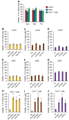
Figure 1
Figure 1
Cytoprotective activity of PAs in normal CD4+ T cells. (A) Results of the MTT test for cells incubated with 10 µM of each PA for 72 h. The results of the MTT test for cancer (BD) Jurkat, (EG) K562, or (HJ) normal CD4+ T cells incubated with genotoxic agents: 1 µM doxorubicin (Dox), 0.5 µM cisplatin (Cis), or 1 µM irinotecan (Irt) in the presence or absence of each PA. n = 8. * p ≤ 0.05 vs. cells not treated with PA. Spd: spermidine; Spm: spermine; Put: putrescine.
Figure 2
Figure 2
Decreased DNA damage in normal CD4+ T cells pretreated with PAs. Representative terminal deoxynucleotidyl transferase-mediated d-UTP nick end labeling (TUNEL) flow-cytometry diagrams for cancer cell lines (A) Jurkat, (C) K562, or (E) normal CD4+ T lymphocytes pretreated with each PA and incubated with genotoxic agents. The results of the TUNEL assay for flow cytometry for treated (B) Jurkat, (D) K562, or (F) CD4+ T cells. n = 4. * p ≤ 0.05. Con: control intact cells; Cis: cisplatin; Dox: doxorubicin; Irt: irinotecan; Spd: spermidine; Spm: spermine; Put: putrescine. n = 4. * p ≤ 0.05.
Figure 3
Figure 3
Prevention of cisplatin-induced apoptosis by putrescin (Put) in normal CD4+ T cells. Cells were labeled with annexin V-FITC and propidium iodide, and flow cytometry was performed 72 h after incubation with cisplatin (Cis). Representative plots for cancer cell lines (A) Jurkat, (C) K562, or (E) normal CD4+ T lymphocytes pretreated with Put and incubated with Cis. The proportions of live cells (lower left quadrants), apoptotic cells (lower right quadrants), and dead cells (two upper quadrants) are presented. (B,D,F) Histograms of live, apoptotic, and dead cells measured by flow cytometry. n = 4. * p ≤ 0.05.
Figure 4
Figure 4
Induced RAD51A expression in cells treated with cisplatin (Cis). mRNA levels of RAD51 members measured by real-time RT-PCR in cancer cell lines (A) Jurkat, (B) K562, or (C) normal CD4+ T lymphocytes pretreated with putrescin (Put) and incubated with Cis. mRNA levels were normalized relative to the expression of the reference gene 18S. (DF) Western blotting for RAD51 protein and the reference protein GAPDH in treated cells. (G) Results of RAD51 protein quantification relative to GAPDH. n = 4. * p ≤ 0.05 vs. control intact nontreated cells.
Figure 5
Figure 5
Induction of alternative splicing of RAD51A pre-mRNA by Put. mRNA levels of RAD51 splice variants measured by real-time RT-PCR in cancer cell lines (A) Jurkat, (B) K562, or (C) normal CD4+ T lymphocytes pretreated with Put and incubated with cisplatin (Cis). mRNA levels of splice variants were normalized relative to the expression of the reference gene 18S. n = 4. FL: full-length splice variant. ∆4: mRNA splice variant with the deletion of exon 4. ∆9: mRNA splice variant with the deletion of exon 9.
Figure 6
Figure 6
Schematic presentation of RAD51A alternative splicing. (A) Deletion of exon 4 as a result of alternative splicing and maturation of ∆4RAD51A mRNA. (B) Splicing regulator proteins SF2/ASF (shown as a green ellipse) interact with its binding sites (shown in bold red font) within intron 3 of RAD51A pre-mRNA. (C) Cell transfection with the 26-mer-specific antisense SSO for RAD51A (presented in red italic font) blocks the SF2/ASF proteins from binding to their binding sites. Exons are shown as red boxes, and introns are shown as blue boxes.
Figure 7
Figure 7
Modulation of RAD51A alternative splicing results in CD4+ T cell protection against cisplatin-induced apoptosis. Transfection efficiency of CD4+ T cells. Representative flow cytometry plots of cells transfected with Cy5.5-labeled (A) SSO for RAD51A or (B) a control 26-mer oligonucleotide 72 h posttransfection. (C) Efficiency of transfection. (D) Mean fluorescence intensity (MFI) of Cy5.5-positive cells. (E) mRNA levels of RAD51A measured by real-time RT-PCR in transfected CD4+ T lymphocytes incubated with cisplatin. mRNA levels were normalized relative to the expression of the reference gene 18S. (F) Western blotting for RAD51 protein and the reference protein GAPDH in transfected cells incubated with cisplatin. (G) Results of RAD51 protein quantification relative to GAPDH. (H) mRNA levels of RAD51A splice variants measured by real-time RT-PCR in CD4+ T cells. (I) Representative TUNEL flow cytometry diagrams for cells transfected with oligonucleotides and incubated with cisplatin. (J) Results of TUNEL assay for flow cytometry. (K) Representative flow cytometry plots for cells labeled with annexin V-FITC and propidium iodide after transfection with oligonucleotides and incubation with cisplatin. The proportions of live cells (low left quadrants), apoptotic cells (low right quadrants), and dead cells (two upper quadrants) are presented. (L) Histograms of live, apoptotic, and dead cells measured by flow cytometry. (M) Representative photo of the MTT test for transfected CD4+ T lymphocytes incubated with cisplatin. (N) Results of MTT test quantification. (O) Bright-field optical images of the MTT test for transfected CD4+ T cells exposed to Cis. AU: arbitrary units; Cis: cisplatin; CO: control nonspecific oligonucleotide; FL: full-length splice variant. SSO: splice-switching oligonucleotide. ∆4: mRNA splice variant with the deletion of exon 4. ∆9: mRNA splice variant with the deletion of exon 9. n = 4. * p ≤ 0.05 vs. initial nontransfected cells. # p ≤ 0.05.

Similar articles

Cited by

References

    1. Bronte V., Zanovello P. Regulation of immune responses by L-arginine metabolism. Nat. Rev. Immunol. 2005;5:641–654. doi: 10.1038/nri1668. - DOI - PubMed
    1. Sagar N.A., Tarafdar S., Agarwal S., Tarafdar A., Sharma S. Polyamines: Functions, Metabolism, and Role in Human Disease Management. Med. Sci. 2021;9:44. doi: 10.3390/medsci9020044. - DOI - PMC - PubMed
    1. Zarza X., Van Wijk R., Shabala L., Hunkeler A., Lefebvre M., Rodriguez-Villalón A., Shabala S., Tiburcio A.F., Heilmann I., Munnik T. Lipid kinases PIP5K7 and PIP5K9 are required for polyamine-triggered K(+) efflux in Arabidopsis roots. Plant J. 2020;104:416–432. doi: 10.1111/tpj.14932. - DOI - PMC - PubMed
    1. Dhara M., Matta J.A., Lei M., Knowland D., Yu H., Gu S., Bredt D.S. Polyamine regulation of ion channel assembly and implications for nicotinic acetylcholine receptor pharmacology. Nat. Commun. 2020;11:2799. doi: 10.1038/s41467-020-16629-3. - DOI - PMC - PubMed
    1. Prusov A.N., Smirnova T.A., Kolomijtseva G.Y. Thermodynamic Study of Interactions of Distamycin A with Chromatin in Rat Liver Nuclei in the Presence of Polyamines. Biochemistry (Moscow) 2018;83:1231–1244. doi: 10.1134/S0006297918100085. - DOI - PubMed