. 2022 Feb 7;23(3):1864.
doi: 10.3390/ijms23031864.

miR-132-3p Modulates DUSP9-Dependent p38/JNK Signaling Pathways to Enhance Inflammation in the Amnion Leading to Labor

Affiliations

miR-132-3p Modulates DUSP9-Dependent p38/JNK Signaling Pathways to Enhance Inflammation in the Amnion Leading to Labor

Zhuxia Zhong et al. Int J Mol Sci. .

Abstract

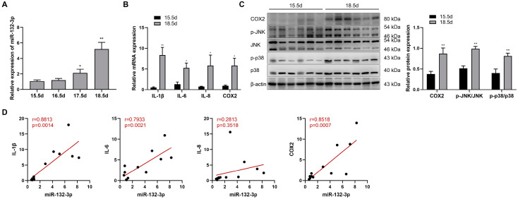
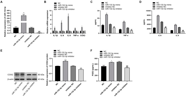
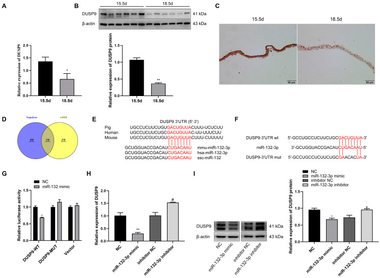
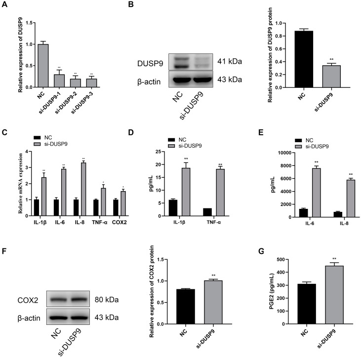
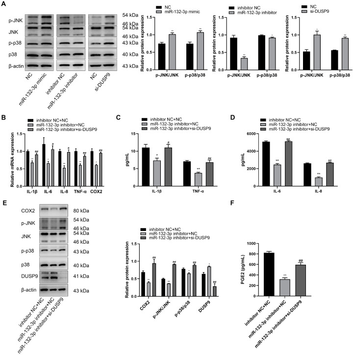
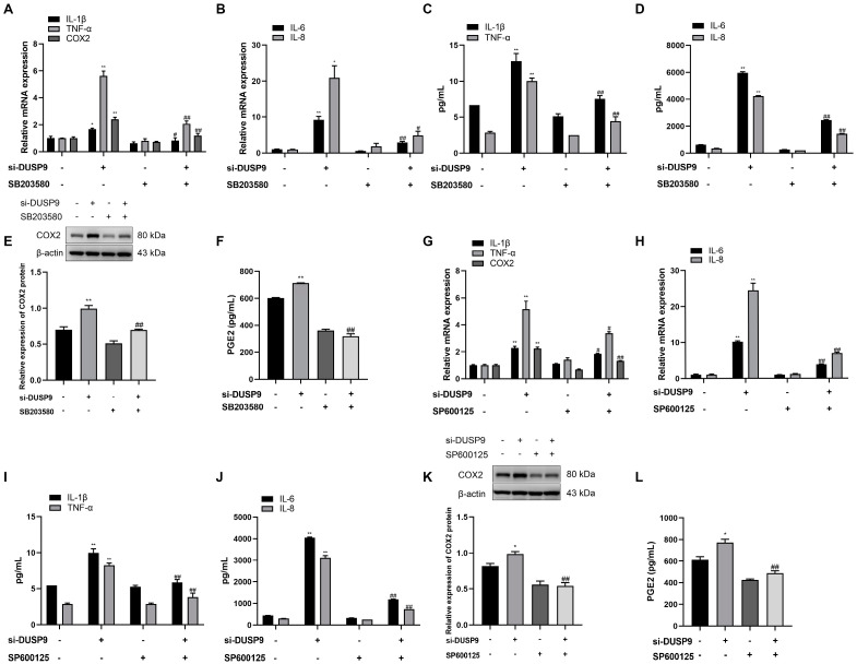
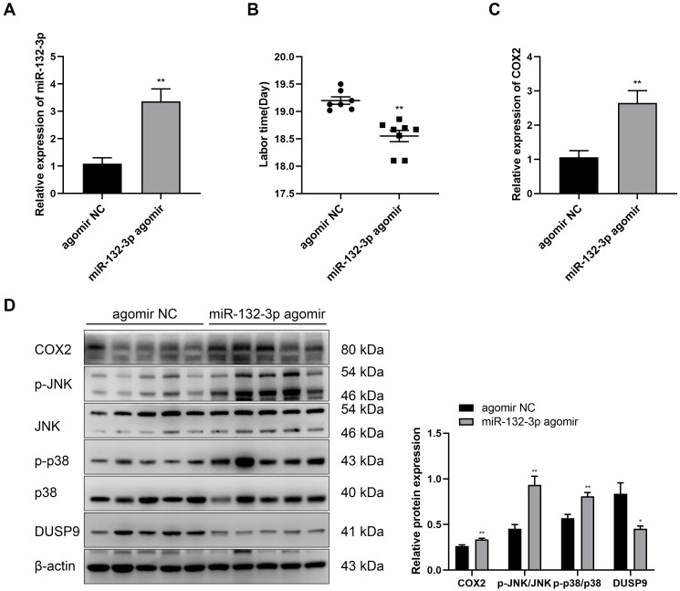
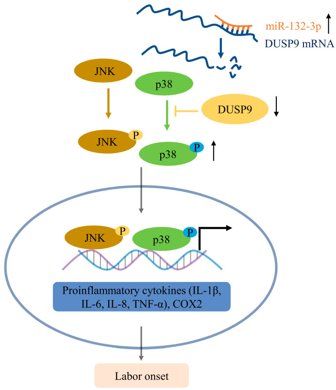
Labor is a process of inflammation and hormonal changes involving both fetal and maternal compartments. MicroRNA-132-3p (miR-132-3p) has been reported to be involved in the development of inflammation-related diseases. However, little is known about its potential role in labor onset. This study aimed to explore the mechanism of miR-132-3p in amnion for labor initiation. In the mouse amnion membranes, the expression of miR-132-3p was found to increase gradually during late gestation. In human amniotic epithelial cell line (WISH), upregulation of miR-132-3p was found to increase proinflammatory cytokines and cyclooxygenase 2 (COX2) as well as prostaglandin E2 (PGE2), which was suppressed by miR-132-3p inhibitor. Dual-specificity phosphatase 9 (DUSP9) was identified as a novel target gene of miR-132-3p, which could be negatively regulated by miR-132-3p. DUSP9 was present in the mouse amnion epithelial cells, with a decrease in its abundance at 18.5 days post coitum (dpc) relative to 15.5 dpc. Silencing DUSP9 was found to facilitate the expression of proinflammatory cytokines and COX2 as well as PGE2 secretion in WISH cells, which could be attenuated by p38 inhibitor SB203580 or JNK inhibitor SP600125. Additionally, intraperitoneal injection of pregnant mice with miR-132-3p agomir not only caused preterm birth, but also promoted the abundance of COX2 as well as phosphorylated JNK and p38 levels, and decreased DUSP9 level in mouse amnion membranes. Collectively, miR-132-3p might participate in inflammation and PGE2 release via targeting DUSP9-dependent p38 and JNK signaling pathways to cause preterm birth.

Keywords: DUSP9; amnion; inflammation; labor; miR-132-3p; p38/JNK.


Conflict of interest statement

The authors declare no conflict of interest.

Figures

Figure 1
Figure 1
Upregulated expressions of miR-132-3p and labor-associated genes are observed in mouse amnion membranes during late gestation. (A) RT-qPCR analysis of miR-132-3p level in the amnion membranes from pregnant mice at 15.5–18.5 days post-coitum (dpc) (n = 6–8). (B) RT-qPCR analysis of the mRNA expression of IL-1β, IL-6, IL-8, and COX2 in mouse amnion membranes at 15.5 and 18.5 dpc (n = 6–8). (C) Western blot analysis of the protein expression of COX2, JNK, and p38 as well as the phosphorylated JNK, and p38 levels in mouse amnion membranes at 15.5 and 18.5 dpc using β-actin as internal control (n = 6). (D) Gene expression correlation analysis between miR-132-3p and IL-1β, IL-6, IL-8, and COX2 mRNA in mouse amnion membranes at 15.5 dpc (n = 6–7) and 18.5 dpc (n = 6). Pearson correlation test and values of r and p are shown for each analysis. Data were shown as mean ± SEM. * p < 0.05, ** p < 0.01 vs. 15.5 dpc.
Figure 2
Figure 2
miR-132-3p promotes the expression of proinflammatory cytokines and COX2 as well as PGE2 in WISH cells. WISH cells were transfected with miR-132-3p mimic, miR-132-3p inhibitor, or the corresponding controls. (A) RT-qPCR analysis of miR-132-3p expression in WISH cells. (B) RT-qPCR analysis of the expression of IL-1β, IL-6, IL-8, TNF-α, and COX2 in WISH cells. Magnetic Luminex Assays of the secretion of IL-1β, TNF-α (C), IL-6, and IL-8 (D) in WISH cells. (E) Western blot analysis of COX2 level in WISH cells. (F) ELISA analysis of PGE2 secretion in WISH cells. Data were shown as mean ± SEM, n = 3. * p < 0.05, ** p < 0.01 vs. NC. # p < 0.05, ## p < 0.01 vs. inhibitor NC.
Figure 3
Figure 3
DUSP9 is a target gene of miR-132-3p. RT-qPCR (A) and Western blot (B) analysis of DUSP9 level in mouse amnion membranes at 15.5 and 18.5 dpc (n = 6–8). (C) Immunohistochemical staining for DUSP9 in the amnion membranes from pregnant mice at 15.5 or 18.5 dpc. Nuclei are stained with hematoxylin. Scale bar, 50 μm. (D) Venn diagram for the overlap of target genes predicted by TargetScan and miRDB. (E) Conservation of the miR-132-3p target sequence in DUSP9 3′UTR and mature miR-132-3p sequence among different species. (F) MiR-132-3p binding site in DUSP9 3′UTR predicted by TargetScan. (G) Luciferase reporter assay of WISH cells co-transfected with miR-132-3p mimic or NC and pmirGLO-DUSP9-3′UTR WT, pmirGLO-DUSP9-3′UTR MUT, or empty vector. RT-qPCR (H) and Western blot (I) analysis of the expression of DUSP9 in WISH cells transfected with miR-132-3p mimic, NC, miR-132-3p inhibitor, or inhibitor NC. Data were shown as mean ± SEM, n = 3. * p < 0.05, ** p < 0.01 vs. NC. # p < 0.05 vs. inhibitor NC.
Figure 4
Figure 4
Inhibition of DUSP9 promotes proinflammatory cytokines and COX2 as well as PGE2 expression in WISH cells. WISH cells were transfected with DUSP9 siRNA fragment and negative control, respectively. (A) RT-qPCR of DUSP9 expression. (B) Western blot analysis of DUSP9 protein level. (C) RT-qPCR analysis of the expression of IL-1β, IL-6, IL-8, TNF-α, and COX2. Magnetic Luminex Assays of the concentrations of IL-1β, TNF-α (D), IL-6, and IL-8 (E). (F) Western blot analysis of the protein expression of COX2. (G) ELISA analysis of PGE2 secretion level. Data were shown as mean ± SEM, n = 3. * p < 0.05, ** p < 0.01 vs. NC.
Figure 5
Figure 5
DUSP9 knockdown attenuates the anti-inflammatory effects of miR-132-3p loss in WISH cells. (A) Western blot analysis of the protein expression level of JNK and p38 as well as the phosphorylated JNK and p38 levels in WISH cells transfected with miR-132-3p mimic, miR-132-3p inhibitor, si-DUSP9, or the corresponding controls. * p < 0.05, ** p < 0.01 vs. the corresponding controls. (B) RT-qPCR analysis of the expression of IL-1β, IL-6, IL-8, TNF-α, and COX2 in WISH cells transfected with both inhibitor NC and NC, both miR-132-3p inhibitor and NC, or both miR-132-3p inhibitor and si-DUSP9. Magnetic Luminex Assays of the concentrations of IL-1β, TNF-α (C), IL-6, and IL-8 (D) in WISH cells transfected with both inhibitor NC and NC, both miR-132-3p inhibitor and NC, or both miR-132-3p inhibitor and si-DUSP9. (E) Western blot analysis of the protein expression level of COX2, JNK, p38, and DUSP9 as well as the phosphorylated JNK and p38 levels in WISH cells transfected with both inhibitor NC and NC, both miR-132-3p inhibitor and NC, or both miR-132-3p inhibitor and si-DUSP9. (F) ELISA analysis of PGE2 secretion in WISH cells transfected with both inhibitor NC and NC, both miR-132-3p inhibitor and NC, or both miR-132-3p inhibitor and si-DUSP9. Data were shown as mean ± SEM, n = 3. * p < 0.05, ** p < 0.01 vs. inhibitor NC + NC, # p < 0.05, ## p < 0.01 vs. miR-132-3p inhibitor + NC.
Figure 6
Figure 6
The role of p38 and JNK in DUSP9 siRNA-induced expression of proinflammatory cytokines and COX2 as well as PGE2 in WISH cells. WISH cells were transfected with si-DUSP9 or NC for 24 h, followed by 24 h of treatment with DMSO, p38 inhibitor SB203580 (10 μM), or JNK inhibitor SP600125 (20 μM). RT-qPCR analysis of the expression of IL-1β, TNF-α, COX2 (A,G), IL-6, and IL-8 (B,H). Magnetic Luminex Assays of the secretion of IL-1β, TNF-α (C,I), IL-6, and IL-8 (D,J). Western blot analysis of COX2 expression (E,K). ELISA analysis of PGE2 level (F,L). Data were shown as mean ± SEM, n = 3. * p < 0.05, ** p < 0.01 vs. control (0), # p < 0.05, ## p < 0.01 vs. si-DUSP9.
Figure 7
Figure 7
Overexpression of miR-132-3p causes preterm labor in mice. At day 15.5 of pregnancy, the pregnant mice were intraperitoneally injected with miR-132-3p agomir or agomir NC. (A) RT-qPCR analysis of the expression of miR-132-3p in mouse amnion membranes (n = 5). (B) Labor time of pregnant mice (n = 7–8). (C) RT-qPCR analysis of COX2 expression in mouse amnion membranes (n = 5). (D) Western blot analysis of the protein levels of COX2, JNK, p38, and DUSP9 as well as the phosphorylated JNK and p38 levels in mouse amnion membranes (n = 5). Data were shown as mean ± SEM. * p < 0.05, ** p < 0.01 vs. agomir NC.
Figure 8
Figure 8
Schematic diagram for the mechanism of miR-132-3p in activating p38 and JNK signaling pathways by downregulating DUSP9, thus promoting inflammation and PGE2 production for parturition initiation, corresponding to the increased expression of IL-1β, IL-6, IL-8, TNF-α, and COX2 in amnion epithelial cells.

Similar articles

Cited by

References

    1. Wang W., Chen Z.J., Myatt L., Sun K. 11β-HSD1 in human fetal membranes as a potential therapeutic target for preterm birth. Endocr. Rev. 2018;39:241–260. doi: 10.1210/er.2017-00188. - DOI - PubMed
    1. Menon R. Human fetal membranes at term: Dead tissue or signalers of parturition? Placenta. 2016;44:1–5. doi: 10.1016/j.placenta.2016.05.013. - DOI - PMC - PubMed
    1. Menon R., Richardson L.S., Lappas M. Fetal membrane architecture, aging and inflammation in pregnancy and parturition. Placenta. 2019;79:40–45. doi: 10.1016/j.placenta.2018.11.003. - DOI - PMC - PubMed
    1. Lundin-Schiller S., Mitchell M.D. The role of prostaglandins in human parturition. Prostaglandins Leukot. Essent. Fatty Acids. 1990;39:1–10. doi: 10.1016/0952-3278(90)90164-G. - DOI - PubMed
    1. Okazaki T., Casey M.L., Okita J.R., MacDonald P.C., Johnston J.M. Initiation of human parturition: XII. Biosynthesis and metabolism of prostaglandins in human fetal membranes and uterine decidua. Am. J. Obstet. Gynecol. 1981;139:373–381. doi: 10.1016/0002-9378(81)90312-4. - DOI - PubMed

MeSH terms