. 2022 Feb 7;23(3):1875.
doi: 10.3390/ijms23031875.

The Competitive Endogenous RNA (ceRNA) Regulation in Porcine Alveolar Macrophages (3D4/21) Infected by Swine Influenza Virus (H1N1 and H3N2)

Affiliations

The Competitive Endogenous RNA (ceRNA) Regulation in Porcine Alveolar Macrophages (3D4/21) Infected by Swine Influenza Virus (H1N1 and H3N2)

Chao-Hui Dai et al. Int J Mol Sci. .

Abstract

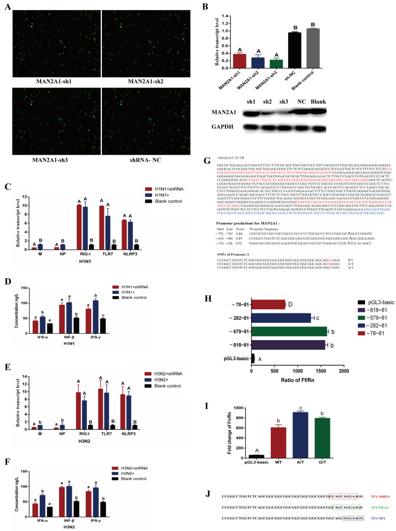
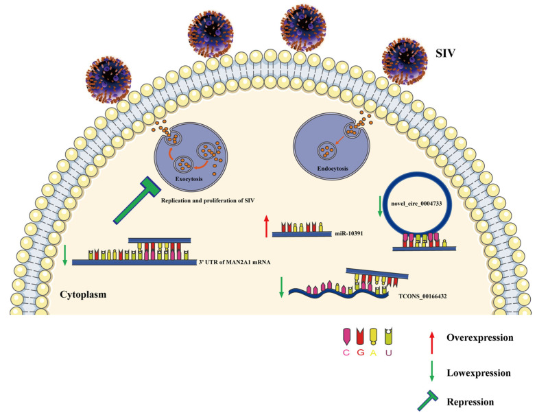
H1N1 and H3N2 are the two most common subtypes of swine influenza virus (SIV). They not only endanger the pig industry, but are also a huge risk of zoonotic diseases. However, the molecular mechanism and regulatory network of pigs (hosts) against influenza virus infection are still unclear. In this study, porcine alveolar macrophage cell (3D4/21) models infected by swine influenza virus (H1N1 and H3N2) were constructed. The expression profiles of miRNAs, mRNAs, lncRNAs and circRNAs after H1N1 and H3N2 infected 3D4/21 cells were revealed in this study. Then, two ceRNAs (TCONS_00166432-miR10391-MAN2A1 and novel_circ_0004733-miR10391-MAN2A1) that regulated H1N1 and H3N2 infection in 3D4/21 cells were verified by the methods of bioinformatics analysis, gene overexpression, gene interference, real-time quantitative PCR (qPCR), dual luciferase activity assay and RNA immunoprecipitation (RIP). In addition, the important candidate molecules (miR-10391, TCONS_00166432, and novel_circ_0004733) were identified by qPCR and enzyme linked immunosorbent assay (ELISA). Finally, the regulatory effect and possible molecular mechanism of the target gene MAN2A1 were identified by the methods of gene interference, qPCR, Western blot and ELISA. The results of this study suggested that TCONS_00166432 and novel_circ_0004733 could competitively bind miR-10391 to target the MAN2A1 gene to regulate swine influenza virus infecting 3D4/21 cells. This study reported for the first time the ceRNA networks involved in the regulation of the swine influenza virus infecting 3D4/21 cells, which provided a new insight into the molecular mechanism of 3D4/21 cells against swine influenza virus infection.

Keywords: ceRNA regulation; influenza virus; pig; porcine alveolar macrophage cell; whole transcriptome sequencing.


Conflict of interest statement

The authors declare no conflict of interest. The funders had no role in the design of the study; in the collection, analyses, or interpretation of data; in the writing of the manuscript or in the decision to publish the results.

Figures

Figure 1
Figure 1
Establishment of alveolar macrophage cell line (3D4/21) models infected by H1N1 and H3N2. (A,B) represent the qPCR results of virus genome genes (M and NP) in 3D4/21 cells infected by H1N1 and H3N2. (C,D) represent the qPCR results of host genes (RIG-I, TLR7 and NLRP3) in 3D4/21 cells infected by H1N1 and H3N2. (E,F) represent the ELISA results of virus-related cytokines in 3D4/21 cells infected by H1N1 and H3N2. (G) represents the microscope observation result of 3D4/21 cells infected by H1N1 and H3N2 (100-fold TCID50). (H) represents expression of NP gene in 3D4/21 cells infected by H1N1 and H3N2. (I) represents expression of NP protein in 3D4/21 cells infected by H1N1 and H3N2. For (AF), * Indicates that there is a significant difference between different virus doses at the same time point (p < 0.05), ** indicates p < 0.01. Different lowercase letters indicate that the dose of the same virus is significantly different at different time points (p < 0.05), and different capital letters indicate p < 0.01. 0, 1, 10, and 100 represent 0-fold TCID50 (no virus infection), 1-fold TCID50, 10-fold TCID50 and 100-fold TCID50, respectively (For H1N1, 1-fold TCID50, 10-fold TCID50 and 100-fold TCID50 mean that 100 µL H1N1 virus diluted 104.7, 103.7, and 102.7 times was used to infect 3D4/21 cells, respectively. For H3N2, 1-fold TCID50, 10-fold TCID50 and 100-fold TCID50 mean that 100 µL H3N2 virus diluted 102.75, 101.75, and 100.75 times was used to infect 3D4/21 cells, respectively). For (H), M represents DL500 DNA marker. 1–4 stands for H1N1 group, 5–8 stands for H3N2 group, and 9–12 stands for non-infected (NC) group.
Figure 2
Figure 2
Fluorescence quantitative PCR results of partially differential RNAs. (AD) represents differentially expressed mRNAs, miRNAs, lncRNAs and circRNAs, respectively. * indicates significant difference (p < 0.05), ** indicates significant difference (p < 0.01).
Figure 3
Figure 3
Verification of ceRNA networks in 3D4/21 cells infected by H1N1 and H3N2. (A) represents the predicted ceRNA networks diagram. The red diamond represents miRNA, the yellow ellipse represents lncRNA, the green hexagon represents circRNA, and the pink rectangle represents mRNA. (B) represents the expression of TCONS_00166432 and novel_circ_0004733 in nuclear and cytoplasmic of 3D4/21 cells. (C) represents the sequence alignment of targeted binding sites. (DF) represent fluorescence expression of 3D4/21 cells transfected by recombinant vectors. (G) represents efficiency detection results of overexpression and interference. Different lowercase letters indicate significant differences (p < 0.05), and different capital letters indicate significant differences (p < 0.01). (H) represents the test result of dual luciferase activity. * indicates significant difference (p < 0.05), ** indicates significant difference (p < 0.01). The same below. (I) represents the results of RNA immunoprecipitation (RIP).
Figure 4
Figure 4
The regulatory role of miR-10391, TCONS_00166432 and novel_circ_0004733 in the process of H1N1 and H3N2 infecting 3D4/21 cells. (AF) represents the effect of miR-10391, TCONS_00166432, and novel_circ_0004733 on the expression levels of viral genes and host genes in 3D4/21 cells infected by H1N1 and H3N2, respectively. (GL) represents the effect of miR-10391, TCONS_00166432 and novel_circ_0004733 on the secretion of cytokines in the supernatant of 3D4/21 cells infected by H1N1 and H3N2, respectively. Different lowercase letters indicate significant differences (p < 0.05), and different capital letters indicate significant differences (p < 0.01).
Figure 5
Figure 5
The regulatory role of MAN2A1 gene in the process of H1N1 and H3N2 infecting 3D4/21 cells. (A) represents fluorescence expression of 3D4/21 cells transfected by MAN2A1 interference vectors. (B) represents the qPCR and Western blot results of the MAN2A1 gene in 3D4/21 cells transfected by MAN2A1 interference vectors. Different lowercase letters indicate significant differences (p < 0.05), and different capital letters indicate significant differences (p < 0.01). The same as below. (C,E) represent the effects of MAN2A1 gene on the expression levels of viral genes and host genes in 3D4/21 cells infected by H1N1 and H3N2. (D,F) represent the effects of MAN2A1 gene on the secretion of cytokines in the supernatant of 3D4/21 cells infected by H1N1 and H3N2. (G) represents prediction of MAN2A1 gene promoter. The red letters represent the predicted core promoter sequence, and the blue letters represent the transcript sequence of the MAN2A1 gene. (H) represents the result of dual luciferase detection of MAN2A1 gene promoter. (I) represents the effect of MAN2A1 gene promoter SNP on transcriptional activity. (J) represents the transcription factor bound by the SNP sequence in the core promoter region of MAN2A1 gene. TF represents the abbreviation of transcription factor.
Figure 6
Figure 6
A schematic model displaying the role of TCONS_00166432/novel_circ_0004733-miR-10391-MAN2A1 in 3D4/21 cells infected by SIV.

Similar articles

Cited by

References

    1. Sun Y., Ma S., Wei W., Xiao A., Xu Y., Dong J., Wang X. Epidemiological characteristics, diagnosis and prevention measures of swine influenza virus. Swine Ind. Sci. 2014;31:94–95. (In Chinese)
    1. Wang L., Bi Y., Cao Y., Li H. Isolation, identification and biological characterization of swine influenza virus Guangdong isolates. Chin. J. Vet. Sci. Technol. 2003;2:17–21. (In Chinese)
    1. Thacker E., Janke B. Swine influenza virus: Zoonotic potential and vaccination strategies for the control of avian and swine influenzas. J. Infect. Dis. 2008;197:S19–S24. doi: 10.1086/524988. - DOI - PubMed
    1. Sun H., Xiao Y., Liu J., Wang D., Li F., Wang C., Li C., Zhu J., Song J., Sun H., et al. Prevalent Eurasian avian-like H1N1 swine influenza virus with 2009 pandemic viral genes facilitating human infection. Proc. Natl. Acad. Sci. USA. 2020;117:17204–17210. doi: 10.1073/pnas.1921186117. - DOI - PMC - PubMed
    1. Dhakal S., Hiremath J., Bondra K., Lakshmanappa Y.S., Shyu D., Ouyang K., Kang K., Binjawadagi B., Goodman J., Tabynov K., et al. Biodegradable nanoparticle delivery of inactivated swine influenza virus vaccine provides heterologous cell-mediated immune response in pigs. J. Control. Release. 2017;247:194–205. doi: 10.1016/j.jconrel.2016.12.039. - DOI - PubMed

LinkOut - more resources