

Regulatory T Cell Apoptosis during Preeclampsia May Be Prevented by Gal-2
- PMID: 35163802
- PMCID: PMC8836599
- DOI: 10.3390/ijms23031880
Regulatory T Cell Apoptosis during Preeclampsia May Be Prevented by Gal-2
Abstract
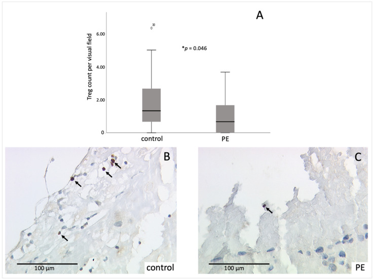
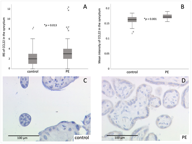
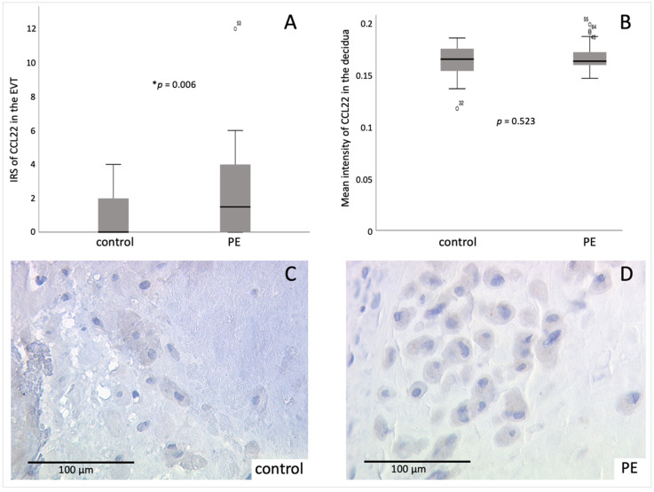
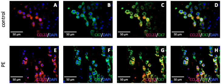
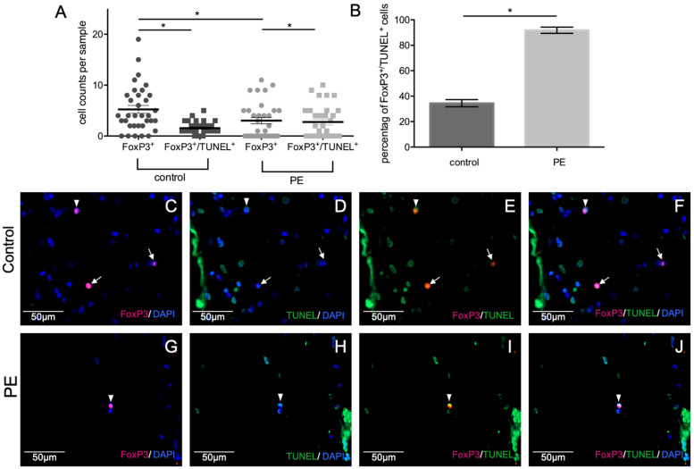
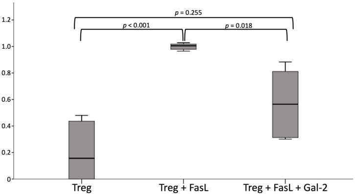
There are several open questions to be answered regarding the pathophysiology of the development of preeclampsia (PE). Numerous factors are involved in its genesis, such as defective placentation, vascular impairment, and an altered immune response. The activation of the adaptive and innate immune system represents an immunologic, particularity during PE. Proinflammatory cytokines are predominantly produced, whereas immune regulatory and immune suppressive factors are diminished in PE. In the present study, we focused on the recruitment of regulatory T cells (Tregs) which are key players in processes mediating immune tolerance. To identify Tregs in the decidua, an immunohistochemical staining of FoxP3 of 32 PE and 34 control placentas was performed. A clearly reduced number of FoxP3-positive cells in the decidua of preeclamptic women could be shown in our analysis (p = 0.036). Furthermore, CCL22, a well-known Treg chemoattractant, was immunohistochemically evaluated. Interestingly, CCL22 expression was increased at the maternal-fetal interface in PE-affected pregnancies (psyncytiotrophoblast = 0.035, pdecidua = 0.004). Therefore, the hypothesis that Tregs undergo apoptosis at the materno-fetal interface during PE was generated, and verified by FoxP3/TUNEL (TdT-mediated dUTP-biotin nick end labeling) staining. Galectin-2 (Gal-2), a member of the family of carbohydrate-binding proteins, which is known to be downregulated during PE, seems to play a pivotal role in T cell apoptosis. By performing a cell culture experiment with isolated Tregs, we could identify Gal-2 as a factor that seems to prevent the apoptosis of Tregs. Our findings point to a cascade of apoptosis of Tregs at the materno-fetal interface during PE. Gal-2 might be a potential therapeutic target in PE to regulate immune tolerance.
Keywords: Galectin-2; apoptosis; preeclampsia; regulatory T cells.
Conflict of interest statement
S.M. (Sven Mahner) received research support, honoraria, and travel expenses from AbbVie, AstraZeneca, Clovis, Eisai, GlaxoSmithKline, Medac, MSD, Novartis, Olympus, PharmaMar, Roche, Sensor Kinesis, Teva, and Tesaro. He is also on relevant advisory boards. T.K. holds stock in Roche, and a relative is employed by Roche.
Figures
Similar articles
-
Galectin 2 (gal-2) expression is downregulated on protein and mRNA level in placentas of preeclamptic (PE) patients.Placenta. 2015 Apr;36(4):438-45. doi: 10.1016/j.placenta.2015.01.198. Epub 2015 Feb 7. Placenta. 2015. PMID: 25707742
-
Abnormal lymphatic vessel development is associated with decreased decidual regulatory T cells in severe preeclampsia.Am J Reprod Immunol. 2018 Jul;80(1):e12970. doi: 10.1111/aji.12970. Epub 2018 May 14. Am J Reprod Immunol. 2018. PMID: 29756666
-
Decidual memory T-cell subsets and memory T-cell stimulatory cytokines in early- and late-onset preeclampsia.Am J Reprod Immunol. 2020 Oct;84(4):e13293. doi: 10.1111/aji.13293. Epub 2020 Jul 8. Am J Reprod Immunol. 2020. PMID: 32572999 Free PMC article.
-
New immune horizons in therapeutics and diagnostic approaches to Preeclampsia.Am J Reprod Immunol. 2023 Feb;89(2):e13670. doi: 10.1111/aji.13670. Epub 2023 Jan 2. Am J Reprod Immunol. 2023. PMID: 36565013 Review.
-
Regulatory T Cells in Pregnancy: It Is Not All About FoxP3.Front Immunol. 2020 Jun 23;11:1182. doi: 10.3389/fimmu.2020.01182. eCollection 2020. Front Immunol. 2020. PMID: 32655556 Free PMC article. Review.
Cited by
-
Galectins: Important Regulators in Normal and Pathologic Pregnancies.Int J Mol Sci. 2022 Sep 3;23(17):10110. doi: 10.3390/ijms231710110. Int J Mol Sci. 2022. PMID: 36077508 Free PMC article. Review.
-
Placental galectins regulate innate and adaptive immune responses in pregnancy.Front Immunol. 2022 Dec 28;13:1088024. doi: 10.3389/fimmu.2022.1088024. eCollection 2022. Front Immunol. 2022. PMID: 36643922 Free PMC article.
-
Investigation of LGALS2 expression in the TCGA database reveals its clinical relevance in breast cancer immunotherapy and drug resistance.Sci Rep. 2023 Oct 14;13(1):17445. doi: 10.1038/s41598-023-44777-1. Sci Rep. 2023. PMID: 37838802 Free PMC article.
-
The regulated cell death at the maternal-fetal interface: beneficial or detrimental?Cell Death Discov. 2024 Feb 26;10(1):100. doi: 10.1038/s41420-024-01867-x. Cell Death Discov. 2024. PMID: 38409106 Free PMC article. Review.
-
A comprehensive review of the roles of T-cell immunity in preeclampsia.Front Immunol. 2025 Feb 6;16:1476123. doi: 10.3389/fimmu.2025.1476123. eCollection 2025. Front Immunol. 2025. PMID: 39981257 Free PMC article. Review.
References
-
- Park Y., Lee H.J., Jung Y.J., Kwon H.Y., Kim H., Lee J., Kim Y.H., Maeng Y.S., Kwon J.Y. CD133+/C-kit+Lin(-) endothelial progenitor cells in fetal circulation demonstrate impaired differentiation potency in severe preeclampsia. Pregnancy Hypertens. 2019;15:146–153. doi: 10.1016/j.preghy.2018.12.005. - DOI - PubMed
MeSH terms
Substances
Grants and funding
LinkOut - more resources
Full Text Sources
Molecular Biology Databases
