Review
. 2022 Feb 9;23(3):1912.
doi: 10.3390/ijms23031912.

Mitochondrial-Targeted Therapy for Doxorubicin-Induced Cardiotoxicity

Affiliations
Review

Mitochondrial-Targeted Therapy for Doxorubicin-Induced Cardiotoxicity

Bin Bin Wu et al. Int J Mol Sci. .

Abstract

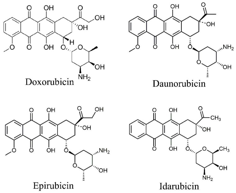
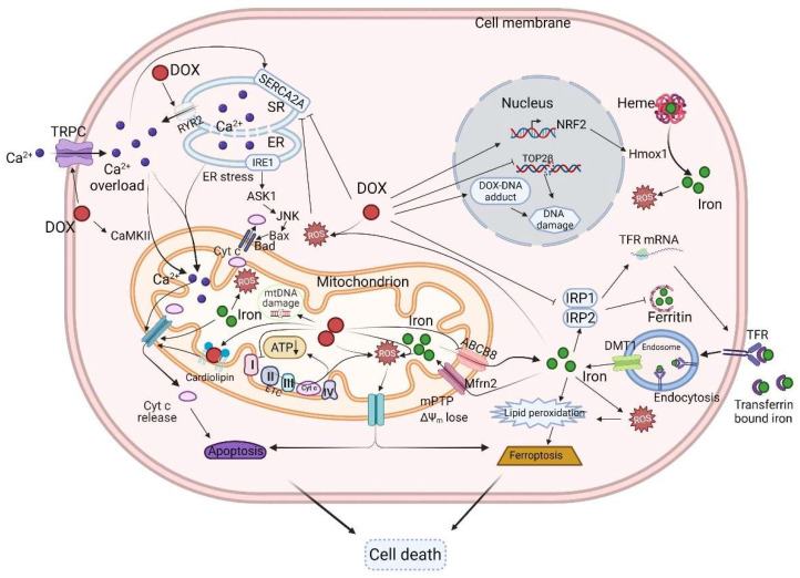
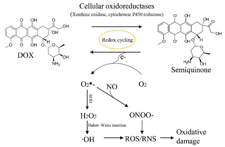
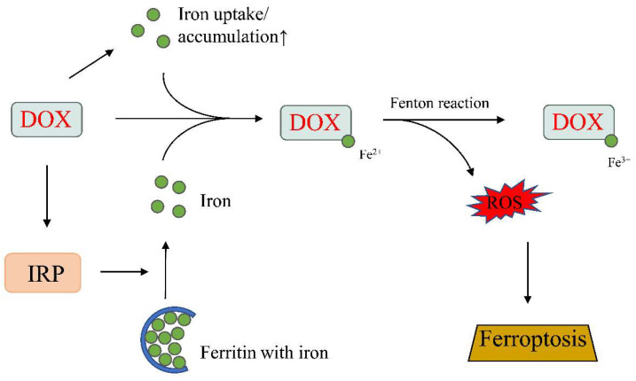
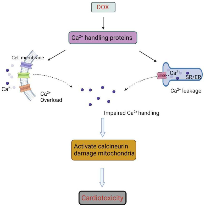
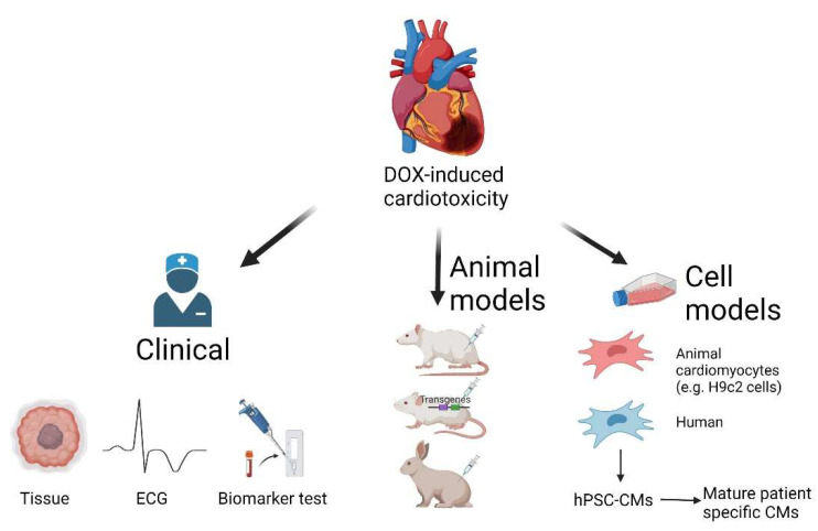
Anthracyclines, such as doxorubicin, are effective chemotherapeutic agents for the treatment of cancer, but their clinical use is associated with severe and potentially life-threatening cardiotoxicity. Despite decades of research, treatment options remain limited. The mitochondria is commonly considered to be the main target of doxorubicin and mitochondrial dysfunction is the hallmark of doxorubicin-induced cardiotoxicity. Here, we review the pathogenic mechanisms of doxorubicin-induced cardiotoxicity and present an update on cardioprotective strategies for this disorder. Specifically, we focus on strategies that can protect the mitochondria and cover different therapeutic modalities encompassing small molecules, post-transcriptional regulators, and mitochondrial transfer. We also discuss the shortcomings of existing models of doxorubicin-induced cardiotoxicity and explore advances in the use of human pluripotent stem cell derived cardiomyocytes as a platform to facilitate the identification of novel treatments against this disorder.

Keywords: anthracyclines; cardiotoxicity; doxorubicin (DOX); hPSC-cardiomyocytes; human pluripotent stem cells (hPSC); mitochondria.


Conflict of interest statement

The authors declare no conflict of interest.

Figures

Figure 1
Figure 1
The chemical structure of anthracyclines.
Figure 2
Figure 2
Schematic diagram of the mechanisms of DOX-induced cardiotoxicity. DOX can suppress the function of the ETC, and thereby reduce ATP levels. Increased ROS production induces the opening of the mPTP and the release of pro-apoptotic proteins such as cyt C. DOX can also induce calcium overload by altering the levels and activities of Ca2+ handling proteins such as TRPC, RYR2, and SERCA2A. DOX can disturb iron uptake and storage, leading to iron overload, apoptosis, and ferroptosis. DOX can complex with topoisomerase to induce DNA damage. DOX, doxorubicin; ER, endoplasmic reticulum; TRPC, transient receptor potential canonical; IRP, iron-regulatory protein; TfR, transferrin receptor; DMT1, divalent metal transporter 1; mPTP, mitochondrial permeability transition pore; Δψm, mitochondrial membrane potential; TOP2β, topoisomerase 2β; RYR2, ryanodine receptor 2; SERCA2A, sarcoplasmic/endoplasmic reticulum calcium ATPase 2; Mfrn2, mitoferrin-2; ABCB8, ABC protein B8; ETC, electron transport chain; Cyt C, cytochrome C. Created with BioRender.com (accessed on 28 December 2021).
Figure 3
Figure 3
Schematic diagram of DOX-induced intracellular ROS generation. DOX was reduced to semiquinone by cellular oxidoreductases, and then parent quinone was regenerated by transferring an electron to oxygen (O2). The resulting superoxide (O2) radical initiates the formation of ROS. NO, nitric oxide; RNS, reactive nitrogen species; SOD, superoxide dismutase.
Figure 4
Figure 4
DOX-induced ferroptosis. DOX promotes iron uptake and accumulation, increases ROS production, and induces ferroptosis. IRP, iron-regulatory protein. Up arrow, increase. Created with BioRender.com (accessed on 28 December 2021).
Figure 5
Figure 5
DOX can induce calcium overload by increasing calcium influx and intracellular calcium release. SR, sarcoplasmic reticulum; ER, endoplasmic reticulum. Created with BioRender.com (accessed on 28 December 2021).
Figure 6
Figure 6
Research models for DOX-induced cardiotoxicity. Created with BioRender.com (accessed on 28 December 2021).

Similar articles

Cited by

References

    1. Harake D., Franco V.I., Henkel J.M., Miller T.L., Lipshultz S.E. Cardiotoxicity in Childhood Cancer Survivors: Strategies for Prevention and Management. Future Cardiol. 2012;8:647–670. doi: 10.2217/fca.12.44. - DOI - PMC - PubMed
    1. Lipshultz S.E., Rifai N., Dalton V.M., Levy D.E., Silverman L.B., Lipsitz S.R., Colan S.D., Asselin B.L., Barr R.D., Clavell L.A., et al. The Effect of Dexrazoxane on Myocardial Injury in Doxorubicin-Treated Children with Acute Lymphoblastic Leukemia. N. Engl. J. Med. 2004;351:145–153. doi: 10.1056/NEJMoa035153. - DOI - PubMed
    1. Minotti G., Menna P., Salvatorelli E., Cairo G., Gianni L. Anthracyclines: Molecular Advances and Pharmacologie Developments in Antitumor Activity and Cardiotoxicity. Pharmacol. Rev. 2004;56:185–229. doi: 10.1124/pr.56.2.6. - DOI - PubMed
    1. Kolaric K., Bradamante V., Cervek J., Cieslinska A., Cisarz-Filipcak E., Denisov L.E., Donat D., Drosik K., Gershanovic M., Hudziec P., et al. A Phase II Trial of Cardioprotection with Cardioxane (ICRF-187) in Patients with Advanced Breast Cancer Receiving 5-Fluorouracil, Doxorubicin and Cyclophosphamide. Oncology. 1995;52:251–255. doi: 10.1159/000227467. - DOI - PubMed
    1. Swain S.M., Whaley F.S., Gerber M.C., Weisberg S., York M., Spicer D., Jones S.E., Wadler S., Desai A., Vogel C., et al. Cardioprotection with Dexrazoxane for Doxorubicin-Containing Therapy in Advanced Breast Cancer. J. Clin. Oncol. 1997;15:1318–1332. doi: 10.1200/JCO.1997.15.4.1318. - DOI - PubMed

LinkOut - more resources