. 2022 Jan 26;27(3):812.
doi: 10.3390/molecules27030812.

Spin Trapping Hydroxyl and Aryl Radicals of One-Electron Reduced Anticancer Benzotriazine 1,4-Dioxides

Affiliations

Spin Trapping Hydroxyl and Aryl Radicals of One-Electron Reduced Anticancer Benzotriazine 1,4-Dioxides

Wen Qi et al. Molecules. .

Abstract

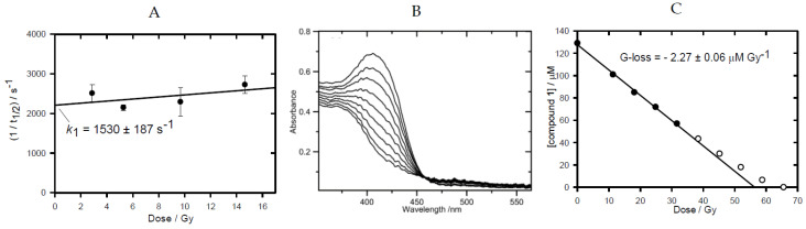
Hypoxia in tumors results in resistance to both chemotherapy and radiotherapy treatments but affords an environment in which hypoxia-activated prodrugs (HAP) are activated upon bioreduction to release targeted cytotoxins. The benzotriazine 1,4-di-N-oxide (BTO) HAP, tirapazamine (TPZ, 1), has undergone extensive clinical evaluation in combination with radiotherapy to assist in the killing of hypoxic tumor cells. Although compound 1 did not gain approval for clinical use, it has spurred on the development of other BTOs, such as the 3-alkyl analogue, SN30000, 2. There is general agreement that the cytotoxin(s) from BTOs arise from the one-electron reduced form of the compounds. Identifying the cytotoxic radicals, and whether they play a role in the selective killing of hypoxic tumor cells, is important for continued development of the BTO class of anticancer prodrugs. In this study, nitrone spin-traps, combined with electron spin resonance, give evidence for the formation of aryl radicals from compounds 1, 2 and 3-phenyl analogues, compounds 3 and 4, which form carbon C-centered radicals. In addition, high concentrations of DEPMPO (5-(diethoxyphosphoryl)-5-methyl-1-pyrroline N-oxide) spin-trap the •OH radical. The combination of spin-traps with high concentrations of DMSO and methanol also give evidence for the involvement of strongly oxidizing radicals. The failure to spin-trap methyl radicals with PBN (N-tert-butylphenylnitrone) on the bioreduction of compound 2, in the presence of DMSO, implies that free •OH radicals are not released from the protonated radical anions of compound 2. The spin-trapping of •OH radicals by high concentrations of DEPMPO, and the radical species arising from DMSO and methanol give both direct and indirect evidence for the scavenging of •OH radicals that are involved in an intramolecular process. Hypoxia-selective cytotoxicity is not related to the formation of aryl radicals from the BTO compounds as they are associated with high aerobic cytotoxicity.

Keywords: aryl radical; benzotriazine 1,4-dioxide; cytochrome P450 oxidoreductase; cytotoxicity; electron spin resonance; hydroxyl radical; hypoxia-activated prodrug; tirapazamine.


Conflict of interest statement

The authors declare no conflict of interest. The funders had no role in the design of the study, in the collection, analyses, or interpretation of data, in the writing of the manuscript, or in the decision to publish the results.

Figures

Figure 1
Figure 1
(A) Structures of compounds used in this study. (B) Scheme showing the activation of TPZ following oxygen-sensitive one-electron reduction to produce the radical anion intermediate and possible unimolecular routes of dehydration and homolytic fragmentation of the N-OH bond of its protonated form to produce aryl, BTZ and •OH radicals.
Figure 2
Figure 2
Radiation chemistry data. (A) Dependence of the elimination rate on the radiation dose, measured at 530 nm (k1 is the value of the intercept). The N2O-saturated solution (pH 7) contained compound 3 (100 µM) and sodium formate (0.1 M). (B) Stepwise changes in absorption spectrum with accumulated radiation dose in N2-saturated solution, containing compound 1 (129 μM), sodium formate (0.1 M) and phosphate buffer (2.5 mM) at pH 7. (C) Changes in absorption at 407 nm upon stepwise irradiation of compound 1, as displayed in (B). The G-loss value is calculated from the linear regression fit to the initial points.
Figure 3
Figure 3
EPR spectra obtained for compound 2 and their simulation. (A) (a) EPR spectra (100 scans) obtained on reduction of compound 2 (17 mM) by POR (14 ng/mL) in anaerobic solutions at 37 °C containing phosphate buffer (50 mM, pH 7), DTPA (100 µM), SOD (300 units/mL), catalase (1500 units/mL), glucose-6-phosphate (10 mM), glucose-6-phosphate-dehydrogenase (13 units/mL), and NADPH (1 mM) in presence of PBN (250 mM); (b) simulated spectrum of PBN-aryl (0.91) and PBN-C-centered species (0.07), r = 0.94. (B) (a) EPR spectra (70 scans) obtained on reduction of compound 2 (16 mM) under the same conditions as for A with DMSO (2 M) added; (b) simulated spectrum of PBN-CH2(CH3)SO (0.74) and PBN-aryl (0.26), r = 0.99. (C) (a) EPR spectra (50 scans) obtained on reduction of 2 (18 mM) under the same conditions as for A with DEPMPO (250 mM); (b) simulated spectrum of DEPMPO-aryl (0.49), DEPMPO-OH (0.39) and DEPMPO-C-centered species (0.12), r = 0.97. (D) (a) EPR spectra (149 scans) obtained on reduction of compound 2 (16 mM) under the same conditions as for A with methanol (2.5 M); (b) simulated spectrum of DEPMPO-CH2OH, r = 0.98. (E) (a) EPR spectra (110 scan) of radicals obtained on reduction of compound 2 (15 mM) under the same conditions as for A with DMSO (2 M) added; (b) simulated spectrum of DEPMPO-carbon, r = 0.98.
Figure 4
Figure 4
EPR spectra obtained for compound 3 and their simulation. (A) (a) EPR spectra (150 scans) obtained on reduction of compound 3 (10 mM) under the same conditions as Figure 3A with POBN (100 mM); (b) simulated spectrum of POBN-aryl (0.75) and POBN-C-centered species (0.25), r = 0.96. (B) (a) EPR spectra (125 scans) obtained on reduction of compound 3 (14 mM) under the same conditions as Figure 3A with DEPMPO (100 mM); (b) simulated spectrum of DEPMPO-C-centered species (0.65) and DEPMPO-OH (0.35), r = 0.99.
Figure 5
Figure 5
Scheme showing the formation of the aryl radical and BTZ radicals from the protonated radical anion of compound 2, their possible reactions with DMSO to form a C-centered radical on DMSO and its spin-trapping by PBN to form the •CH2(CH3)SO-PBN adduct.
Figure 6
Figure 6
Synthesis of dioxide compound 3. Reagents: (a) Phenylboronic acid, PdCl2.dppf.DCM, K2HPO4, DMF, water; (b) 3-(4-morpholinyl)propanol, NaH, THF; (c) H2O2, (CF3CO)2O, CF3CO2H, CHCl3.

References

    1. Brown J.M. SR 4233 (tirapazamine): A new anticancer drug exploiting hypoxia in solid tumours. Br. J. Cancer. 1993;67:1163–1170. doi: 10.1038/bjc.1993.220. - DOI - PMC - PubMed
    1. Brown J.M., Wilson W.R. Exploiting tumour hypoxia in cancer treatment. Nat. Rev. Cancer. 2004;4:437–447. doi: 10.1038/nrc1367. - DOI - PubMed
    1. Zeman E.M., Brown J.M., Lemmon M.J., Hirst V.K., Lee W.W. SR 4233: A new bioreductive agent with high selective toxicity for hypoxic mammalian cells. Int. J. Radiat. Oncol. Biol. Phys. 1986;12:1239–1242. doi: 10.1016/0360-3016(86)90267-1. - DOI - PubMed
    1. Baker M.A., Zeman E.M., Hirst V.K., Brown J.M. Metabolism of SR 4233 by chinese hamster ovary cells: Basis of selective hypoxic cytotoxicity. Cancer Res. 1988;48:5947–5952. - PubMed
    1. Anderson R.F., Shinde S.S., Hay M.P., Gamage S.A., Denny W.A. Activation of 3-amino-1,2,4-benzotriazine 1,4-dioxide antitumor agents to oxidizing species following their one-electron reduction. J. Am. Chem. Soc. 2003;125:748–756. doi: 10.1021/ja0209363. - DOI - PubMed

LinkOut - more resources