. 2022 Jan 27;27(3):880.
doi: 10.3390/molecules27030880.

Conjugation of Palbociclib with MHI-148 Has an Increased Cytotoxic Effect for Breast Cancer Cells and an Altered Mechanism of Action

Affiliations

Conjugation of Palbociclib with MHI-148 Has an Increased Cytotoxic Effect for Breast Cancer Cells and an Altered Mechanism of Action

Peter Jaein Choi et al. Molecules. .

Abstract

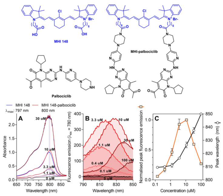
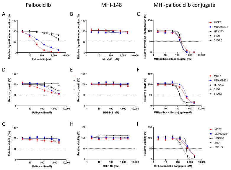
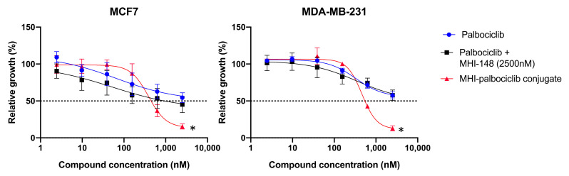
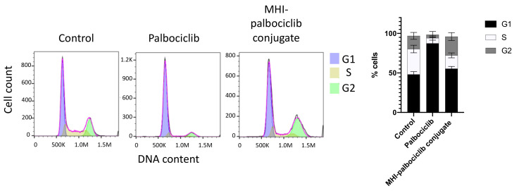
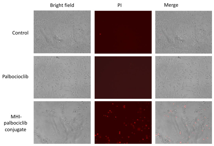
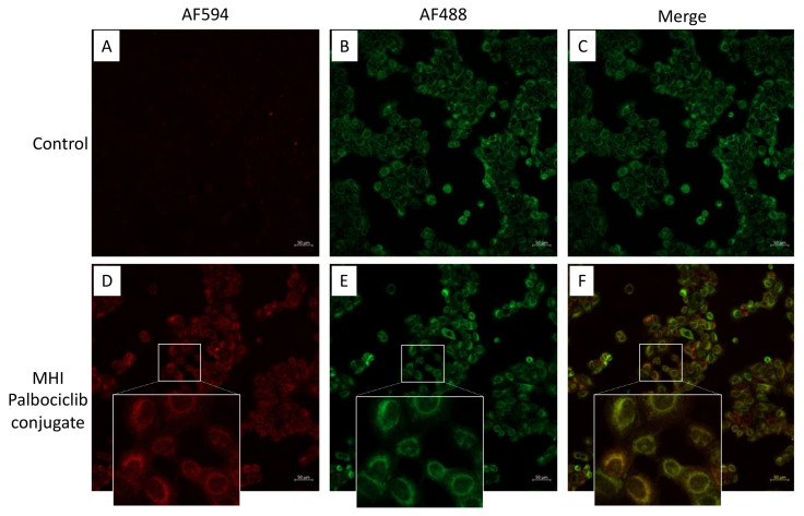
The CDK4/6 inhibitor palbociclib, combined with endocrine therapy, has been shown to be effective in postmenopausal women with estrogen receptor-positive, HER2-negative advanced or metastatic breast cancer. However, palbociclib is not as effective in the highly aggressive, triple-negative breast cancer that lacks sensitivity to chemotherapy or endocrine therapy. We hypothesized that conjugation of the near-infrared dye MHI-148 with palbociclib can produce a potential theranostic in triple-negative, as well as estrogen receptor-positive, breast cancer cells. In our study, the conjugate was found to have enhanced activity in all mammalian cell lines tested in vitro. However, the conjugate was cytotoxic and did not induce G1 cell cycle arrest in breast cancer cells, suggesting its mechanism of action differs from the parent compound palbociclib. The study highlights the importance of investigating the mechanism of conjugates of near-infrared dyes to therapeutic compounds, as conjugation can potentially result in a change of mechanism or target, with an enhanced cytotoxic effect in this case.

Keywords: CDK4/6 inhibitor palbociclib; MHI-148; breast cancer; cell cycle arrest; near-infrared fluorescent (NIRF).


Conflict of interest statement

All authors declare no conflict of interest.

Figures

Figure 1
Figure 1
The conjugation of palbociclib to MHI-148 does not significantly influence the absorbance and fluorescence properties of the parent MHI-148 dye. (A) Absorbance spectra of MHI-148 and MHI-palbociclib in DMSO. A representative experiment of two independent repeats. Vertical lines denote λmax, which indicates the wavelength at the peak maximum. (B) Fluorescence spectra of MHI-palbociclib in DMSO. Each fluorescence emission spectrum represents an average of three measurements. Only MHI-palbociclib is displayed, as MHI-148 showed essentially identical normalised spectra. (C) Impact of fluorophore concentration on fluorescence intensity and peak wavelength. Each data point represents a mean and standard deviation of respective values for MHI-148 and MHI-palbociclib obtained from data in panel (B). Increasing MHI-palbociclib’s concentration redshifts the fluorescence and induces self-quenching.
Figure 2
Figure 2
Effect of palbociclib, MHI-148 and MHI-palbociclib on (AC) cell proliferation ([3H]-thymidine incorporation assay), (DF) cell growth (SRB assay), and (GI) cell viability (WST-1 assay). Effects on breast cancer cell lines and non-cancerous cell lines were measured after 3 days of treatment with the compounds. Data presented are an average of at least three independent experiments.
Figure 3
Figure 3
Inhibition of relative growth by MHI-palbociclib is significantly higher than the parent compound palbociclib, or the combination of palbociclib with unconjugated MHI-148 in breast cancer MCF-7 and MDA-MB-231 cell lines. Effects on breast cancer cell lines were measured after 3 days exposure to the compounds. Data presented are an average of at least three independent experiments, * p < 0.05.
Figure 4
Figure 4
The cell cycle proportions of MDA-MB-231 breast cancer cells after exposure to 500 nM palbociclib or MHI-palbociclib for 24 h, followed by flow cytometry cell cycle analysis. Control: no drugs administered. Representative cell cycle profiles are shown on the left, and the average result of three independent experiments are shown in the bar graph.
Figure 5
Figure 5
Cytotoxicity in MDA-MB-231 cells after MHI-palbociclib treatment is significantly greater than after treatment with palbociclib. After exposure to 1 µM of indicated compound for 24 h, dying cells were stained by propidium iodide (red). Photographs were taken by a Floid imaging station (460× magnification). Representative images of three independent experiments are shown.
Figure 6
Figure 6
MHI-palbociclib localized in mitochondria. Red fluorescence (MHI-palbociclib) (A,D) and green fluorescence (Mitotracker Green) (B,E) were captured using confocal microscopy. DMSO was used as control. (C) Merged image of (A,B) using DMSO as control. (F) Merged image showing MHI-palbociclib (red) co-localized in mitochondria (Mitotracker Green). Scale bar: 50 µm.

References

    1. [(accessed on 23 November 2021)]. Available online: https://www.who.int/news-room/fact-sheets/detail/breast-cancer.
    1. Dai X., Li T., Bai Z., Yang Y., Liu X., Zhan J., Shi B. Breast cancer intrinsic subtype classification, clinical use and future trends. Am. J. Cancer Res. 2015;5:2929–2943. - PMC - PubMed
    1. Hanahan D., Weinberg R.A. The hallmarks of cancer. Cell. 2000;100:57–70. doi: 10.1016/S0092-8674(00)81683-9. - DOI - PubMed
    1. Trotter E.W., Hagan I.M. Release from cell cycle arrest with Cdk4/6 inhibitors generates highly synchronized cell cycle progression in human cell culture. Open Biol. 2020;10:200200. doi: 10.1098/rsob.200200. - DOI - PMC - PubMed
    1. Otto T., Sicinski P. Cell cycle proteins as promising targets in cancer therapy. Nat. Rev. Cancer. 2017;17:93–115. doi: 10.1038/nrc.2016.138. - DOI - PMC - PubMed

MeSH terms