Review
. 2022 Jan 30;27(3):951.
doi: 10.3390/molecules27030951.

Metabolic Features of Brain Function with Relevance to Clinical Features of Alzheimer and Parkinson Diseases

Affiliations
Review

Metabolic Features of Brain Function with Relevance to Clinical Features of Alzheimer and Parkinson Diseases

David Allan Butterfield et al. Molecules. .

Abstract

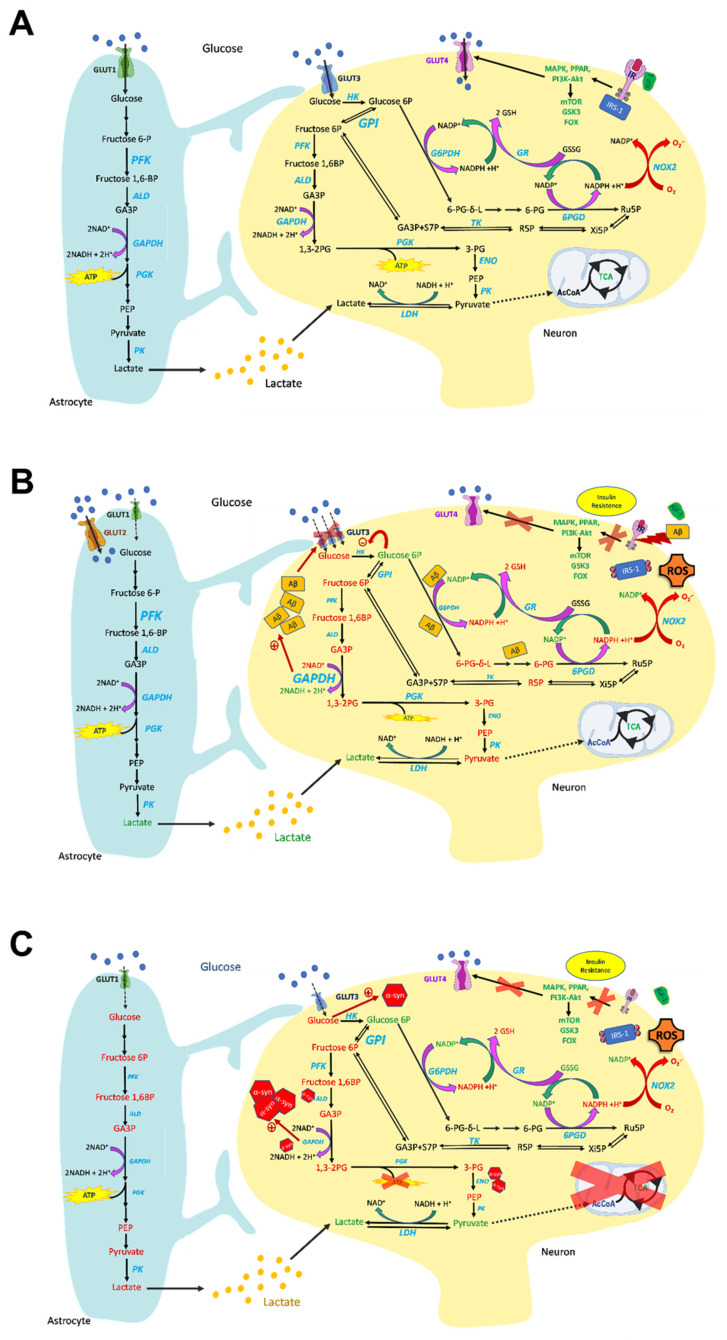
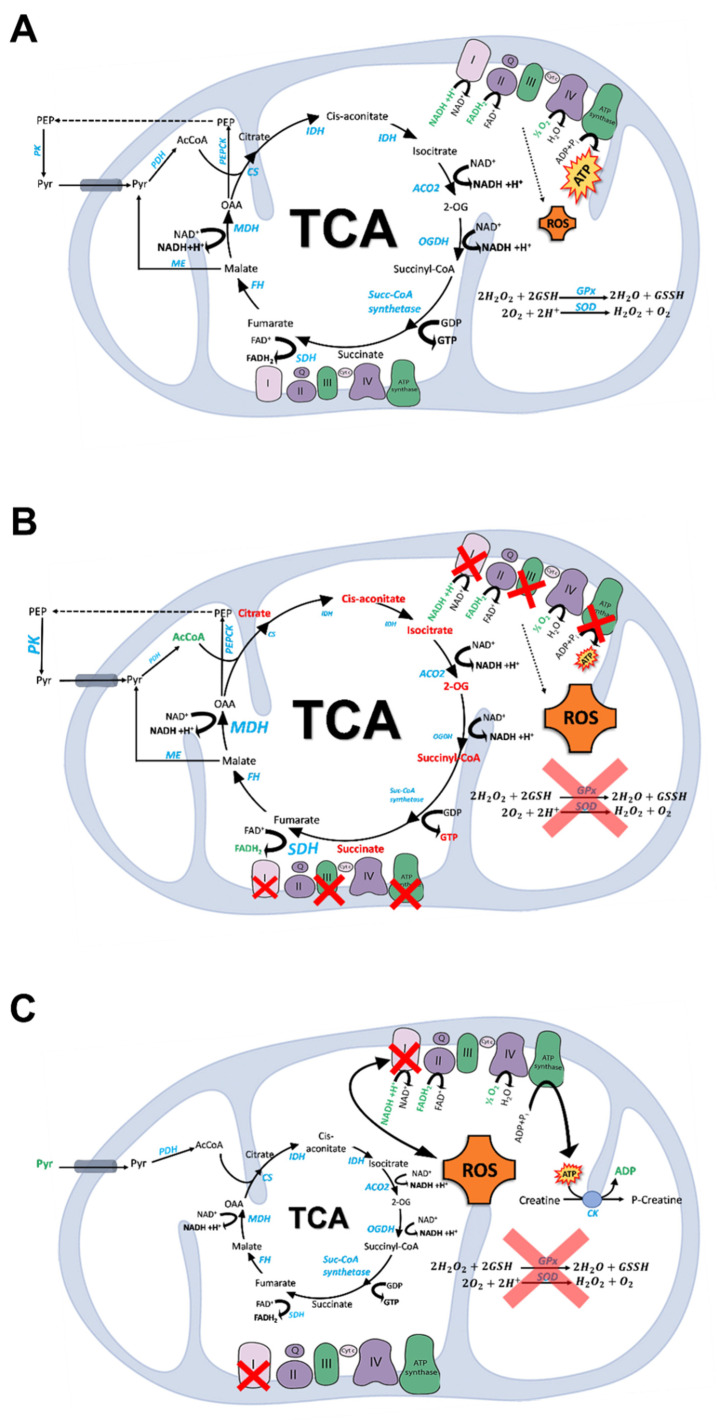
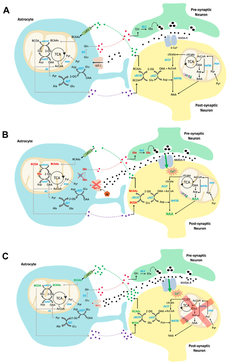
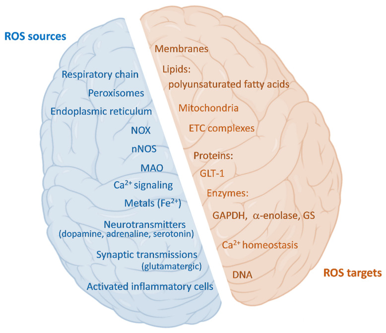
Brain metabolism is comprised in Alzheimer's disease (AD) and Parkinson's disease (PD). Since the brain primarily relies on metabolism of glucose, ketone bodies, and amino acids, aspects of these metabolic processes in these disorders-and particularly how these altered metabolic processes are related to oxidative and/or nitrosative stress and the resulting damaged targets-are reviewed in this paper. Greater understanding of the decreased functions in brain metabolism in AD and PD is posited to lead to potentially important therapeutic strategies to address both of these disorders, which cause relatively long-lasting decreased quality of life in patients.

Keywords: AD; Alzheimer’s disease; PD brain metabolism; Parkinson’s disease; glucose; metabolic reprogramming; neurodegeneration; oxidative stress.


Conflict of interest statement

The authors declare no conflict of interest.

Figures

Figure 1
Figure 1
Overview of glucose metabolism in normal (A), AD (B), and PD (C) brains. (A) Glucose is the main energy substrate in the brain, as it is the principal source of ATP. In the brain, glucose is taken up from the bloodstream by specific glucose transporters, to be metabolized through glycolysis. Pyruvate enters the TCA cycle coupled with OXPHOS and ATP synthesis. Glucose transport in the brain occurs via GLUT1 in astrocytes and GLUT3 in neurons. Insulin-dependent GLUT4 transport also occurs in the brain, and its activation leads to insulin receptor signaling, occurring through the PI3K/Akt and MAPK pathways, regulating the brain’s main cellular functions. Glucose metabolism is prominent in the brain—especially in astrocytes—and strongly interconnected among the different cell types. Glucose 6-phosphate (G6P) can be channeled into the pentose phosphate pathway (PPP) to support NADPH synthesis, which is necessary to sustain the brain’s antioxidant defense, and this is enhanced by the recycling of fructose 6-phosphate (F6P) originating from the PPP back to G6P due to high GPI activity. (B) Glucose metabolism is impaired in AD brains. Signs of impaired insulin signaling cascade are present in AD brains, with insulin resistance and downregulation of insulin receptors, which contribute to brain glucose hypometabolism. In AD, decreased glucose metabolism impacts the metabolic crosstalk between astrocytes and neurons, as the lactate shuttle is impaired, leading to reduced ATP synthesis. GLUT1 and -3 are decreased in AD brains, and this correlates to glucose hypometabolism, and is a major pathological sign of AD, whereas GLUT2 increases, indicating prominent astroglial activation in AD brains. Glycolysis increases in astrocytes and microglia, and this is associated with neurodegeneration. G6P and fructose 1,6-bisphosphate (F1,6BP) levels are inversely correlated with age. Hexokinase (HK), PK, and PFK are downregulated in neurons, whereas GAPDH is upregulated and can promote Aβ amyloidogenesis. Aβ plays a role in impairing the PPP, leading to G6P accumulation—which inhibits HK activity—and to decreased defense against ROS. Aβ oligomers also reduce the IR and promote synaptic spine deterioration. (C) Glucose metabolism is dysfunctional in PD brains, and this mirrors a significant loss of IR. Furthermore, IRS phosphorylation deactivates insulin signaling, leading to insulin resistance. Moreover, the glucose transporters GLUT1 and GLUT3 are downregulated. The decrease in glucose metabolism, prominent both in neurons and in astrocytes, is associated with pyruvate and lactate accumulation and deleterious ATP depletion. Depletion of the ATP-generating enzyme PGK is associated with neuronal deficits with PD-like symptoms. Low glucose promotes α-synuclein (α-syn) aggregation. α-Syn fibrils interact with GAPDH, aldolase (ALD), and enolase (ENO), and their activities are consequently decreased. Furthermore, GAPDH directly regulates α-syn aggregation and apoptotic neuronal cell death. Increased metabolite levels are reported in green, whereas decreased levels are reported in red.
Figure 2
Figure 2
Overview of the TCA and OXPHOS functions in normal (A), AD (B), and PD (C) brains. (A) The TCA cycle oxidizes acetyl-CoA to two molecules of CO2, leading to the production of ATP and the reduction of NAD+ and FAD+ to NADH and FADH2, which enter the electron transport chain (ETC) complexes I and II, respectively. The electron flux from complexes I and II through the ETC leads to production of ATP by means of OXPHOS, which is coupled with the TCA cycle as it reoxidizes the coenzymes necessary for TCA function. ROS produced by OXPHOS are counteracted by Cu,Zn-superoxide dismutase, Mn-superoxide dismutase, peroxiredoxin, and glutathione. In the brain, pyruvate is recycled from malate and oxaloacetate through malic enzyme and phosphoenolpyruvate carboxykinase (PEPCK), respectively. (B) Aging is characterized by dysfunctions of the TCA cycle. In both aging and AD murine models, the levels of acetyl-CoA and NADH were increased, whereas the levels of succinic acid, 2-OG, citric acid, cis-aconitic acid, fumaric acid are decreased. This mirrors the reduction of IDH, 2-OGDH, PDH complexes, and CS with the increase in MDH and SDH in AD brains. Complex I, complex III, and complex V proteins are reduced in different regions of AD brains, leading to compromised OXPHOS. The activity of cytochrome c oxidase (complex IV) is lower in many brain regions, but is increased in the hippocampus. The damage to mitochondrial respiratory function in AD patients is associated with ROS formation, enhanced by the cellular inability to cope with the oxidative surge due to a lower antioxidant defense. (C) PD is associated with mitochondrial dysfunctions. In PD brains the lactate/pyruvate ratio is high, and this leads to TCA dysregulation. Mitochondrial complex I deficiency and oxidative stress are key factors in PD’s pathogenesis, and they are interconnected in a vicious cycle, in which a weakened antioxidant defense plays a role. Energy failure of PD brains leads to a creatine kinase (CK)-mediated increase in ADP phosphorylation at the expense of phosphocreatine, which is linked to upregulated creatine synthesis. Increased metabolite levels are shown in green, whereas decreased levels are shown in red.
Figure 3
Figure 3
Overview of amino acid (AA) metabolism in normal (A), AD (B), and PD (C) brains. (A) BCAAs enter the CNS via the BBB (LAT1) and, mainly in astrocytes (mitochondria), undergo transamination, which yields the corresponding BCOAs and glutamate via BCAT. BCOAs enter the TCA for energy production; in this way, BCATs are a constant source of glutamate. Astrocytes then release the oxoacids to the neuron, where they are reconverted to BCAAs (cytosol), which are released back into the extracellular space. Glutamate is an excitatory neurotransmitter. Once glutamate’s signaling role is executed, it is taken up by astrocytes (GLT-1, also known as EAAT2), in which glutamate is converted to glutamine in a reaction catalyzed by GS. This prevents the so-called excitotoxic effect of glutamate accumulation in the synapse. Furthermore, de novo glutamate synthesis occurs exclusively in astrocytes following pyruvate-carboxylase-dependent anaplerosis and BCAA transamination. Glutamate transamination also occurs in the brain as a result of the activity of ALT and AST in both astrocytes and neurons. In this way, ammonia transfer can occur. (B) BCAA metabolism is altered in AD brains. Reduced levels of BCAAs (indicated in red) have been found in the blood, CSF, and brains of AD patients, and this reduction is associated with cognitive decline in AD. BCAA diminution might impair glutamate synthesis, leading to impaired neurotransmission and impaired NMDAR function. The glutamate/glutamine cycle is impaired in AD brains, due to GS and GLT-1 oxidation-related loss of activity in astrocytes, exposing neurons to the effect of glutamate excitotoxicity. NAA is accumulated in AD brains, suggesting a cytosolic and mitochondrial metabolic compromise in AD brains. (C) BCAA metabolism is altered in PD brains, with accumulation of these amino acids (in green). Glutamate excitotoxicity is also prominent in PD brains, and this is associated with downregulation of astrocytic GLT-1. Increased metabolite levels are shown in green, whereas decreased levels are shown in red.
Figure 4
Figure 4
ROS sources and targets in the brain. Schematic representation of cerebral ROS sources and targets. Sources: beyond the mitochondrial inner-membrane-resident respiratory chain, peroxisomes, endoplasmic reticulum, NADPH oxidases (NOX), and activated inflammatory cells, there are also specific brain sources such as neuronal nitric oxide synthase (nNOS), monoamine oxidases (MAO), Ca2+ signaling, redox-active metal ions, neurotransmitters, and synaptic transmission. Targets of ROS include lipids present in membranes, in particular polyunsaturated fatty acids; mitochondria, and especially the electron transport chain (ETC) complexes; proteins such as the glutamate transporter (GLT-1), or enzymes, for example (among others), glyceraldehyde 3-phosphate dehydrogenase (GAPDH), α-enolase, or glutamine synthetase (GS) in AD brains; Ca2+ homeostasis; DNA.

Similar articles

Cited by

References

    1. Hyder F., Rothman D.L., Bennett M.R. Cortical energy demands of signaling and nonsignaling components in brain are conserved across mammalian species and activity levels. Proc. Natl. Acad. Sci. USA. 2013;110:3549–3554. doi: 10.1073/pnas.1214912110. - DOI - PMC - PubMed
    1. Harris M.E., Hensley K., Butterfield D.A., Leedle R.A., Carney J.M. Direct evidence of oxidative injury produced by the Alzheimer’s β-Amyloid peptide (1–40) in cultured hippocampal neurons. Exp. Neurol. 1995;131:193–202. doi: 10.1016/0014-4886(95)90041-1. - DOI - PubMed
    1. Attwell D., Laughlin S.B. An energy budget for signaling in the grey matter of the brain. J. Cereb. Blood Flow Metab. 2001;21:1133–1145. doi: 10.1097/00004647-200110000-00001. - DOI - PubMed
    1. Pathak D., Shields L.Y., Mendelsohn B.A., Haddad D., Lin W., Gerencser A.A., Kim H., Brand M.D., Edwards R.H., Nakamura K. The Role of Mitochondrially Derived ATP in Synaptic Vesicle Recycling. J. Biol. Chem. 2015;290:22325. doi: 10.1074/jbc.M115.656405. - DOI - PMC - PubMed
    1. Silverman D.H.S., Chen W., Czernin J., Kowell A.P., Gambhir S.S., Phelps M.E., Lu C.S., Kung de Aburto M.A.K., Chang C.Y., Small G.W., et al. Positron Emission Tomography in Evaluation of Dementia: Regional Brain Metabolism and Long-term Outcome. JAMA. 2001;286:2120–2127. doi: 10.1001/jama.286.17.2120. - DOI - PubMed