

Stretching prior to resistance training promotes adaptations on the postsynaptic region in different myofiber types
- PMID: 35164481
- PMCID: PMC8875788
- DOI: 10.4081/ejh.2022.3356
Stretching prior to resistance training promotes adaptations on the postsynaptic region in different myofiber types
Abstract
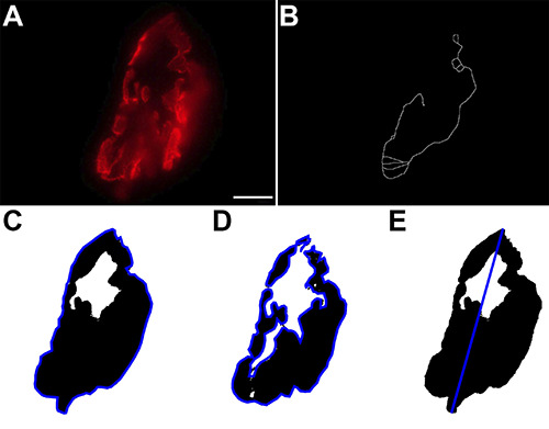
The morphology of the neuromuscular junction adapts according to changes in its pattern of use, especially at the postsynaptic region according to the myofibrillar type and physical exercise. This investigation revealed the morphological adaptations of the postsynaptic region after static stretching, resistance training, and their association in adult male Wistar rats. We processed the soleus and plantaris muscles for histochemical (muscle fibers) and postsynaptic region imaging techniques. We observed muscle hypertrophy in both groups submitted to resistance training, even though the cross-section area is larger when there is no previous static stretching. The soleus postsynaptic region revealed higher compactness and fragmentation index in the combined exercise. The resistance training promoted higher adaptations in the postsynaptic area of plantaris; moreover, the previous static stretching decreased this area. In conclusion, the neuromuscular system's components responded according to the myofiber type even though it is the same physical exercise. Besides, static stretching (isolated or combined) plays a crucial role in neuromuscular adaptations.
Figures


Similar articles
-
Divergent effects of resistance training and anabolic steroid on the postsynaptic region of different skeletal muscles of aged rats.Exp Gerontol. 2017 Nov;98:80-90. doi: 10.1016/j.exger.2017.08.018. Epub 2017 Aug 12. Exp Gerontol. 2017. PMID: 28811140
-
Effect of resistance training on neuromuscular junctions of young and aged muscles featuring different recruitment patterns.J Neurosci Res. 2015 Mar;93(3):504-13. doi: 10.1002/jnr.23495. Epub 2014 Oct 7. J Neurosci Res. 2015. PMID: 25287122 Free PMC article.
-
Neuromuscular adaptability of male and female rats to muscle unloading.J Neurosci Res. 2018 Feb;96(2):284-296. doi: 10.1002/jnr.24129. Epub 2017 Jul 31. J Neurosci Res. 2018. PMID: 28759131 Free PMC article.
-
Muscle mechanics: adaptations with exercise-training.Exerc Sport Sci Rev. 1996;24:427-73. Exerc Sport Sci Rev. 1996. PMID: 8744258 Review.
-
Long-term metabolic and skeletal muscle adaptations to short-sprint training: implications for sprint training and tapering.Sports Med. 2001;31(15):1063-82. doi: 10.2165/00007256-200131150-00003. Sports Med. 2001. PMID: 11735686 Review.
Cited by
-
Training modalities for elder sarcopenic obesity: a systematic review and network meta-analysis.Front Nutr. 2025 Feb 19;12:1537291. doi: 10.3389/fnut.2025.1537291. eCollection 2025. Front Nutr. 2025. PMID: 40046765 Free PMC article.
References
-
- Deschenes MR, Li S, Adan MA, Oh JJ, Ramsey HC. Muscle fibers and their synapses differentially adapt to aging and endurance training. Exp Gerontol 2018;106:183-91. - PubMed
-
- Deschenes MR. Adaptations of the neuromuscular junction to exercise training. Curr Opin Physiol 2019;10:10-6.
-
- Deschenes MR, Maresh CM, Crivello JF, Armstrong LE, Kraemer WJ, Covault J. The effects of exercise training of different intensities on neuromuscular junction morphology. J Neurocytol 1993;22:603–15. - PubMed
-
- Fahim MA. Endurance exercise modulates neuromuscular junction of C57BL/6NNia aging mice. J Appl Physiol 1997;83:59–66. - PubMed
MeSH terms
LinkOut - more resources
Full Text Sources
