. 2021 Feb 22;13(1):e0016922.
doi: 10.1128/mbio.00169-22. Epub 2022 Feb 15.

Genome-Wide Characterization of SARS-CoV-2 Cytopathogenic Proteins in the Search of Antiviral Targets

Affiliations

Genome-Wide Characterization of SARS-CoV-2 Cytopathogenic Proteins in the Search of Antiviral Targets

Jiantao Zhang et al. mBio. .

Abstract

Therapeutic inhibition of critical viral functions is important for curtailing coronavirus disease 2019 (COVID-19). We sought to identify antiviral targets through the genome-wide characterization of severe acute respiratory syndrome coronavirus 2 (SARS-CoV-2) proteins that are crucial for viral pathogenesis and that cause harmful cytopathogenic effects. All 29 viral proteins were tested in a fission yeast cell-based system using inducible gene expression. Twelve proteins, including eight nonstructural proteins (NSP1, NSP3, NSP4, NSP5, NSP6, NSP13, NSP14, and NSP15) and four accessory proteins (ORF3a, ORF6, ORF7a, and ORF7b), were identified that altered cellular proliferation and integrity and induced cell death. Cell death correlated with the activation of cellular oxidative stress. Of the 12 proteins, ORF3a was chosen for further study in mammalian cells because it plays an important role in viral pathogenesis and its activities are linked to lung tissue damage and a cytokine storm. In human pulmonary and kidney epithelial cells, ORF3a induced cellular oxidative stress associated with apoptosis and necrosis and caused activation of proinflammatory response with production of the cytokines tumor necrosis factor alpha (TNF-α), interleukin-6 (IL-6), and IFN-β1, possibly through the activation of nuclear factor kappa B (NF-κB). To further characterize the mechanism, we tested a natural ORF3a Beta variant, Q57H, and a mutant with deletion of the highly conserved residue, ΔG188. Compared with wild-type ORF3a, the ΔG188 variant yielded more robust activation of cellular oxidative stress, cell death, and innate immune response. Since cellular oxidative stress and inflammation contribute to cell death and tissue damage linked to the severity of COVID-19, our findings suggest that ORF3a is a promising, novel therapeutic target against COVID-19. IMPORTANCE The ongoing COVID-19 pandemic caused by SARS-CoV-2 has claimed over 5.5 million lives with more than 300 million people infected worldwide. While vaccines are effective, the emergence of new viral variants could jeopardize vaccine protection. Treatment of COVID-19 by antiviral drugs provides an alternative to battle against the disease. The goal of this study was to identify viral therapeutic targets that can be used in antiviral drug discovery. Utilizing a genome-wide functional analysis in a fission yeast cell-based system, we identified 12 viral candidates, including ORF3a, which cause cellular oxidative stress, inflammation, apoptosis, and necrosis that contribute to cytopathogenicity and COVID-19. Our findings indicate that antiviral agents targeting ORF3a could have a great impact on COVID-19.

Keywords: ORF3a; SARS-CoV-2; Schizosaccharomyces pombe; apoptosis and necrosis; fission yeast; oxidative stress; proinflammatory response; viral therapeutic target.


Conflict of interest statement

The authors declare no conflict of interest.

Figures

FIG 1
FIG 1
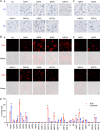
Effect of SARS-CoV-2 expression on fission yeast colony formation (A), cellular growth (B), and summary of the relative cellular growth (15%, red; 25% blue) of each of the SARS-CoV-2 protein-expressing cells (C). The name of each SARS-CoV-2 protein is labeled above each agar plate. An empty pYZ1N vector (Ctr) was used as a control. Gene-off, no SARS-CoV-2 protein production; gene-on, the specific SARS-CoV-2 protein production was induced by triggering the nmt1 promoter-mediated gene transcription. Fission yeast colony formation was measured by growing SARS-CoV-2 protein-expressing fission yeast cells on the selective EMM agar plates and incubated at 30°C for 3 to 5 days before the pictures were taken. The cell proliferation analysis was carried out by comparing cellular growth between the SARS-CoV-2 protein-producing cells and the SARS-CoV-2 protein-suppressing cells over time. Cell growth was measured by spectrophotometry (OD650). Only the effect of those SARS-CoV-2 proteins that showed complete (NSP1, NSP4, NSP6, NSP13, NSP14, ORF3a, ORF6, and ORF7a) or nearly complete (NSP3, NSP5, and NSP15) inhibition of yeast colony formation is shown in A and thereafter. Complete data on cell proliferation are included in Fig. S2. Each experiment was repeated at least three times, and the standard errors of each time point were calculated.
FIG 2
FIG 2
The effect of SARS-CoV-2 protein on fission yeast cellular morphology. Only those SARS-CoV-2 proteins that affected cell proliferation presented in Fig. 1 are shown here. Complete SARS-CoV-2 genome-wide data on fission yeast cellular morphology are included in Fig. S3. (A) Shows the effect of individual SARS-CoV-2 proteins on fission yeast cell morphology. Each image was taken 48 h agi using bright field microscopy. Scale bar = 10 μM. (B) Overall cell morphology as shown by the forward-scattered analysis. A total of 10,000 cells were measured 48 h agi. The forward-scatter light (FSC) measures the distribution of all cell sizes. The side-scatter light (SSC) determines intracellular complexity. Gene-off, no SARS-CoV-2 protein production; gene-on, SARS-CoV-2 protein produced.
FIG 3
FIG 3
Correlation of SARS-CoV-2 protein-mediated cell death with the induction of oxidative stress in fission yeast. (A) SARS-CoV-2 protein-induced cell death was measured 48 h agi by trypan blue staining. (B) SARS-CoV-2 protein induces oxidative stress, as indicated by the DHE staining showing the production of ROS. Images were taken 48 h agi. Scale bar = 10 μM. BF, bright field; ROS, reactive oxidative species. DHE, an oxidative stress-specific dye (8, 67). (C) Quantitative correlation of SARS-CoV-2 protein-induced cell death (blue bars) and the production of ROS (red bars). Data represent mean ± SE from three independent experiments. Complete SARS-CoV-2 genome-wide data on fission yeast cell death and ROS production are included in Fig. S4.
FIG 4
FIG 4
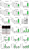
SARS-CoV-2 ORF3a-induced apoptosis and necrosis are correlated with the induction of cellular oxidative stress and innate immune proinflammatory responses in mammalian cells. Expression of SARS-CoV-2 ORF3a induces cellular growth reduction and cell death 72 hpt in human lung epithelial A549 cells (A) and human kidney epithelial 293T cells (B). (C) ORF3a induces apoptosis and necrosis 48 hpt measured by Annexin V (a), necrosis (b), and caspase-3 cleavage (c). (D) ORF3a triggers the induction of oxidative stress 48 hpt measured by the DHE straining. The ORF3a was cloned in a lentiviral constitutive expression vector (4). Scale bar = 20 μM. (E) ORF3a elevates NF-κB-mediated transcriptional activities. (F) ORF3a triggers elevated production of TNF-α, IL-6, and NF-κB in Calu-3 (a) and 293T (b) cells. Data are presented as mean ± SE from three independent experiments. Statistical differences between ORF3a and mock (indicated with #) or empty vector control (indicated with *) were evaluated. * or #, P < 0.05; ** or ##, P < 0.01; *** or ###, P < 0.001 (pair-wise t test).
FIG 5
FIG 5
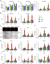
ORF3a-induced apoptosis by natural and artificial mutant variants correlate with cellular oxidative stress and innate immune proinflammatory responses. 293T cells were transfected with plasmids harboring ORF3a wild type (WT), ΔG188, or Q57H mutant variant. Effects of ORF3a mutant variants on cytopathic effects (A), as measured by cellular growth (a), cell viability (b), and cell death (c), at the indicated times. (B) Effects of ORF3a mutant variants on apoptosis (a) and necrosis (b). (C) Induction of oxidative stress. The images were taken at 72 hpt. Scale bar = 20 μM. (D) ORF3a elevates NF-κB-mediated transcriptional activities. (E) Activation of cellular innate immune proinflammatory responses, as measured by qRT-PCR. Data are presented as mean ± SE from three independent experiments. Statistical differences between control and ORF3a WT or mutants (indicated with #) or between WT and mutants (indicated with *) were evaluated. * or #, P < 0.05; ** or ##, P < 0.01; *** or ###, P < 0.001 (one-way ANOVA).

Update of

Similar articles

Cited by

References

    1. Cameroni E, Bowen JE, Rosen LE, Saliba C, Zepeda SK, Culap K, Pinto D, VanBlargan LA, De Marco A, di Iulio J, Zatta F, Kaiser H, Noack J, Farhat N, Czudnochowski N, Havenar-Daughton C, Sprouse KR, Dillen JR, Powell AE, Chen A, Maher C, Yin L, Sun D, Soriaga L, Bassi J, Silacci-Fregni C, Gustafsson C, Franko NM, Logue J, Iqbal NT, Mazzitelli I, Geffner J, Grifantini R, Chu H, Gori A, Riva A, Giannini O, Ceschi A, Ferrari P, Cippà PE, Franzetti-Pellanda A, Garzoni C, Halfmann PJ, Kawaoka Y, Hebner C, Purcell LA, Piccoli L, Pizzuto MS, Walls AC, Diamond MS, et al. . 2021. Broadly neutralizing antibodies overcome SARS-CoV-2 Omicron antigenic shift. Nature doi:10.1038/s41586-021-04386-2. - DOI - PMC - PubMed
    1. Coronaviridae Study Group of the International Committee on Taxonomy of Viruses. 2020. The species severe acute respiratory syndrome-related coronavirus: classifying 2019-nCoV and naming it SARS-CoV-2. Nat Microbiol 5:536–544. doi:10.1038/s41564-020-0695-z. - DOI - PMC - PubMed
    1. Sola I, Almazan F, Zuniga S, Enjuanes L. 2015. Continuous and discontinuous RNA synthesis in coronaviruses. Annu Rev Virol 2:265–288. doi:10.1146/annurev-virology-100114-055218. - DOI - PMC - PubMed
    1. Gordon DE, Jang GM, Bouhaddou M, Xu J, Obernier K, White KM, O’Meara MJ, Rezelj VV, Guo JZ, Swaney DL, Tummino TA, Hüttenhain R, Kaake RM, Richards AL, Tutuncuoglu B, Foussard H, Batra J, Haas K, Modak M, Kim M, Haas P, Polacco BJ, Braberg H, Fabius JM, Eckhardt M, Soucheray M, Bennett MJ, Cakir M, McGregor MJ, Li Q, Meyer B, Roesch F, Vallet T, Mac Kain A, Miorin L, Moreno E, Naing ZZC, Zhou Y, Peng S, Shi Y, Zhang Z, Shen W, Kirby IT, Melnyk JE, Chorba JS, Lou K, Dai SA, Barrio-Hernandez I, Memon D, Hernandez-Armenta C, et al. . 2020. A SARS-CoV-2 protein interaction map reveals targets for drug repurposing. Nature 583:459–468. doi:10.1038/s41586-020-2286-9. - DOI - PMC - PubMed
    1. Zhao RY. 2017. Yeast for virus research. Microb Cell 4:311–330. doi:10.15698/mic2017.10.592. - DOI - PMC - PubMed

Substances

Supplementary concepts