. 2021 Feb 22;13(1):e0004422.
doi: 10.1128/mbio.00044-22. Epub 2022 Feb 15.

CRP-Like Transcriptional Regulator MrpC Curbs c-di-GMP and 3',3'-cGAMP Nucleotide Levels during Development in Myxococcus xanthus

Affiliations

CRP-Like Transcriptional Regulator MrpC Curbs c-di-GMP and 3',3'-cGAMP Nucleotide Levels during Development in Myxococcus xanthus

Sofya Kuzmich et al. mBio. .

Abstract

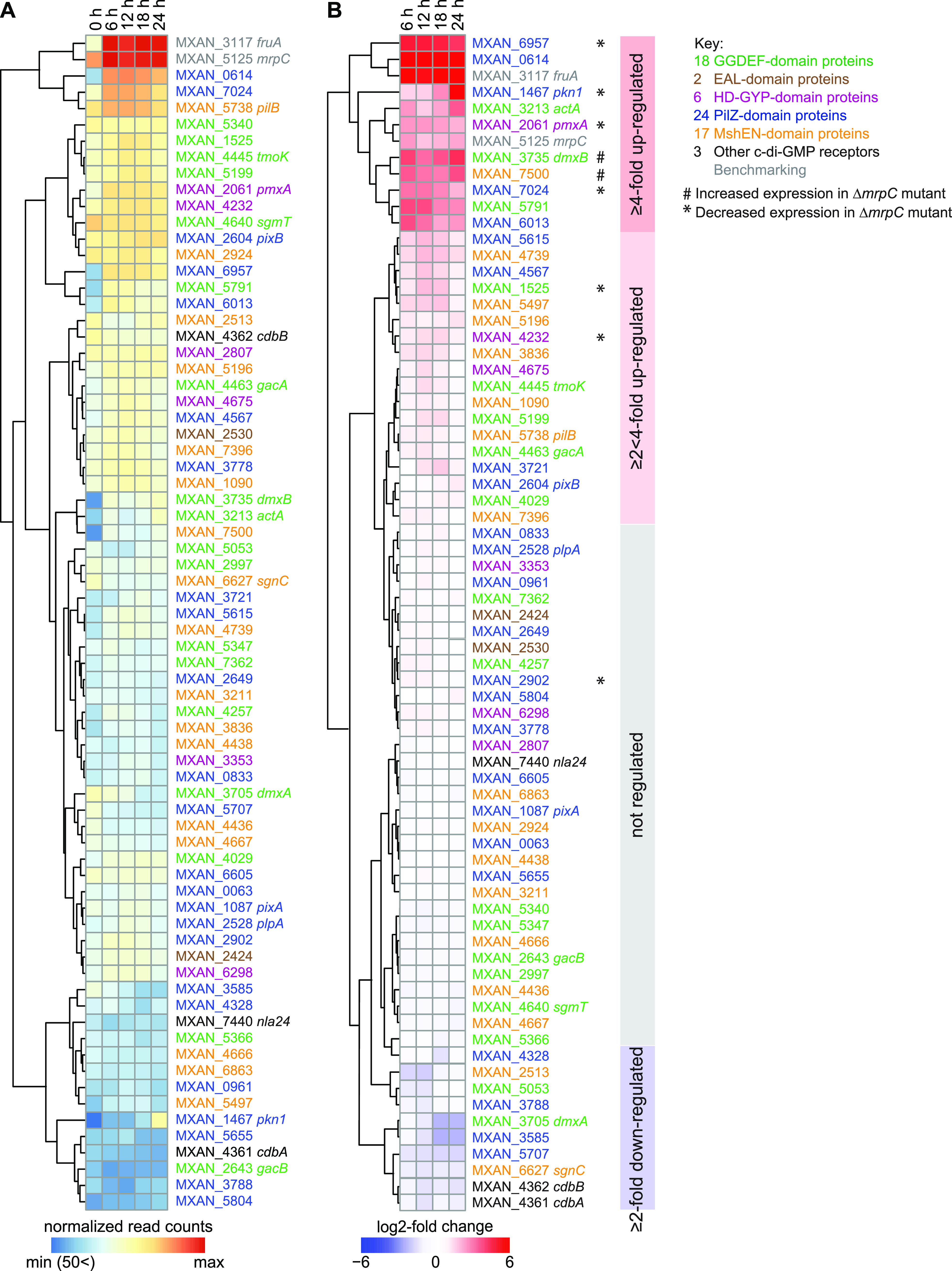
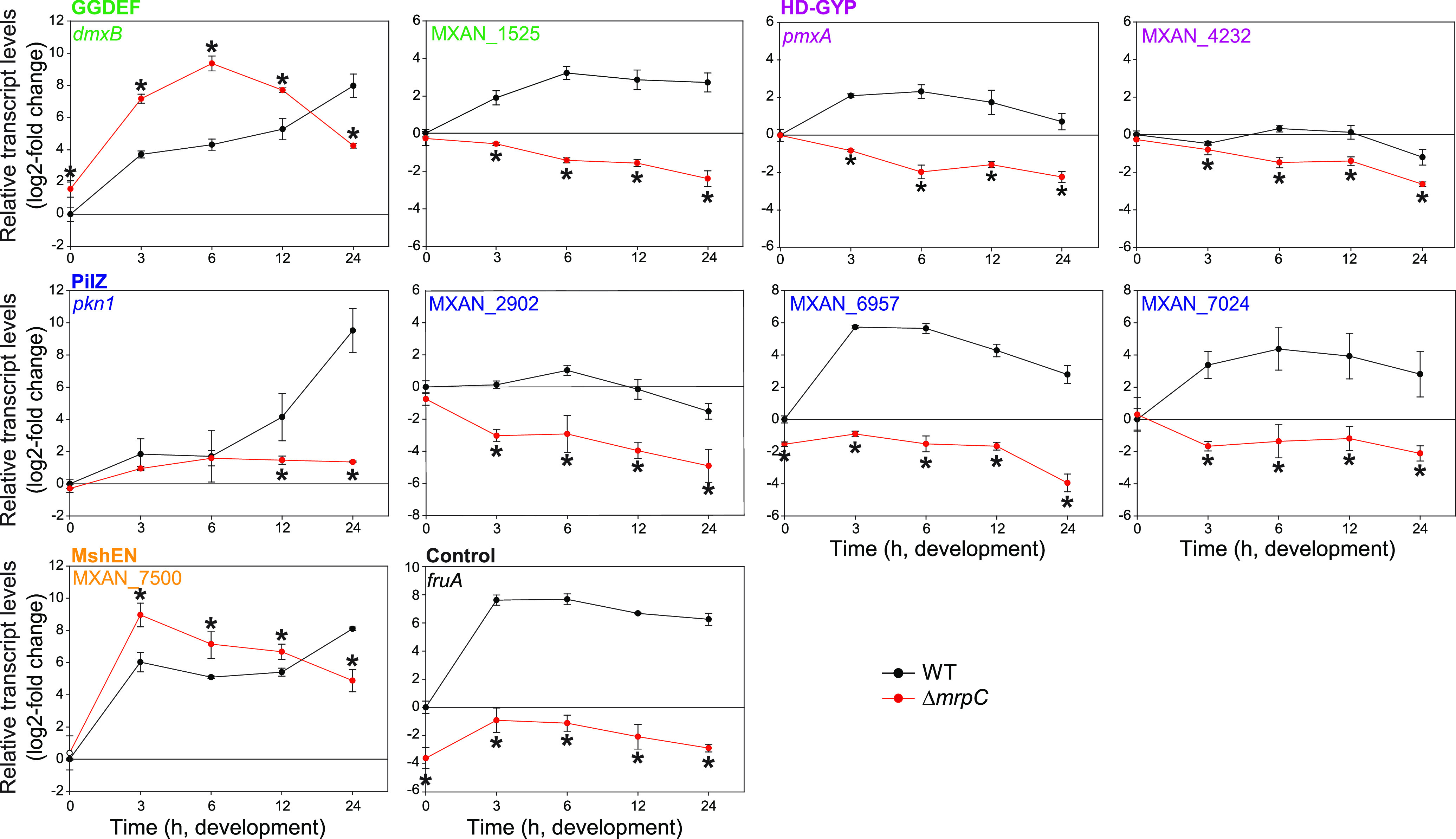
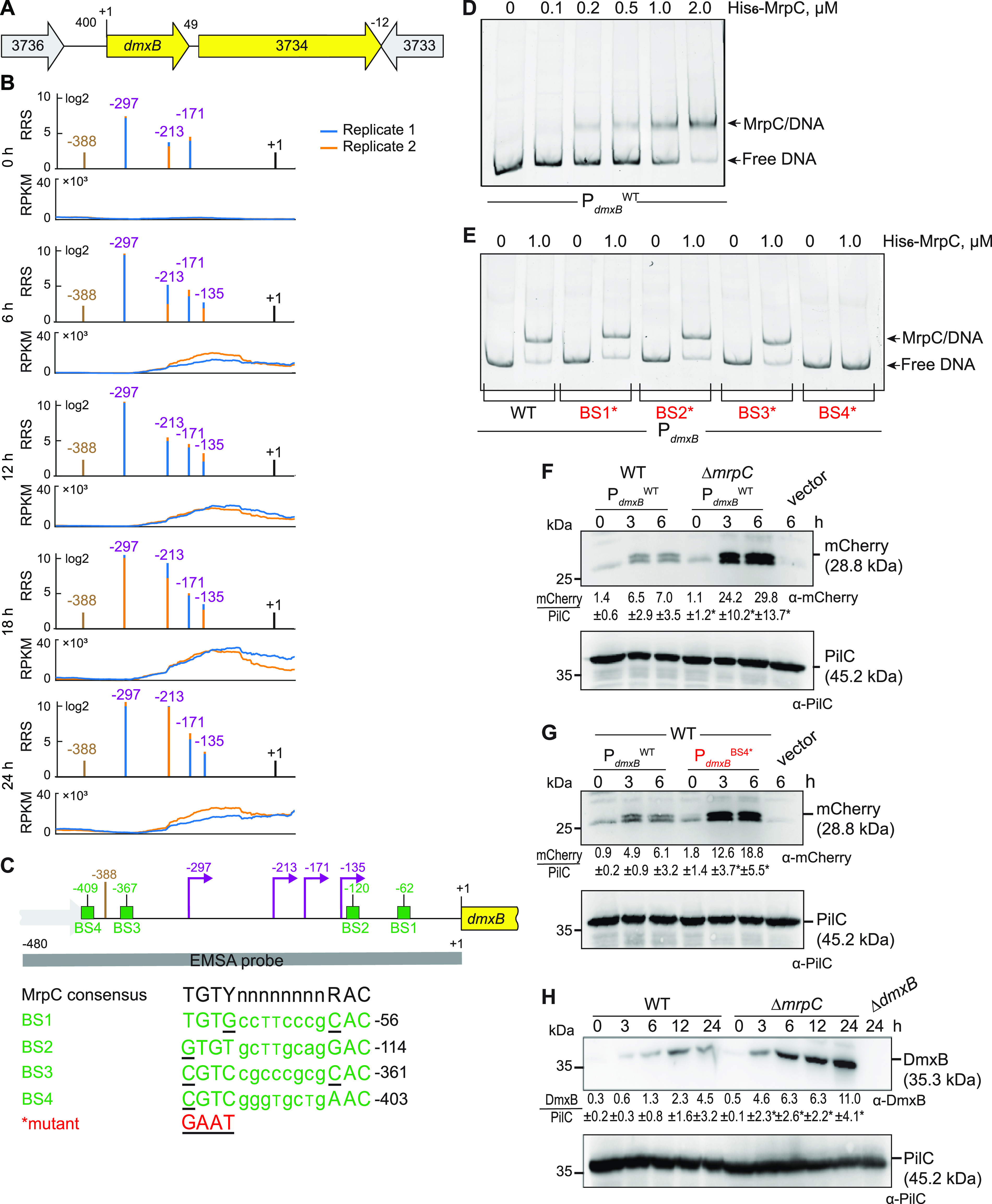
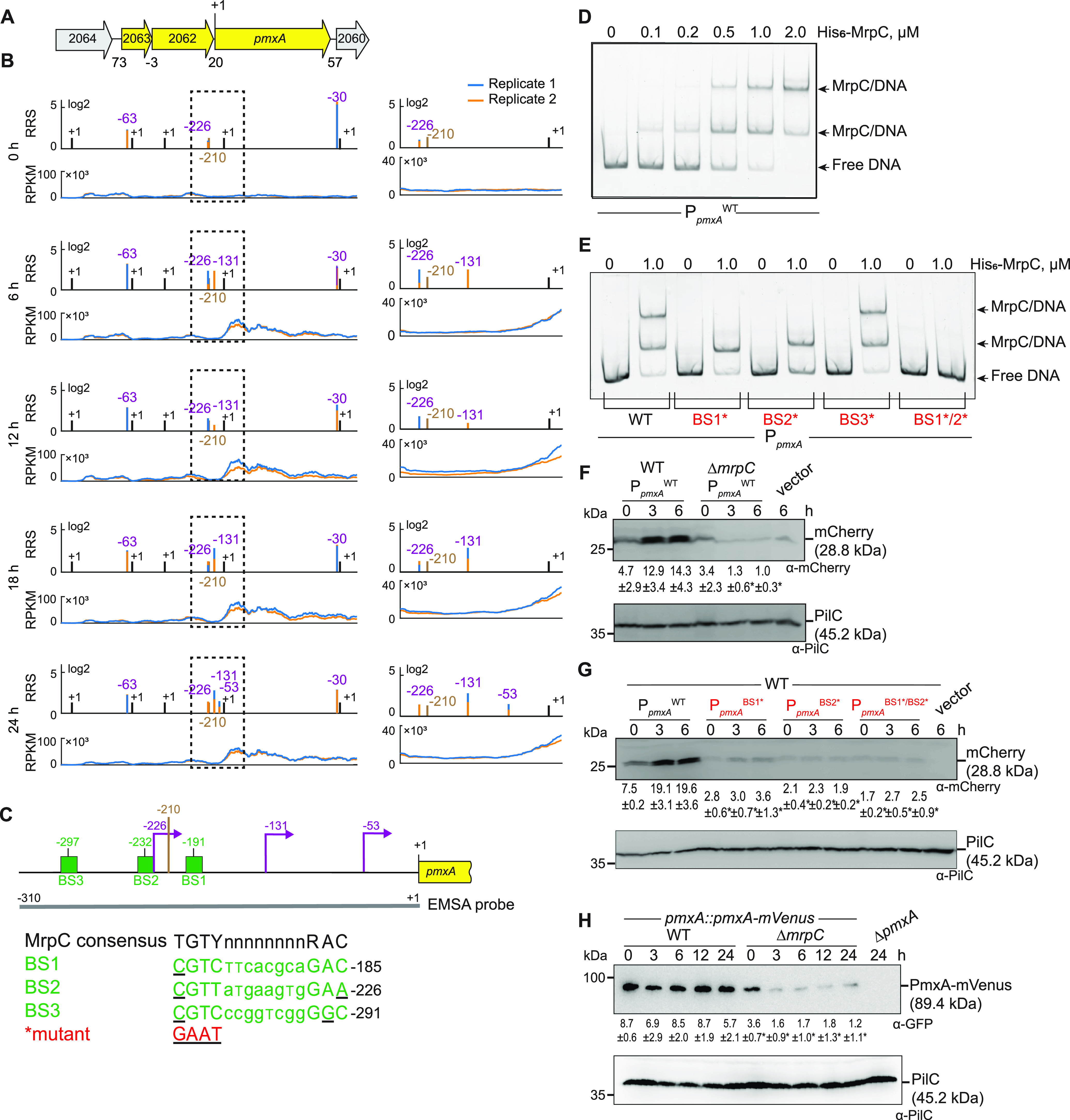
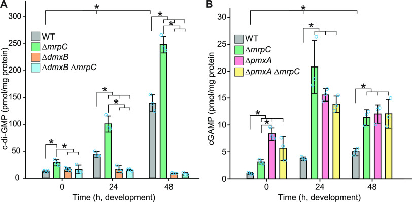
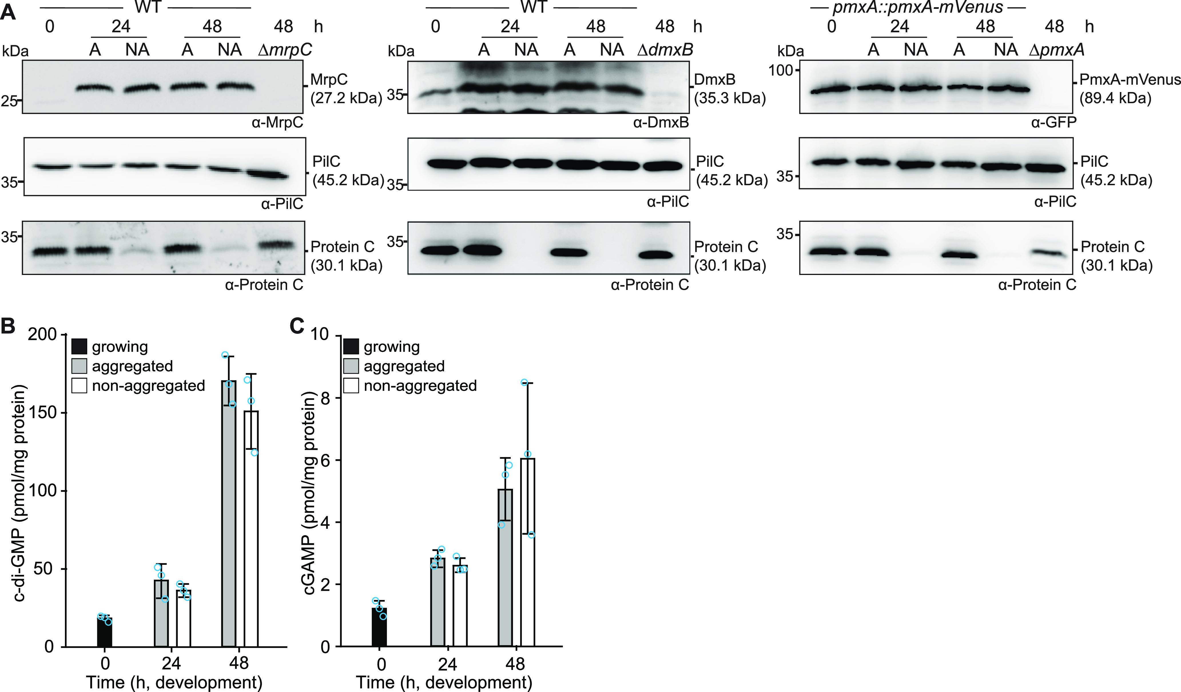
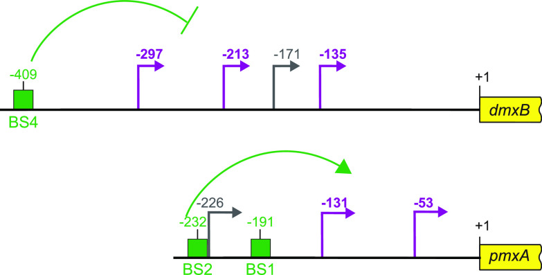
Myxococcus xanthus has a nutrient-regulated biphasic life cycle forming predatory swarms in the presence of nutrients and spore-filled fruiting bodies in the absence of nutrients. The second messenger 3'-5', 3'-5 cyclic di-GMP (c-di-GMP) is essential during both stages of the life cycle; however, different enzymes involved in c-di-GMP synthesis and degradation as well as several c-di-GMP receptors are important during distinct life cycle stages. To address this stage specificity, we determined transcript levels using transcriptome sequencing (RNA-seq) and transcription start sites using Cappable sequencing (Cappable-seq) during growth and development genome wide. All 70 genes encoding c-di-GMP-associated proteins were expressed, with 28 upregulated and 10 downregulated during development. Specifically, the three genes encoding enzymatically active proteins with a stage-specific function were expressed stage specifically. By combining operon mapping with published chromatin immunoprecipitation sequencing (ChIP-seq) data for MrpC (M. Robinson, B. Son, D. Kroos, L. Kroos, BMC Genomics 15:1123, 2014, http://dx.doi.org/10.1186/1471-2164-15-1123), the cAMP receptor protein (CRP)-like master regulator of development, we identified nine developmentally regulated genes as regulated by MrpC. In particular, MrpC directly represses the expression of dmxB, which encodes the diguanylate cyclase DmxB that is essential for development and responsible for the c-di-GMP increase during development. Moreover, MrpC directly activates the transcription of pmxA, which encodes a bifunctional phosphodiesterase that degrades c-di-GMP and 3',3'-cGAMP in vitro and is essential for development. Thereby, MrpC regulates and curbs the cellular pools of c-di-GMP and 3',3'-cGAMP during development. We conclude that temporal regulation of the synthesis of proteins involved in c-di-GMP metabolism contributes to c-di-GMP signaling specificity. MrpC is important for this regulation, thereby being a key regulator of developmental cyclic di-nucleotide metabolism in M. xanthus. IMPORTANCE The second messenger c-di-GMP is important during both stages of the nutrient-regulated biphasic life cycle of Myxococcus xanthus with the formation of predatory swarms in the presence of nutrients and spore-filled fruiting bodies in the absence of nutrients. However, different enzymes involved in c-di-GMP synthesis and degradation are important during distinct life cycle stages. Here, we show that the three genes encoding enzymatically active proteins with a stage-specific function are expressed stage specifically. Moreover, we find that the master transcriptional regulator of development MrpC directly regulates the expression of dmxB, which encodes the diguanylate cyclase DmxB that is essential for development, and of pmxA, which encodes a bifunctional phosphodiesterase that degrades c-di-GMP and 3',3'-cGAMP in vitro and is essential for development. We conclude that temporal regulation of the synthesis of proteins involved in c-di-GMP metabolism contributes to c-di-GMP signaling specificity and that MrpC plays an important role in this regulation.

Keywords: 3'; 3'-cGAMP; CRP; CRP-like proteins; Cappable-seq; PilZ; c-di-GMP; cGAMP; cyclic nucleotides; development; diguanylate cyclase; fruiting body formation; phosphodiesterase; second messenger; sporulation.


Conflict of interest statement

The authors declare no conflict of interest.

Figures

FIG 1
FIG 1
Expression of genes for “c-di-GMP-associated proteins.” (A) Expression of the genes encoding c-di-GMP-associated proteins. The heatmap shows normalized read counts at the indicated time points. Genes are color-coded according to the key on the right. MXAN_2807 is indicated as a protein with an HD-GYP domain; this protein also contains a MshEN domain. (B) Relative transcript levels during development for genes encoding c-di-GMP-associated proteins. The heatmap shows the log2-fold change at 6, 12, 18, or 24 h of development relative to 0 h calculated from the normalized read counts (Table S1B). Genes marked * or # were expressed at lower and higher levels, respectively, in the ΔmrpC mutant compared with those in the WT, as determined using RT-qPCR (see also Fig. 2 and Fig. S3). Colored boxes on the right indicate the four clusters with distinct expression profiles.
FIG 2
FIG 2
Regulation of the expression of genes encoding c-di-GMP-associated proteins by MrpC. Total RNA was isolated from cells developed in MC7 submerged cultures at the indicated time points from WT (black) and the ΔmrpC mutant (red). Transcript levels were determined using RT-qPCR and are shown as mean ± standard deviation (SD) from two biological replicates, each with two technical replicates, relative to WT at 0 h. *, P < 0.05; Student’s t test in which samples from the ΔmrpC mutant were compared with the samples from WT at the same time point. fruA served as a positive control. Based on protein sequence analysis, MXAN_1525 and MXAN_4232 are predicted to have DGC and PDE activity, respectively; however, neither a ΔMXAN_1525 nor a ΔMXAN_4232 mutant has defects during growth or development (37, 40). pkn1, MXAN_2902, MXAN_6957, and MXAN_7024 are PilZ domain proteins; however, none contain the conserved motifs for c-di-GMP binding (27, 36). Except for Pkn1, a lack of any of these four proteins does not cause defects during growth or development (36, 60). MXAN_7500 is a MshEN domain protein with the sequence motifs for c-di-GMP binding (17); however, it is not known whether this protein binds c-di-GMP or whether it is important during growth and development.
FIG 3
FIG 3
MrpC negatively regulates the expression of dmxB. (A) Schematic of the dmxB locus. The direction of transcription is indicated by the arrows. +1 indicates TSC of dmxB. Numbers above indicate the distance between start and stop codons of flanking genes. MXAN_3734 encodes a 577-amino acid residues protein with a C-terminal receiver domain of response regulators; the remainder of the protein does not contain known domains; MXAN_3734 is not important for development (40). (B) RNA-seq (bottom) and Cappable-seq (top) data at different time points. For each time point, data for both biological replicates are shown in blue and orange. For Cappable-seq, the RRS is indicated for each TSS on a log2 scale; for RNA-seq, reads per kilo base per million mapped reads (RPKM) values were calculated for each nucleotide position. Data from RNA-seq and Cappable-seq are from different samples. +1 indicates the dmxB TSC. TSSs as mapped by Cappable-seq are indicated in purple relative to the TSC of dmxB. The center of the MrpC ChIP-seq peak is indicated in brown. (C) Feature map of dmxB promoter region. +1 and color code is as in panel B. Green boxes labeled BS1 to 4 indicate potential MrpC binding sites based on the consensus sequence as defined by reference ; sequences of BS1 to 4 are shown below and in which underlined text indicates a mismatch. Red indicates the sequence used to generate the mutant binding sites. The gray line indicates the EMSA probe and contains all four predicted MrpC binding sites. (D and E) MrpC binds to the dmxB promoter region using BS4. The indicated Hex-labeled probes were mixed with the indicated concentrations of His6-MrpC EMSA and analyzed by EMSA. (F) MrpC represses dmxB promoter(s). Total cell lysates from the indicated strains expressing mcherry from PdmxBWT were harvested from cells developed in MC7 submerged cultures at the indicated time points. A total of 10 μg of protein was loaded per lane, and samples were separated by SDS-PAGE. Top and bottom blots were probed with α-mCherry and α-PilC antibodies, respectively. The PilC blot served as a loading control. Numbers below the top panel indicate in the accumulation of mCherry relative to PilC as mean ± SD as measured in three biological replicates (Materials and Methods). *, P < 0.05; in Student’s t test in which samples from the ΔmrpC mutant were compared with samples from WT at the same time point. Vector with mCherry but without the dmxB promoter served as a negative control (vector). mCherry separates into two bands; the reason for this result is not known. (G) BS4 is important for MrpC-dependent repression of dmxB promoter(s). Total cell lysates from the indicated WT strains expressing mCherry from the two indicated promoters were prepared and analyzed as in panel F. (H) DmxB accumulates at increased levels in the ΔmrpC mutant. Total cell lysates of the indicated strains were harvested from cells developed in MC7 submerged conditions at indicated time points and analyzed as in panel F except that the accumulation of DmxB relative to PilC was calculated.
FIG 4
FIG 4
MrpC positively regulates the expression of pmxA. (A) Schematic of the pmxA locus. The direction of transcription is indicated by the arrows. +1 indicates TSC of pmxA. Numbers above indicate the distance between start and stop codons of flanking genes. MXAN_2063 encodes a FecR domain-containing protein with a lipoprotein signal peptide and MXAN_2062 encodes a protein with a type I signal peptide, an N-terminal LysM domain, and a C-terminal extracellular fibronectin type III domain. The function of these two proteins is not known. (B) RNA-seq (bottom) and Cappable-seq (top) data at different time points for genes at pmxA locus. For each time point, data for both biological replicates are shown in blue and orange. For Cappable-seq, the RRS is indicated for each TSS on a log2 scale; for RNA-seq, RPKM values were calculated for each nucleotide position. The data from RNA-seq and Cappable-seq were obtained from different samples. Left, +1 indicates TSCs of MXAN_2064-_2060; right, zoom of region indicated in the hatched box in left panels immediately upstream of pmxA and where +1 indicates the TSC of pmxA. In both sets of panels, TSSs as mapped by Cappable-seq are indicated in purple relative to the nearest TSC. The center of the MrpC ChIP-seq peak is indicated in brown. (C) Feature map of pmxA promoter region. +1 and color code is as in panel B. Green boxes labeled BS1 to 3 indicate potential MrpC binding sites based on the consensus sequence as defined by reference ; sequences of BS1 to 3 are shown below and in which underlined text indicates a mismatch. Red indicates the sequence used to generate the mutant binding sites. The gray line indicates the EMSA probe and contains all three predicted MrpC binding sites. (D and E) MrpC binds to the pmxA promoter region using BS1 and BS2. The indicated Hex-labeled probes were mixed with the indicated concentrations of His6-MrpC EMSA and analyzed by EMSA. (F) MrpC activates pmxA promoter(s). Total cell lysates from the indicated strains expressing mCherry from PpmxAWT were harvested from cells developed in MC7 submerged cultures at the indicated time points and then analyzed as in Fig. 3F. (G) BS1 and BS2 are important for MrpC-dependent activation of the pmxA promoter(s). Total cell lysates from the indicated WT strains expressing mCherry from the indicated promoters were prepared and analyzed as in Fig. 3F. (H) PmxA accumulates at reduced levels in the ΔmrpC mutant. Total cell lysates of the indicated strains were harvested from cells developed in MC7 submerged conditions at indicated time points and analyzed as in Fig. 3F except that the accumulation of PmxA-mVenus relative to PilC was calculated.
FIG 5
FIG 5
c-di-GMP and cGAMP accumulation during growth and development. (A and B) Cells were harvested at the indicated time points of development, and nucleotide levels and protein concentrations were determined. Levels are shown as mean ± SD calculated from three biological replicates. Individual data points are in light blue. *, P < 0.05; in Student’s t test. At each specific time point, only pairwise comparisons with significant differences are indicated.
FIG 6
FIG 6
MrpC, DmxA, PmxA-mVenus, c-di-GMP, and cGAMP accumulation in aggregated and nonaggregated cells. (A) MrpC, DmxB, and PmxA-mVenus accumulate at the same levels in aggregated (A) and nonaggregated (NA) cells. Cells were harvested at the indicated time points of development and separated into the two cell fractions. A total of 10 μg of protein was loaded per lane, and samples were separated by SDS-PAGE. Top blots were probed with α-MrpC, α-DmxB, or α-GFP; middle blots with α-PilC; and bottom blots with α-Protein C antibodies. The PilC blots served as loading controls and the Protein C blots as cell separation controls. (B and C) c-di-GMP (B) and cGAMP (C) accumulate at the same levels in aggregated and nonaggregated cells of WT. Samples were generated as in panel A. Levels are shown as mean ± SD calculated from three biological replicates. Individual data points are in light blue. At each specific time point, no significant differences were identified in pairwise comparisons in Student’s t test.
FIG 7
FIG 7
Schematic of dmxB and pmxA promoter regions. +1 indicate TSC of dmxB or pmxA; TSSs are indicated in purple and gray and with developmentally regulated TSSs in purple; green boxes indicate verified MrpC binding sites named as in Fig. 3C and 4C. All coordinates are relative to the TSC (+1).

Similar articles

Cited by

References

    1. Römling U, Galperin MY, Gomelsky M. 2013. Cyclic di-GMP: the first 25 years of a universal bacterial second messenger. Microbiol Mol Biol Rev 77:1–52. doi:10.1128/MMBR.00043-12. - DOI - PMC - PubMed
    1. Jenal U, Reinders A, Lori C. 2017. Cyclic di-GMP: second messenger extraordinaire. Nat Rev Microbiol 15:271–284. doi:10.1038/nrmicro.2016.190. - DOI - PubMed
    1. Yin W, Cai X, Ma H, Zhu L, Zhang Y, Chou S-H, Galperin MY, He J. 2020. A decade of research on the second messenger c-di-AMP. FEMS Microbiol Rev 44:701–724. doi:10.1093/femsre/fuaa019. - DOI - PMC - PubMed
    1. Irving SE, Choudhury NR, Corrigan RM. 2021. The stringent response and physiological roles of (pp)pGpp in bacteria. Nat Rev Microbiol 19:256–271. doi:10.1038/s41579-020-00470-y. - DOI - PubMed
    1. Yoon SH, Waters CM. 2021. The ever-expanding world of bacterial cyclic oligonucleotide second messengers. Curr Opin Microbiol 60:96–103. doi:10.1016/j.mib.2021.01.017. - DOI - PMC - PubMed

MeSH terms

Substances