. 2021 Feb 22;13(1):e0387321.
doi: 10.1128/mbio.03873-21. Epub 2022 Feb 15.

Plasma Membrane Phosphatidylinositol-4-Phosphate Is Not Necessary for Candida albicans Viability yet Is Key for Cell Wall Integrity and Systemic Infection

Affiliations

Plasma Membrane Phosphatidylinositol-4-Phosphate Is Not Necessary for Candida albicans Viability yet Is Key for Cell Wall Integrity and Systemic Infection

Rocio Garcia-Rodas et al. mBio. .

Abstract

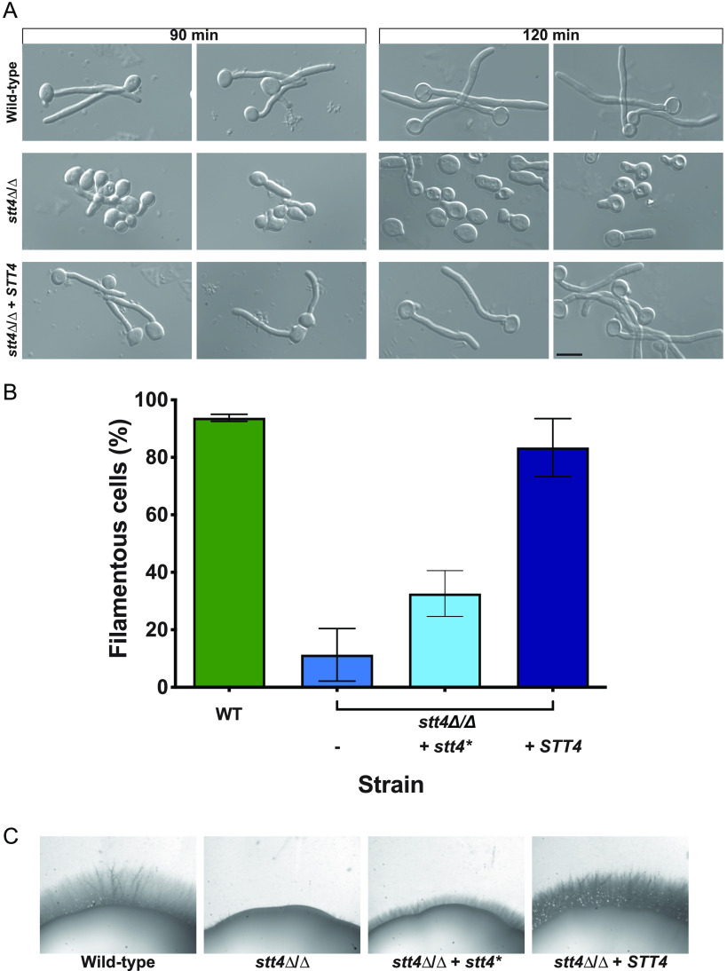
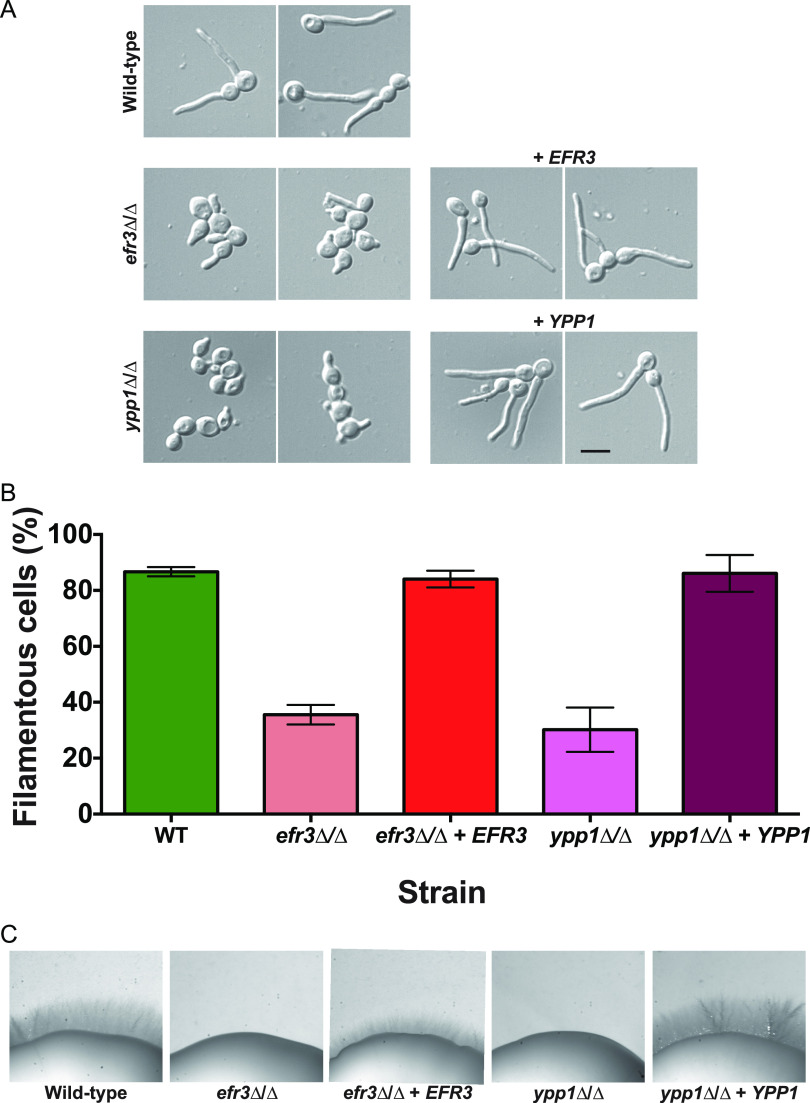
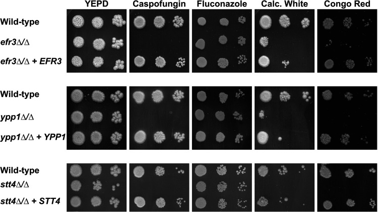
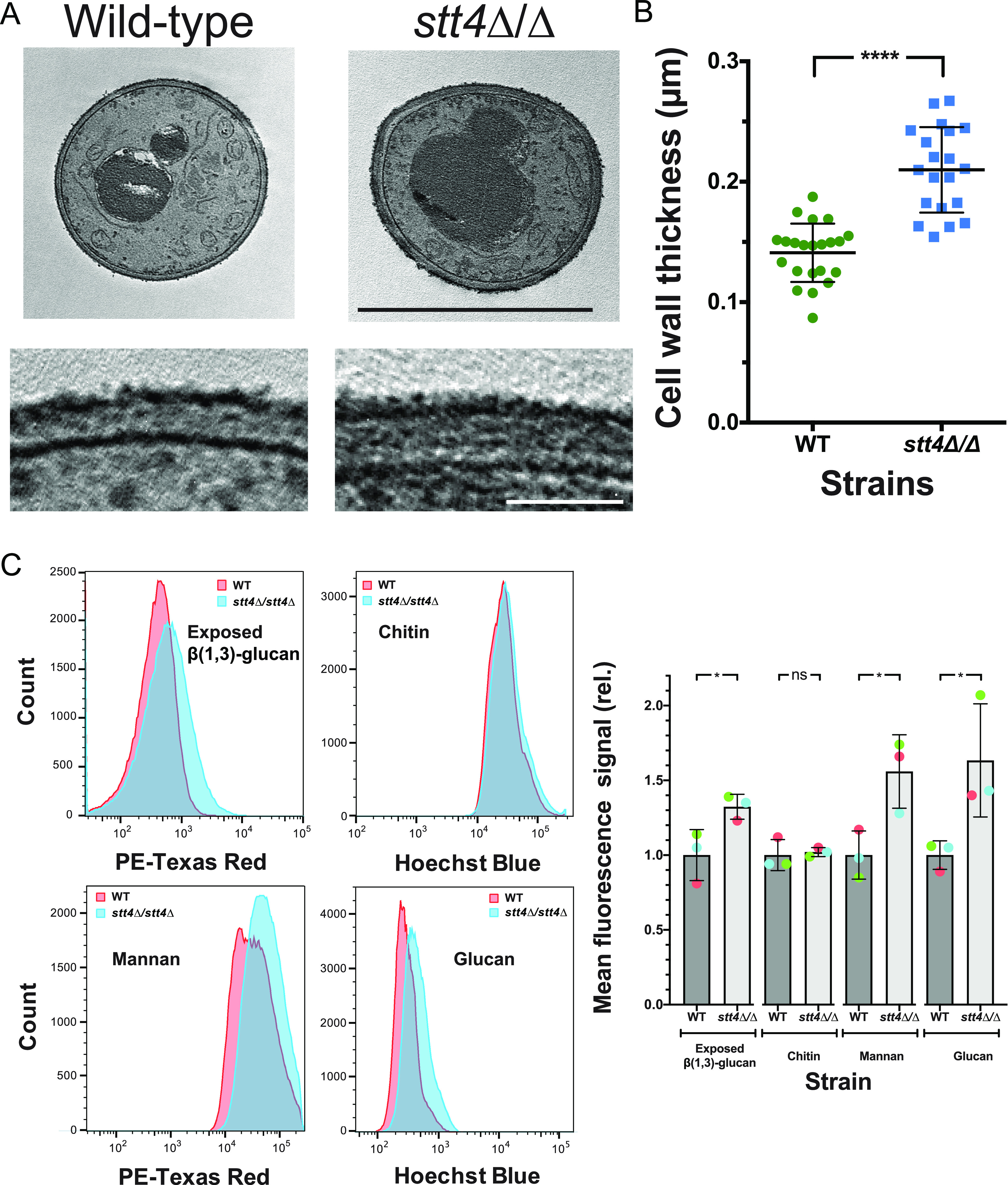
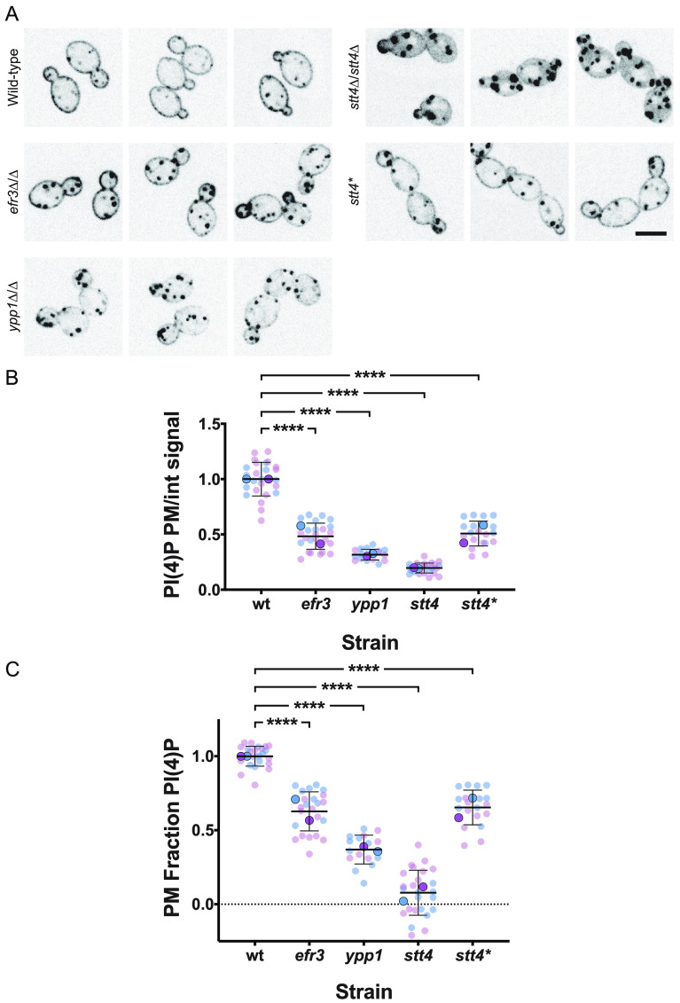
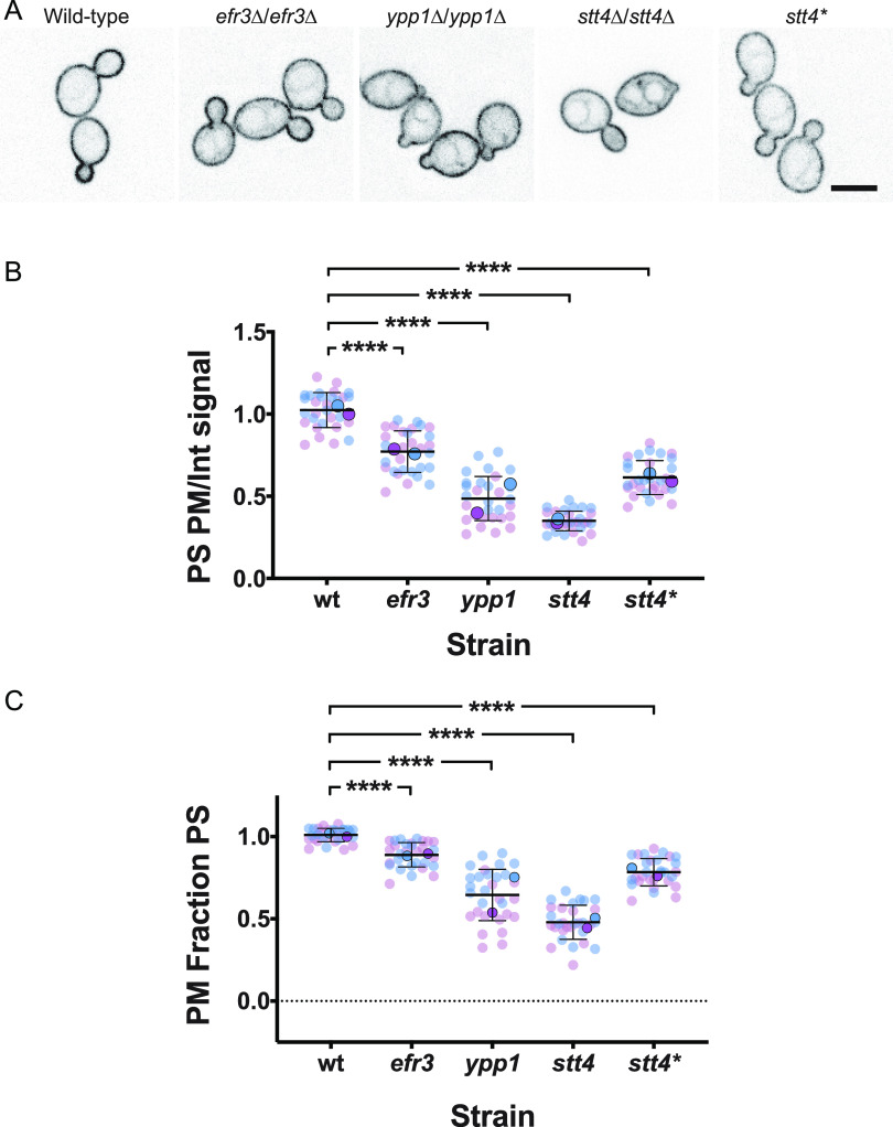
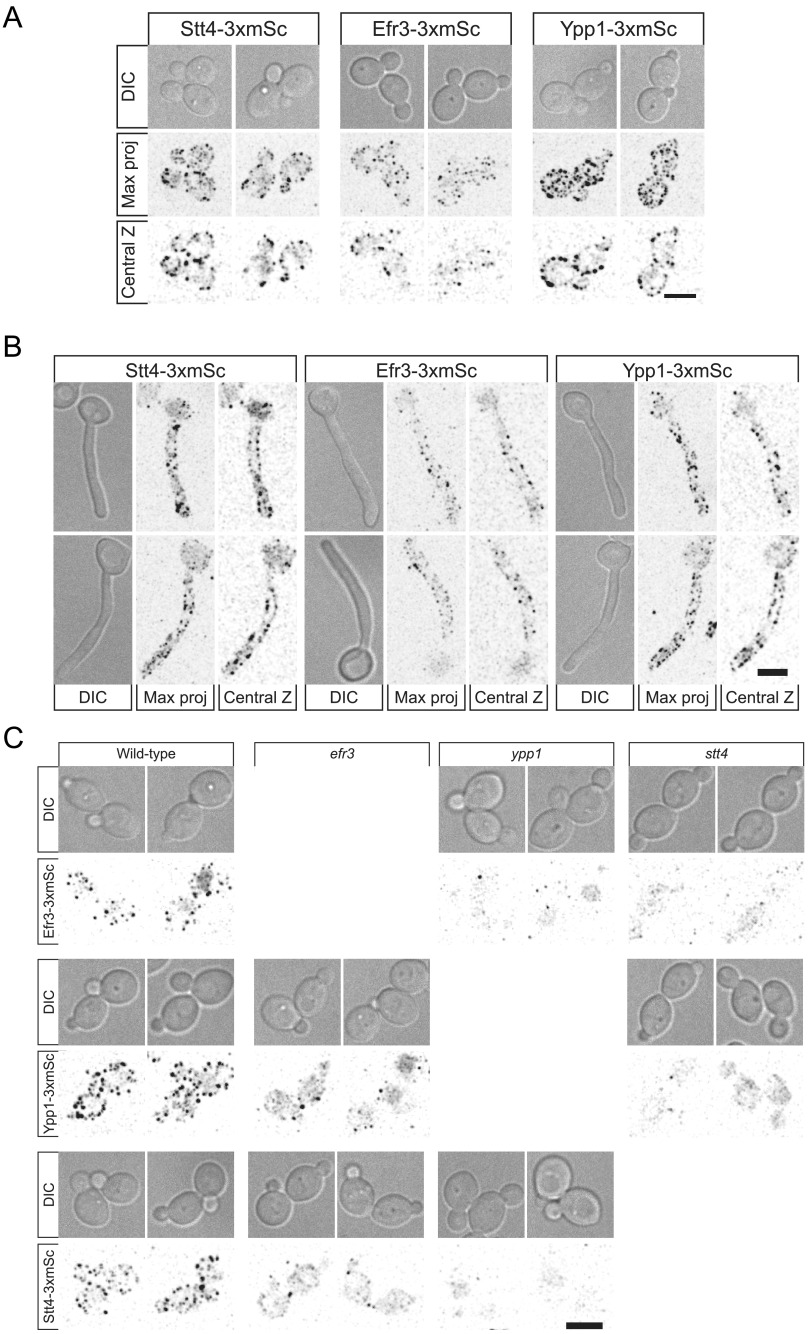
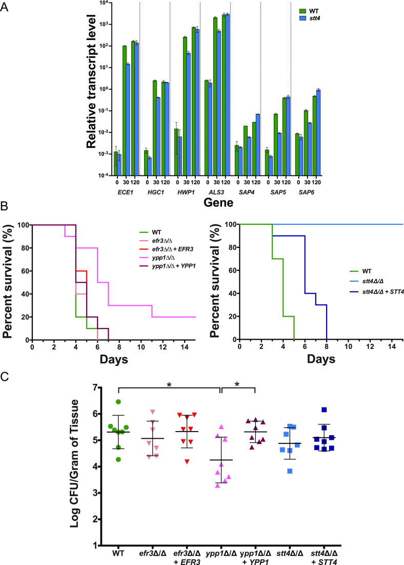
Phosphatidylinositol phosphates are key phospholipids with a range of regulatory roles, including membrane trafficking and cell polarity. Phosphatidylinositol-4-phosphate [PI(4)P] at the Golgi apparatus is required for the budding-to-filamentous-growth transition in the human-pathogenic fungus Candida albicans; however, the role of plasma membrane PI(4)P is unclear. We have investigated the importance of this phospholipid in C. albicans growth, stress response, and virulence by generating mutant strains with decreased levels of plasma membrane PI(4)P, via deletion of components of the PI-4-kinase complex, i.e., Efr3, Ypp1, and Stt4. The amounts of plasma membrane PI(4)P in the efr3Δ/Δ and ypp1Δ/Δ mutants were ∼60% and ∼40%, respectively, of that in the wild-type strain, whereas it was nearly undetectable in the stt4Δ/Δ mutant. All three mutants had reduced plas7ma membrane phosphatidylserine (PS). Although these mutants had normal yeast-phase growth, they were defective in filamentous growth, exhibited defects in cell wall integrity, and had an increased exposure of cell wall β(1,3)-glucan, yet they induced a range of hyphal-specific genes. In a mouse model of hematogenously disseminated candidiasis, fungal plasma membrane PI(4)P levels directly correlated with virulence; the efr3Δ/Δ mutant had wild-type virulence, the ypp1Δ/Δ mutant had attenuated virulence, and the stt4Δ/Δ mutant caused no lethality. In the mouse model of oropharyngeal candidiasis, only the ypp1Δ/Δ mutant had reduced virulence, indicating that plasma membrane PI(4)P is less important for proliferation in the oropharynx. Collectively, these results demonstrate that plasma membrane PI(4)P levels play a central role in filamentation, cell wall integrity, and virulence in C. albicans. IMPORTANCE While the PI-4-kinases Pik1 and Stt4 both produce PI(4)P, the former generates PI(4)P at the Golgi apparatus and the latter at the plasma membrane, and these two pools are functionally distinct. To address the importance of plasma membrane PI(4)P in Candida albicans, we generated deletion mutants of the three putative plasma membrane PI-4-kinase complex components and quantified the levels of plasma membrane PI(4)P in each of these strains. Our work reveals that this phosphatidylinositol phosphate is specifically critical for the yeast-to-hyphal transition, cell wall integrity, and virulence in a mouse systemic infection model. The significance of this work is in identifying a plasma membrane phospholipid that has an infection-specific role, which is attributed to the loss of plasma membrane PI(4)P resulting in β(1,3)-glucan unmasking.

Keywords: Candida albicans; cell wall; filamentous growth; opportunistic fungi; phosphatidylinositol phosphates; phospholipids; virulence.


Conflict of interest statement

The authors declare no conflict of interest.

Figures

FIG 1
FIG 1
The PI-4-kinase Stt4 is required for filamentous growth. (A) Strains (wild type, PY4861; stt4Δ/Δ, PY5111; stt4Δ/Δ+STT4, PY5131) were incubated with serum at 37°C for 90 and 120 min. Bar, 5 μm. (B) Percentages of filamentous cells were determined from three independent experiments (n ≥ 120 in each) with the abovementioned strains in addition to PY5757, an stt4Δ/Δ+stt4* strain encoding Stt4[G1810D]. Cells were considered filamentous if germ tubes were twice length of the mother cell or longer. Error bars indicate standard deviations (SD). (C) Stt4 is required for invasive filamentous growth. Strains were incubated for 4 days at 30°C on serum agar plates. Similar results were observed in three independent experiments.
FIG 2
FIG 2
Efr3 and Ypp1 are required for filamentous growth. (A) Indicated strains (wild type, PY4861; efr3Δ/Δ, PY4036; efr3Δ/Δ+EFR3, PY4039; ypp1Δ/Δ, PY4033; ypp1Δ/Δ+YPP1, PY4040) were induced with serum at 37°C for 90 min. Bar, 5 μm. (B) The percentage of filamentous cells was determined from three independent experiments (n ≥ 120 in each). Error bars indicate SD. (C) Efr3 and Ypp1 are required for invasive filamentous growth. Strains were incubated for 4 days at 30°C on serum agar plates. Similar results were observed in three independent experiments.
FIG 3
FIG 3
Plasma membrane PI(4)P is important for cell wall integrity. The indicated strains (wild type, PY4861; efr3Δ/Δ, PY4036; efr3Δ/Δ+EFR3, PY4039; ypp1Δ/Δ, PY4033; ypp1Δ/Δ+YPP1, PY4040; stt4Δ/Δ, PY5111; stt4Δ/Δ+STT4, PY5131) were incubated on YEPD with or without caspofungin (125 ng/mL), fluconazole (10 μg/mL), calcofluor white (25 μg/mL), or Congo red (400 μg/mL) for 3 days at 30°C. Similar results were observed in two independent experiments.
FIG 4
FIG 4
The stt4 deletion mutant has a thicker cell wall with increased mannan, glucan, and exposed β(1,3)-glucan. (A) Transmission electron micrographs of the indicated strains (wild type, PY4861; stt4Δ/Δ, PY5111) (top), with higher magnification of the cell wall (bottom). Bars, 5 μm (top) and 1 μm (bottom). (B) Quantitation of cell wall thickness from electron micrographs. (C) The stt4 mutant has increased exposure of β(1,3)-glucan together with increased levels of mannan and glucan. Flow cytometry analyses of cells (wild type, PY4861; stt4Δ/Δ, PY5111) labeled with anti-β(1,3)-glucan antibodies and a fluorescently labeled secondary antibody, calcofluor white, fluorescently labeled concanavalin A, and aniline blue. Flow cytometry profiles from one biological replicate (105 gated events; left) and means from three biological replicates normalized to each wild-type mean (right). Error bars indicate standard deviations. * P < 0.05; ****, P < 0.0001; ns, not significant.
FIG 5
FIG 5
Efr3, Ypp1, and Stt4 are critical for plasma membrane PI(4)P. (A) Strains expressing the plasma membrane PI(4)P reporter, GFP-Osh2PH-Osh2PH-GFP (wild type, PY2626; efr3Δ/Δ, PY4947; ypp1Δ/Δ, PY3950; stt4Δ/Δ, PY5169; stt4Δ/Δ+stt4*, PY5838) were imaged, and central z-sections of representative cells are shown with an inverted lookup table (LUT). Bar, 5 μm. (B and C) Quantitation of plasma membrane and internal signals reveals little to no plasma membrane PI(4)P in the stt4 mutant. The ratio of plasma membrane to internal signal (normalized to the wild type) and the relative plasma membrane signal (normalized plasma membrane/total signal) is shown. Quantitation of plasma membrane and internal signals was carried out excluding Golgi cisternae. For the wild type, the mean ratio of plasma membrane to internal signal was 3.8 and the ratio of plasma membrane to total signal was 0.8. We were able to detect ∼1.5% of wild-type plasma membrane PI(4)P levels. Smaller symbols are values from two experiments (6 to 15 cells per experiment), and larger symbols are means for each experiment, with bars indicating overall means and standard deviations. ****, P < 0.0001.
FIG 6
FIG 6
The number of Golgi cisternae is not affected by a decrease in plasma membrane PI(4)P. (A and B) Strains expressing Golgi PI(4)P reporter, FAPP1-GFP (wild type, PY2578; efr3Δ/Δ, PY3933; ypp1Δ/Δ, PY3951; stt4Δ/Δ, PY5552) were imaged, and maximum projections of representative cells are shown with an inverted LUT (left). Quantitation of the number of Golgi cisternae per cell (right) in the indicated strains (24 to 34 cells per strain). Bar, 5 μm.
FIG 7
FIG 7
Plasma membrane PI(4,5)P2 is not substantially affected by a decrease in PI(4)P. (A) Strains expressing the PI(4,5)P2 reporter GFP-PHPlcδ-PHPlcδ-GFP (wild type, PY1206; efr3Δ/Δ, PY3935; ypp1Δ/Δ, PY3958; stt4Δ/Δ, PY555) were imaged, and central z-sections of representative cells are shown with an inverted LUT. Bar, 5 μm. (B and C) Quantitation of plasma membrane and internal signals reveals that plasma membrane PI(4,5)P2 is largely unaffected in the absence of Efr3, Ypp1, and Stt4. The ratio of plasma membrane to internal signal and the relative plasma membrane signal were determined as for Fig. 5B and C (15 to 20 cells; 2 experiments for the wild type [WT]). For the wild type, the mean ratio of plasma membrane to internal signal was 2.8 and the ratio of plasma membrane to total signal was 0.7. *, P < 0.02; **, P < 0.01; ***, P < 0.0005.
FIG 8
FIG 8
A reduction in plasma membrane PI(4)P results in an increase in PS at the ER. (A) Strains expressing PS reporter GFP-LactC2 (wild type, PY3239; efr3Δ/Δ, PY4124; ypp1Δ/Δ, PY4131; stt4Δ/Δ, PY5174; stt4Δ/Δ+stt4*, PY5903) were imaged, and central z-sections of representative cells are shown with an inverted LUT. Bar, 5 μm. (B and C) Quantitation of plasma membrane and internal signals reveals a progressive decrease in plasma membrane PS in efr3, ypp1, and stt4 strains. The ratio of plasma membrane to internal signal and the relative plasma membrane signal were determined as for Fig. 5B and C. For the wild type, the mean ratio of plasma membrane to internal signal was 4.5 and the ratio of plasma membrane to total signal was 0.8. Smaller symbols are values from two experiments (15 cells each); larger symbols are means from each experiment, with bars indicating overall means and standard deviations. ****, P < 0.0001.
FIG 9
FIG 9
Plasma membrane PS is proportional to PI(4)P levels. Average levels of plasma membrane PS and PI(4)P in indicated strains were normalized to 1 in the wild type. Linear curve fit: y=1.316x−0.70; r2 = 0.96. Bars show standard deviations for 16 to 40 cells for each determination.
FIG 10
FIG 10
Efr3, Ypp1, and Stt4 localize as cortical patches, with Ypp1 and Stt4 being critical for each other’s localization. (A and B) Strains expressing indicated 3xmScarlet fusions (Stt4-3xmSc, PY6193; Efr3-3xmSc, PY6197; Ypp1-3xmSc, PY6195) were imaged during budding (A) and hyphal (B) growth. Differential interference contrast (DIC) images, central z-sections, and maximum projections of 17 0.5-μm z-sections are shown. (C) Strains (WT, PY6197, PY6195, and PY6193; efr3, PY6136 and PY6142; ypp1, PY6138 and PY6144; stt4, PY6140 and PY6134) expressing respective 3xmScarlet fusions were imaged during budding growth, and maximum projections of 17 0.5-μm z-sections are shown. Bars, 5 μm.
FIG 11
FIG 11
Plasma membrane PI(4)P is specifically required for hematogenously disseminated candidiasis. (A) Hyphal-specific genes are induced in an stt4 deletion mutant. The transcript level of indicated hyphal specific genes was determined in wild-type (PY4861) and stt4Δ/Δ (PY5111) strains at indicated times (in minutes) with serum at 37°C by qRT-PCR and normalized to the levels of TDH3 transcript. Means of 3 determinations from an experiment are shown, with bars indicating standard deviations. Similar results were observed in two additional biological replicates. (B) Stt4 is required for virulence in a murine systemic infection model. Survival of mice (n = 10) over time following injection of strains (wild type, PY4861; efr3Δ/Δ, PY4036; efr3Δ/Δ+EFR3, PY4039; ypp1Δ/Δ, PY4033; ypp1Δ/Δ+YPP1, PY4040; stt4Δ/Δ, PY5111; stt4Δ/Δ+STT4, PY5131). Similar results were observed in two independent experiments; the difference between the WT and the ypp1Δ/Δ mutant was statistically significant (P = 0.002) (left), and that between the WT and the stt4Δ/Δ mutant was statistically significant (P < 0.0001) (right). (C) Stt4 and Efr3 are not required for virulence in a murine oropharyngeal infection model. CFU per gram of tongue tissue was determined 5 days after oropharyngeal infection (8 mice per strain) with the indicated strains (wild type, PY4861; efr3Δ/Δ, PY4036; efr3Δ/Δ+EFR3, PY4039; ypp1Δ/Δ, PY4033; ypp1Δ/Δ+YPP1, PY4040; stt4Δ/Δ, PY4414; stt4Δ/Δ+STT4, PY4433). *, P < 0.05.

Similar articles

Cited by

References

    1. Bochud A, Conzelmann A. 2015. The active site of yeast phosphatidylinositol synthase Pis1 is facing the cytosol. Biochim Biophys Acta 1851:629–640. doi:10.1016/j.bbalip.2015.02.006. - DOI - PubMed
    1. Boura E, Nencka R. 2015. Phosphatidylinositol 4-kinases: function, structure, and inhibition. Exp Cell Res 337:136–145. doi:10.1016/j.yexcr.2015.03.028. - DOI - PubMed
    1. Dornan GL, McPhail JA, Burke JE. 2016. Type III phosphatidylinositol 4 kinases: structure, function, regulation, signalling and involvement in disease. Biochem Soc Trans 44:260–266. doi:10.1042/BST20150219. - DOI - PubMed
    1. De Souza CP, Hashmi SB, Osmani AH, Andrews P, Ringelberg CS, Dunlap JC, Osmani SA. 2013. Functional analysis of the Aspergillus nidulans kinome. PLoS One 8:e58008. doi:10.1371/journal.pone.0058008. - DOI - PMC - PubMed
    1. Lee KT, So YS, Yang DH, Jung KW, Choi J, Lee DG, Kwon H, Jang J, Wang LL, Cha S, Meyers GL, Jeong E, Jin JH, Lee Y, Hong J, Bang S, Ji JH, Park G, Byun HJ, Park SW, Park YM, Adedoyin G, Kim T, Averette AF, Choi JS, Heitman J, Cheong E, Lee YH, Bahn YS. 2016. Systematic functional analysis of kinases in the fungal pathogen Cryptococcus neoformans. Nat Commun 7:12766. doi:10.1038/ncomms12766. - DOI - PMC - PubMed

Substances