. 2022 Feb 14;24(1):13.
doi: 10.1186/s13058-022-01506-2.

Microtubule disruption reduces metastasis more effectively than primary tumor growth

Affiliations

Microtubule disruption reduces metastasis more effectively than primary tumor growth

Keyata N Thompson et al. Breast Cancer Res. .

Abstract

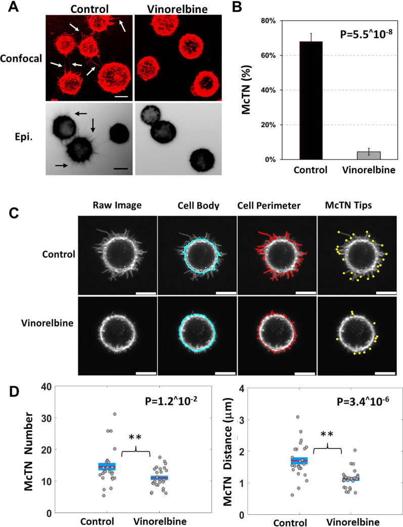
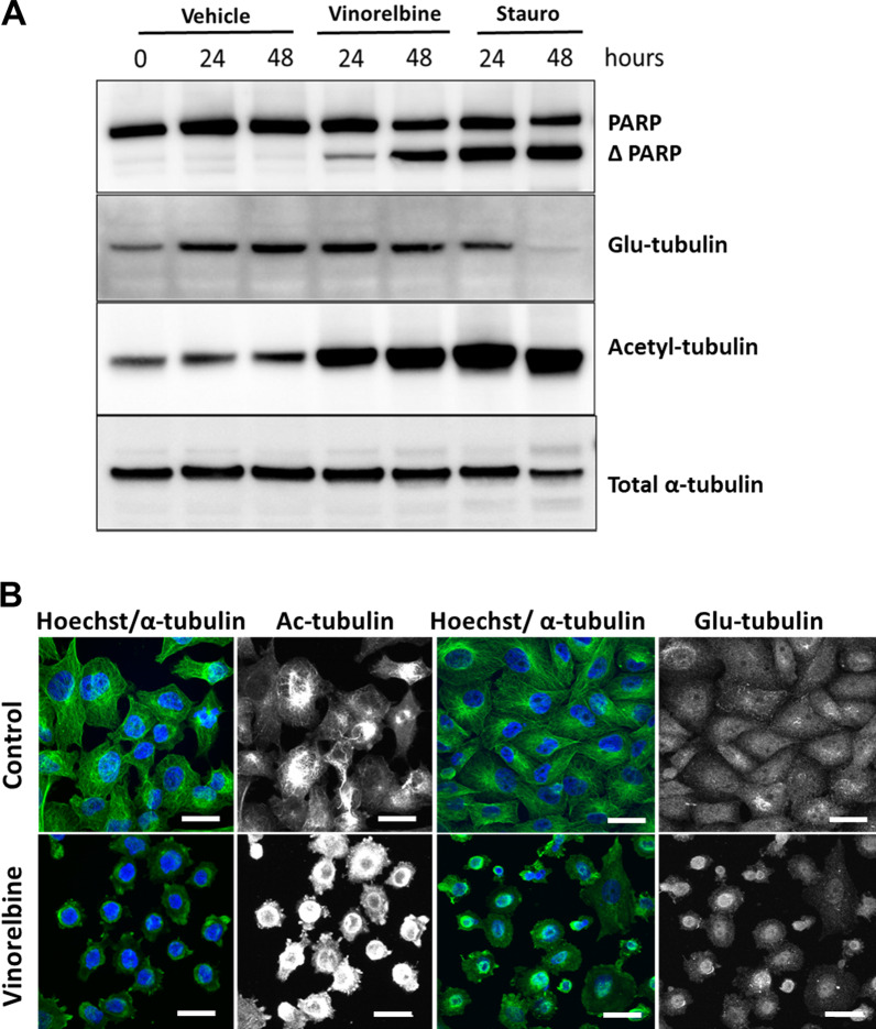
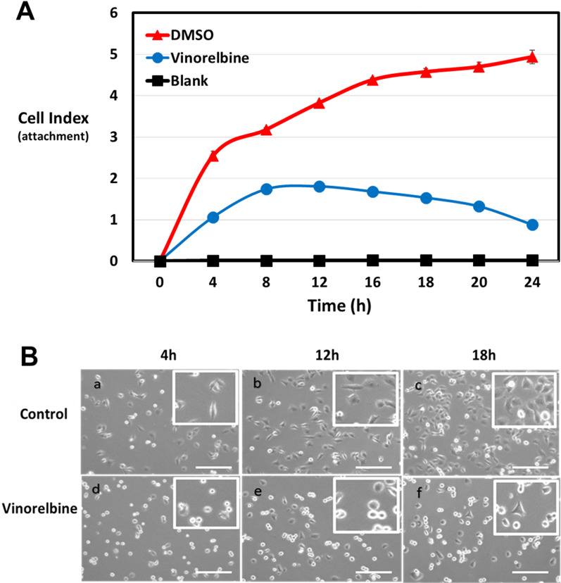
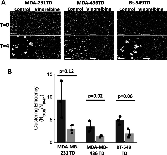
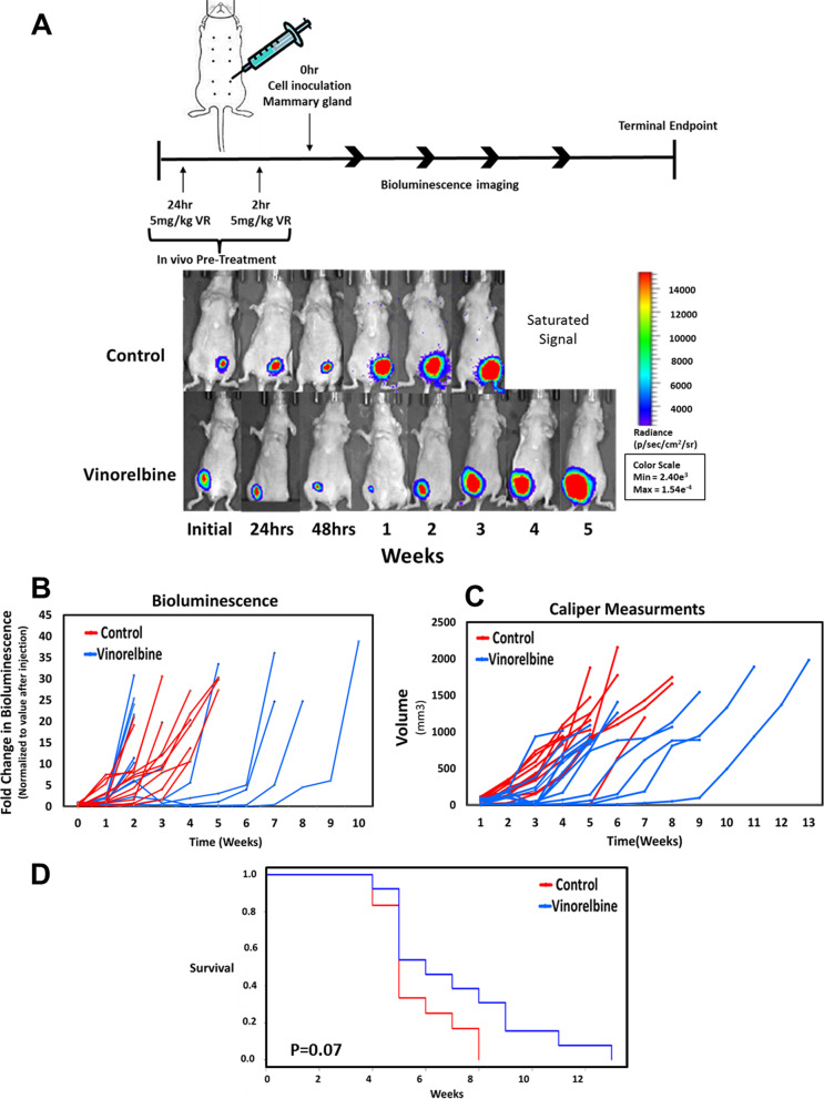
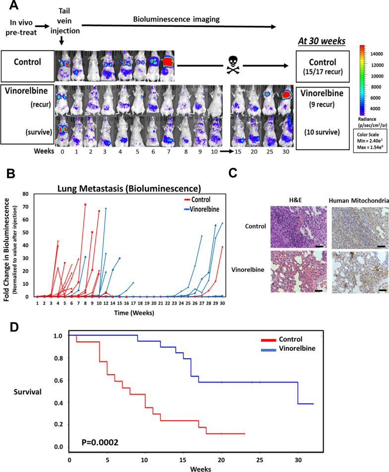
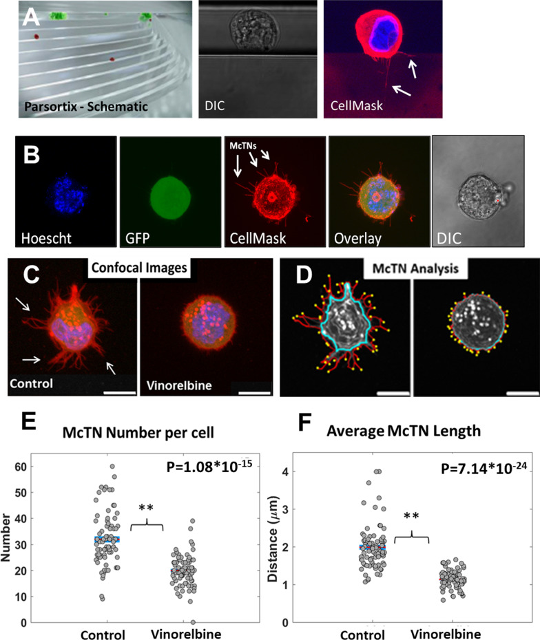
Clinical cancer imaging focuses on tumor growth rather than metastatic phenotypes. The microtubule-depolymerizing drug, Vinorelbine, reduced the metastatic phenotypes of microtentacles, reattachment and tumor cell clustering more than tumor cell viability. Treating mice with Vinorelbine for only 24 h had no significant effect on primary tumor survival, but median metastatic tumor survival was extended from 8 to 30 weeks. Microtentacle inhibition by Vinorelbine was also detectable within 1 h, using tumor cells isolated from blood samples. As few as 11 tumor cells were sufficient to yield 90% power to detect this 1 h Vinorelbine drug response, demonstrating feasibility with the small number of tumor cells available from patient biopsies. This study establishes a proof-of-concept that targeted microtubule disruption can selectively inhibit metastasis and reveals that existing FDA-approved therapies could have anti-metastatic actions that are currently overlooked when focusing exclusively on tumor growth.

Keywords: Breast cancer; Circulating tumor cells; MDA-MB-231; Metastasis; Microtentacles; Reattachment; Vinorelbine.


Conflict of interest statement

The University of Maryland has patents pending on the microfluidic cell tethering technology on which K.R.C, C.M.J. and S.S.M. are listed as inventors. S.S.M and C.M.J. are employees of the VA Maryland Health Care System. The views reported in this paper do not reflect the views of the Department of Veterans Affairs or the United States Government.

Figures

Fig. 1
Fig. 1
Vinorelbine decreases McTNs on breast tumor cells. MDA-MB-231 TD cells stained with CellMask Orange cell membrane dye, detached and suspended in media containing vehicle (0.1% DMSO) or Vinorelbine (10 μM) for 1 h. a Representative confocal images (top) and inverted epifluorescence images (bottom) of free-floating cells on a low attach plate. Scale bars correspond to 10 μm. b Vinorelbine caused a significant decrease in McTN frequency (%) compared to vehicle control. McTN scoring consists of mean values from four independent experiments where 100 cells were blindly counted and averaged. c Representative raw images of tethered cells and computer determined cell body outline (blue), cell perimeter (red), and McTNs tips (yellow). d Live cell analysis measuring an average number of McTN tips for cells treated with vehicle (0.1% DMSO) or Vinorelbine (10 μM) for 1 h. McTN number is the number of McTNs per cell. McTN distance is average distance of McTN tips from cell body boundary. For live cell image analysis, a population of 25 cells per condition was analyzed from 3 independent experimental replicate
Fig. 2
Fig. 2
Vinorelbine increases acetylated α-tubulin while decreasing filamentous microtubule network. a Immunoblot analysis of Vinorelbine (10 μM) treated MDA-MB-231 TD cells results in an increase in acetylated α-tubulin (acetyl-tubulin) whereas detyrosinated tubulin (Glu-tubulin) and total α-tubulin remain unchanged at 24 h and 48 h, compared with vehicle control (0.1% DMSO). Vinorelbine (10 μM) yielded minimal PARP cleavage at 24 h. However, by 48 h., approximately 50% PARP cleavage is observed. b Immunofluorescence shows filamentous tubulin (green) is destroyed within 15 min of 10 μM Vinorelbine treatment, as well as the organization of acetylated (Ac) and detyrosinated (Glu) microtubules. Analysis at additional times (data not shown) reveals the continued absence of filamentous tubulin. Hoechst was used to visualize the nuclei (blue). Images taken at 60 × magnification. Scale bar = 20 µm
Fig. 3
Fig. 3
Vinorelbine treatment decreases tumor cell reattachment. A Reattachment efficiency of the MDA-MB-231-TD cells treated with Vinorelbine (10 μM) is significantly lower than vehicle control treated cells (0.1% DMSO). Changes in impedance are apparent as early as 1 h and significant differences continue for 24 h after initial seeding. Representative experiment from three independent experiments; each performed in quadruplicate. B Representative phase contrast images of vehicle control (a–c) and Vinorelbine-treated (d–f) MDA-MB-231-TD cells over time to visualize cell attachment or lack of attachment (rounding). Panels, 4 × magnification; Insets, 10 × magnification. Scale bar = 100 µm
Fig. 4
Fig. 4
Vinorelbine treatment decreases homotypic cluster aggregation in vitro. MDA-MB-231 TD, MDA-MB-436 TD and BT-549 TD cells were treated in attached conditions for 1 h in Vinorelbine (10 μM). Cells were trypsinized, counted, and plated in a low attach plates for 4 h to test aggregation. At given timepoints, cells were tethered onto a TetherChip surface and stained with the nuclear dye Hoechst 33258 (1:5000). a Representative images of control and Vinorelbine-treated MDA-MB-231 TD, MDA-MB-436 TD and BT-549 TD cells over time to visualize cluster formation efficiency. Images taken of tethered and stained cells at t = 0 and 4 h. at a 4 × magnification. Scale bar = 100 µm. b Analysis measuring the efficiency of clustering by comparing the number of individual clusters over time. Individual values at t = 0 h were divided by respective experimental final cluster numbers (t = 4 h) for each condition
Fig. 5
Fig. 5
Primary tumor development is uninhibited in the presence of Vinorelbine. a Representative bioluminescence imaging from mice treated with vehicle control (0.1% DMSO) or 5 mg/kg Vinorelbine during a 24 h and 2 h prior to injection of MDA-MB-231 TD cells. Photon flux color scale is shown to the right. b Graphical representation of the growth curve in each mouse measured as a fold difference of bioluminescence signal over time. To quantitate values for each time point, the background was subtracted from the peak signal, and the difference was normalized to the initial value (first timepoint) for each animal (n = 5 per group). c Graphical representation of the tumor size for each mouse measured by external caliper measurement. Volumetric measurement of xenografted tumors was obtained using the ellipsoid calculation (V = xy2/2). d The probability of overall survival was assessed by Kaplan–Meier analysis (Log-rank test P = .07) for mice treated with Vinorelbine
Fig. 6
Fig. 6
Lung retention and metastatic development after Vinorelbine treatment. a Representative bioluminescence images of mice treated with vehicle control (0.1% DMSO) or 5 mg/kg Vinorelbine at 24 h and 2 h prior to injection of MDA-MB-231 TD cells. The vehicle control population resulted in 15 out of 17 animals (88%) exhibiting tumor formation in lung tissue between 3–12 weeks post inoculation with 2/17 (12%) surviving. The focused Vinorelbine treatment resulted in 10 out of 19 animals (53%) with disease-free survival at 30 weeks and 9 out of 19 animals (47%) with delayed tumor formation. Photon flux color scale is shown to the right. b Fold differences of retained bioluminescence in the lung of each mouse inoculated with MDA-MB-231 TD cells via tail vein. Data represent individual animal examined and measured as a fold change of the initial value of each independent animal. c Representative images of immunohistochemistry for human mitochondria and hematoxylin and eosin (H&E) staining performed at ethical end-points. Images captured at a magnification of 20 × indicate metastatic burden, scale bar, 200 μm. d Overall survival was assessed by Kaplan–Meier analysis (Wilcoxon test P < 0.001 and Log-rank test P < 0.001) for mice treated with Vinorelbine
Fig. 7
Fig. 7
Vinorelbine drug response in tumor cells recovered from blood samples. MDA-MB-231 TD cells captured using ANGLE Parsortix and VTX-1 systems. a Parsortix schematic of CTC staircase capture method from whole blood. Live DIC images to visualize cell capture and CellMask orange stained cells to visualize McTNs (arrows). b MDA-MB-231 TD cells (1 × 103) spiked into 7.5 ml whole blood were recovered in Parsortix cassette, eluted onto microfluidic cell tethering slides and imaged live after DNA staining with Hoechst and CellMask Orange cell membrane dye. c Confocal microscopy of MDA-MB-231 tumor cells (5 × 103) isolated from 7.5 ml whole blood using the VTX-1 system and then treated with Vinorelbine (10 μM, 1 h) or drug vehicle (0.1% DMSO, 1 h) before elution onto microfluidic cell tethering slides, chemical fixation and Hoescht (blue)/WGA (red) staining. Images show an overlay of each color. Arrows show McTN protrusions on isolated tumor cells. d McTN analysis of the cell body outline (blue), cell perimeter (red), and McTNs tips (yellow) of fixed and tethered cells. e McTN analysis measuring average number McTNs/cell after treatment with vehicle or Vinorelbine. f Analysis of average distance of McTN tips from cell body boundary for cells treated with vehicle or Vinorelbine. For cell isolation and fixation analysis a total of 78 cells in the vehicle control population and 80 cells for the Vinorelbine treated population was analyzed from three independent experimental replicates

Similar articles

Cited by

References

    1. Anderson RL, Balasas T, Callaghan J, Coombes RC, Evans J, Hall JA, Kinrade S, Jones D, Jones PS, Jones R, et al. A framework for the development of effective anti-metastatic agents. Nat Rev Clin Oncol. 2019;16(3):185–204. - PMC - PubMed
    1. Li G, Xie H, Ning H, Citrin D, Capala J, Maass-Moreno R, Guion P, Arora B, Coleman N, Camphausen K, et al. Accuracy of 3D volumetric image registration based on CT, MR and PET/CT phantom experiments. J Appl Clin Med Phys. 2008;9(4):2781. - PMC - PubMed
    1. Pantel K, Alix-Panabieres C. Liquid biopsy and minimal residual disease—latest advances and implications for cure. Nat Rev Clin Oncol. 2019;16:409–424. - PubMed
    1. Cristofanilli M, Budd GT, Ellis MJ, Stopeck A, Matera J, Miller MC, Reuben JM, Doyle GV, Allard WJ, Terstappen LW, et al. Circulating tumor cells, disease progression, and survival in metastatic breast cancer. N Engl J Med. 2004;351(8):781–791. - PubMed
    1. Hekimian K, Meisezahl S, Trompelt K, Rabenstein C, Pachmann K. Epithelial cell dissemination and readhesion: analysis of factors contributing to metastasis formation in breast cancer. ISRN Oncol. 2012;2012:601810. - PMC - PubMed

Publication types