. 2022 Feb 14;24(1):44.
doi: 10.1186/s13075-022-02738-5.

Chondrocyte-derived exosomes promote cartilage calcification in temporomandibular joint osteoarthritis

Affiliations

Chondrocyte-derived exosomes promote cartilage calcification in temporomandibular joint osteoarthritis

Qian Liu et al. Arthritis Res Ther. .

Abstract

Backgrounds: Abnormal cartilage calcification is one of the pathological changes of temporomandibular joint (TMJ) osteoarthritis (OA). Recent studies have reported that exosomes can regulate the formation of abnormal calcified nodules in diseases including atherosclerosis and chronic kidney disease. However, the influences of chondrocyte-derived exosomes on abnormal cartilage calcification in TMJ OA are still unclear.

Methods: TMJ OA was induced by unilateral anterior crossbite (UAC) for 4, 8, or 12 weeks in rats to observe abnormal calcification in TMJ condylar cartilage and exosome formation. Concomitantly, GW4869, the inhibitor of exosome formation, was locally injected to the TMJ of rats under stimulation of UAC, while the exosomes extracted from primary condylar chondrocytes stimulated with fluid flow shear stress (FFSS) were locally injected to rats TMJ.

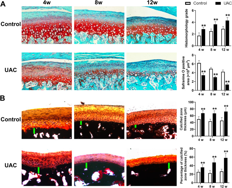
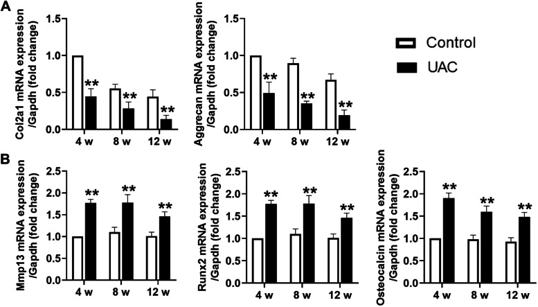
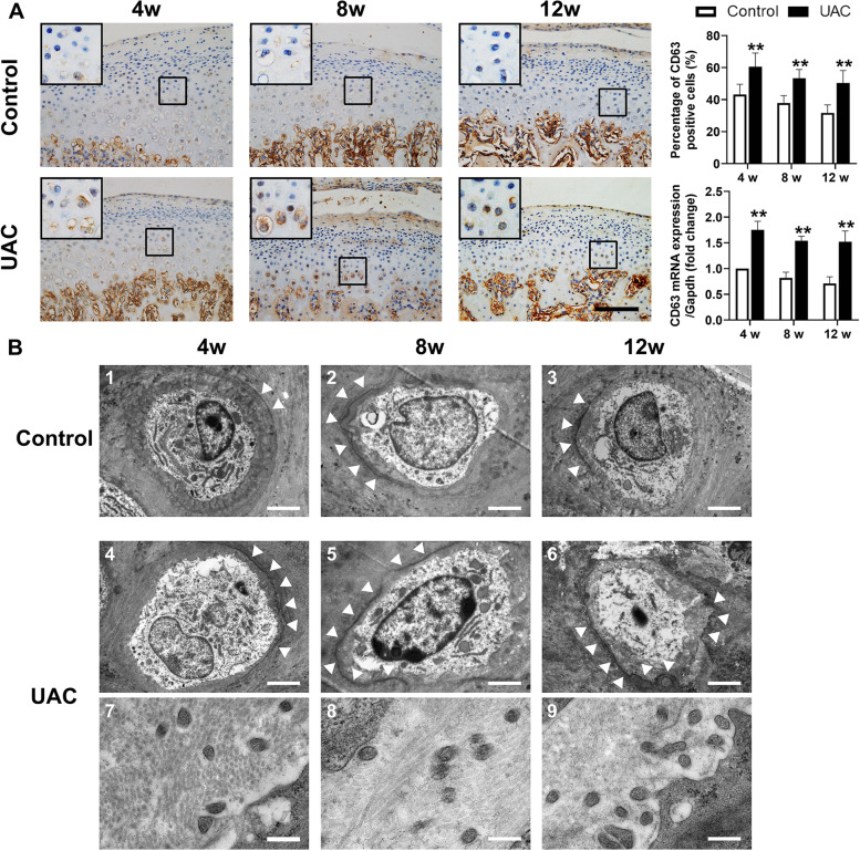
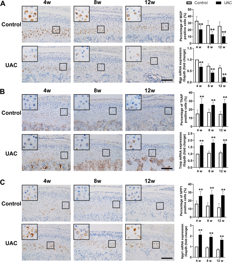
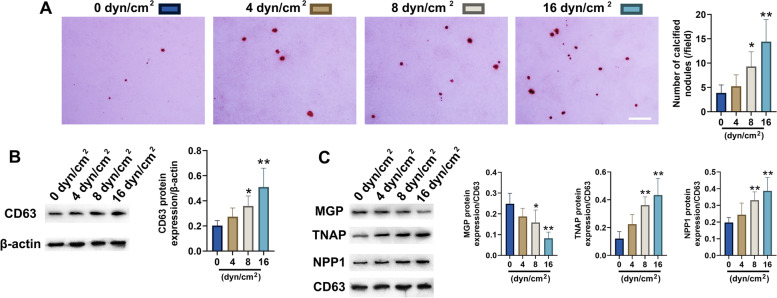
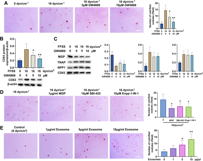
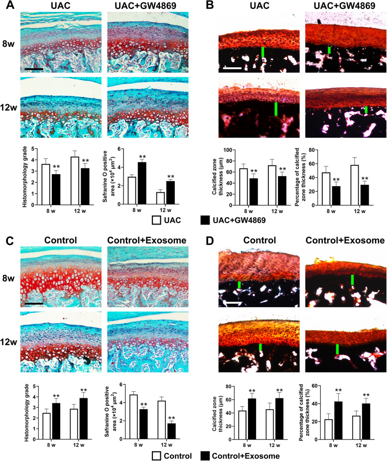
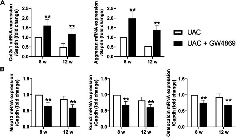
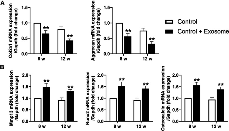
Results: Abnormal calcification was enhanced in the degenerative cartilage of TMJ OA in UAC rats, and a large number of exosome-like structures with diameters of 50-150 nm were found in the calcified cartilage together with decreased expression of matrix Gla protein (MGP) and increased expression of CD63, tissue-nonspecific alkaline phosphatase (TNAP) and nucleotide pyrophosphatase/phosphodiesterase-1 (NPP1). After FFSS stimulation, the number of exosomes secreted by chondrocytes and the numbers of calcified nodules were increased in cultured cells, and the protein levels of MGP, TNAP, and NPP1 in exosomes were changed. Inhibition of exosome formation, TNAP, and NPP1 or supplementation with exogenous MGP effectively alleviated FFSS-induced chondrocyte calcification. Local injection of GW4869, the exosome inhibitor, alleviated TMJ OA-related cartilage degeneration and calcification in UAC rats. Local injection of exosomes obtained from chondrocytes stimulated by FFSS to the TMJs of normal rats induced cartilage degeneration and calcification similar to that in TMJ OA.

Conclusions: Abnormal biomechanical loading leads to enhanced formation of chondrocyte-derived exosomes, in which promoters of calcification increased and inhibitors decreased, resulting in accelerating abnormal cartilage calcification in TMJ OA. The inhibition of degenerative chondrocyte-derived exosomes is expected to be a new way to prevent and treat TMJ OA.

Keywords: Cartilage calcification; Chondrocyte; Exosome; Osteoarthritis; Temporomandibular joint.


Conflict of interest statement

The authors declare that they have no competing interests.

Figures

Fig. 1
Fig. 1
Degeneration and calcification in condylar cartilage of rats with TMJ OA induced by UAC stimulation. A Safranine O staining and the grade of histomorphology presented obvious cartilage degeneration was in the TMJs of UAC rats (n = 6). B Von Kossa staining revealed obvious cartilage calcification was in the TMJs of UAC rats (n = 6). Scale bar = 100 μm. Green bar, calcified cartilage. Control, control group; UAC, unilateral anterior crossbite group; 4w, 4 weeks; 8w, 8 weeks; 12w, 12 weeks. All data are presented as means ± SD. The data of compared groups were from the populations of Gaussian distribution and consistent with homogeneity of variance. Comparisons between the control and UAC groups of the same timepoint were performed by Student’s t test. **< 0.01, compared with the age-matched control group
Fig. 2
Fig. 2
Changed mRNA expression in condylar cartilage of rats with TMJ OA induced by UAC stimulation. A mRNA expression of Col2a1 and Aggrecan was significantly decreased in the TMJs of UAC rats (n = 6). B mRNA expression of Mmp13, Runx2, and Osteocalcin was significantly increased in the TMJs of UAC rats (n = 6). Control, control group; UAC, unilateral anterior crossbite group; 4w, 4 weeks; 8w, 8 weeks; 12w, 12 weeks. All data are presented as means ± SD. The data of compared groups were from the populations of Gaussian distribution and consistent with homogeneity of variance. Comparisons between the control and UAC groups of the same timepoint were performed by Student’s t test. **< 0.01, compared with the age-matched control group
Fig. 3
Fig. 3
Increased formation of exosomes in the TMJ OA cartilage of UAC rats. A The number of CD63-positive chondrocytes and the mRNA expression of CD63 were increased in the condylar cartilage of UAC rats (n = 6). Scale bar = 100 μm. All data are presented as means ± SD. The data of compared groups were from the populations of Gaussian distribution and consistent with homogeneity of variance. Comparisons between the control and UAC groups of the same timepoint were performed by Student’s t test. **< 0.01, compared with the age-matched control group. B TEM observation of the condylar cartilage in age-matched control and UAC rats. The white arrows in 1–6 pointed out the calcification margin around chondrocytes. One to 3 showed the calcification in the control group and 4–6 showed the calcification in the UAC group while 7–9 showed the increased exosome-like structures in the calcified matrix. Scale bar, 1–6 = 5 μm; 7–9 = 100 nm. Control, control group; UAC, unilateral anterior crossbite group; 4w, 4 weeks; 8w, 8 weeks; 12w, 12 weeks
Fig. 4
Fig. 4
The expression of calcification inhibitor was decreased in condylar cartilage, while the expression of calcification promotors was increased. A The number of MGP-positive chondrocytes and the mRNA expression of Mgp were decreased in the condylar cartilage of UAC rats (n = 6). B The number of TNAP-positive chondrocytes and the mRNA expression of Tnap were decreased in the condylar cartilage of UAC rats (n = 6). C The number of NPP1-positive chondrocytes and the mRNA expression of Npp1 were decreased in the condylar cartilage of UAC rats (n = 6). Scale bar = 100 μm. Control, control group; UAC, unilateral anterior crossbite group; 4w, 4 weeks; 8w, 8 weeks; 12w, 12 weeks. All data are presented as means ± SD. The data of compared groups were from the populations of Gaussian distribution and consistent with homogeneity of variance. Comparisons between the control and UAC groups of the same timepoint were performed by Student’s t test. **< 0.01, compared with the age-matched control group
Fig. 5
Fig. 5
FFSS promoted calcification and exosome formation in cultured condylar chondrocytes and changed the protein levels in exosomes. A FFSS promoted the formation of calcified nodules in cultured condylar chondrocytes (n = 6). Scale bar = 100 μm. B FFSS promoted the exosome formation of cultured condylar chondrocytes (n = 6). C FFSS changed the protein levels in exosomes, as demonstrated by decreased expression of MGP and increased expression of TNAP and NPP1 (n = 6). All data are presented as means ± SD. The data of compared groups were from the populations of Gaussian distribution and consistent with homogeneity of variance. Comparisons among groups were performed by one-way ANOVA, and it was followed by Tukey’s post hoc tests to evaluate the statistical significance in groups with the control group (0 dyn/cm2 group) respectively. *< 0.05, **< 0.01, compared with the control group (0 dyn/cm2 group)
Fig. 6
Fig. 6
Inhibition of exosome formation alleviated calcification of condylar chondrocytes induced by FFSS. A Inhibition of exosome formation with GW4869 decreased the number of calcified nodules after FFSS stimulation in cultured condylar chondrocytes (n = 6). B GW4869 decreased exosome formation by condylar chondrocytes stimulated with FFSS (n = 6). C Inhibition of exosome formation did not affect the changes in protein levels in exosomes induced by FFSS (n = 6). Scale bar = 100 μm. All data are presented as means ± SD. Comparisons among groups were performed by one-way ANOVA, and it was followed by Tukey’s post hoc tests to evaluate the statistical significance in groups with the control group (0 dyn/cm2 group), respectively. *P<0.05, **< 0.01, compared with the control group (0 dyn/cm2 group). D Supplementation with exogenous MGP or inhibition of TNAP and NPP1 effectively decreased the numbers of calcified nodules in condylar chondrocytes stimulated with FFSS (n = 6). Scale bar = 100 μm. Comparisons among groups were performed by one-way ANOVA, and it was followed by Tukey’s post hoc tests to evaluate the statistical significance in groups with the 16 dyn/cm2 group respectively. *< 0.05, **< 0.01, compared with the 16 dyn/cm2 group. E Exosomes from condylar chondrocytes stimulated with FFSS promoted the formation of calcified nodules in cultured condylar cartilage (n = 6). Scale bar = 100 μm. Comparisons among groups were performed by one-way ANOVA, and it was followed by Tukey’s post hoc tests to evaluate the statistical significance in groups with the control group (0 dyn/cm2 group), respectively. The data of compared groups were from the populations of Gaussian distribution and consistent with homogeneity of variance. *< 0.05, **< 0.01, compared with the control group (0 dyn/cm2 group)
Fig. 7
Fig. 7
Effect of local injection of GW4869 and exosomes on cartilage degeneration and calcification of condylar cartilage in the TMJs of rats. A Cartilage degeneration of the TMJ was alleviated in UAC rats injected with GW4869 (n = 6). B Cartilage calcification of the TMJ was alleviated in UAC rats injected with GW4869 (n = 6). Scale bar = 100 μm. Green bar, calcified cartilage. UAC, unilateral anterior crossbite group; UAC + GW4869, unilateral anterior crossbite group with GW4869 injection; 8w, 8 weeks; 12w, 12 weeks. All data are presented as means ± SD. Comparisons between the UAC and UAC + GW4869 group of the same timepoint were performed by Student’s t test. **< 0.01, compared with the age-matched UAC group. C Cartilage degeneration of the TMJ was induced in control rats with exosome injection (n = 6). D Cartilage calcification of the TMJ was induced in control rats with exosome injection (n = 6). Scale bar = 100 μm. Green bar, calcified cartilage. Control, control group; Control + Exosome, control group with exosome injection; 8w, 8 weeks; 12w, 12 weeks. All data are presented as means ± SD. The data of compared groups were from the populations of Gaussian distribution and consistent with homogeneity of variance. Comparisons between the control and control + exosome group of the same timepoint were performed by Student’s t test. **< 0.01, compared with the age-matched control group
Fig. 8
Fig. 8
GW4869 injection reversed changed mRNA expression in condylar cartilage of rats with TMJ OA induced by UAC stimulation. A mRNA expression of Col2a1 and Aggrecan was significantly increased in the TMJs of UAC rats with GW4869 injection (n = 6). B mRNA expression of Mmp13, Runx2, and Osteocalcin was significantly decreased in the TMJs of UAC rats with GW4869 injection (n = 6). UAC, unilateral anterior crossbite group; UAC+GW4869, unilateral anterior crossbite group with GW4869 injection; 8w, 8 weeks; 12w, 12 weeks. All data are presented as means ± SD. The data of compared groups were from the populations of Gaussian distribution and consistent with homogeneity of variance. Comparisons between the UAC and UAC + GW4869 group of the same timepoint were performed by Student’s t-test. **< 0.01, compared with the age-matched UAC group
Fig. 9
Fig. 9
Exosome injection-induced degenerative lesion in condylar cartilage of control rats. A mRNA expression of Col2a1 and Aggrecan was significantly decreased in the TMJs of control rats with exosome injection (n = 6). B mRNA expression of Mmp13, Runx2, and Osteocalcin was significantly increased in the TMJs of control rats with exosome injection (n = 6). Control, control group; Control + Exosome, control group with exosome injection; 8w, 8 weeks; 12w, 12 weeks. All data are presented as means ± SD. The data of compared groups were from the populations of Gaussian distribution and consistent with homogeneity of variance. Comparisons between the control and control + exosome group of the same timepoint were performed by Student’s t test. **< 0.01, compared with the age-matched control group

Similar articles

Cited by

References

    1. Pantoja LLQ, de Toledo IP, Pupo YM, Porporatti AL, De Luca CG, Zwir LF, Guerra ENS. Prevalence of degenerative joint disease of the temporomandibular joint: a systematic review. Clin Oral Investig. 2019;23(5):2475–2488. doi: 10.1007/s00784-018-2664-y. - DOI - PubMed
    1. Zhao Y-p, Zhang Z-y, Wu Y-t, Zhang W-l, Ma X-c. Investigation of the clinical and radiographic features of osteoarthrosis of the temporomandibular joints in adolescents and young adults. Oral Surg Oral Med Oral Pathol Oral Radiol Endod. 2011;111(2):e27–e34. doi: 10.1016/j.tripleo.2010.09.076. - DOI - PubMed
    1. Stack J, McCarthy G. Basic calcium phosphate crystals and osteoarthritis pathogenesis: novel pathways and potential targets. Curr Opin Rheumatol. 2016;28(2):122–126. doi: 10.1097/BOR.0000000000000245. - DOI - PubMed
    1. Kourí JB, Aguilera JM, Reyes J, Lozoya KA, González S. Apoptotic chondrocytes from osteoarthrotic human articular cartilage and abnormal calcification of subchondral bone. J Rheumatol. 2000;27(4):1005–1019. - PubMed
    1. Zhang M, Wang H, Zhang J, Zhang H, Yang H, Wan X, Jing L, Lu L, Liu X, Yu S, et al. Unilateral anterior crossbite induces aberrant mineral deposition in degenerative temporomandibular cartilage in rats. Osteoarthr Cartil. 2016;24(5):921–931. doi: 10.1016/j.joca.2015.12.009. - DOI - PMC - PubMed

Publication types