

Role of strain echocardiography in patients with hypertension
- PMID: 35164856
- PMCID: PMC8845306
- DOI: 10.1186/s40885-021-00186-y
Role of strain echocardiography in patients with hypertension
Abstract
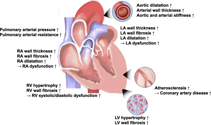
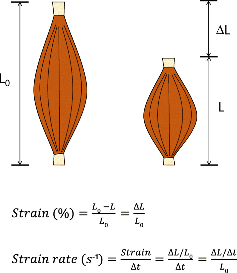
Hypertension is a well-recognized risk factor for the development of cardiovascular disease, and the early detection of cardiac changes from hypertension can allow reversing these. Hypertensive heart diseases (HHD) refer to the complex and diverse change of the cardiac structure and function secondary to hypertension. Although conventional echocardiography is the most common imaging modality in detecting HHD, it cannot detect subtle changes of cardiac structure in subclinical states. Because strain echocardiography is another echocardiographic modality can detect subclinical myocardial dysfunction by measuring intrinsic myocardial deformation, it became more and more popular in clinical and research fields. In this review article, we described the basic concept of strain echocardiography and summarized several clinical studies showing its clinical utilities in the detection of HHD.
Keywords: Echocardiography; Hypertension; Hypertrophy; Left ventricular; Strain echocardiography.
© 2022. The Author(s).
Conflict of interest statement
Jin Kyung Oh has nothing to declare. Jae-Hyeong Park serves on the editorial boards as a deputy editor in the Clinical Hypertension.
Figures

Similar articles
-
Multimodality imaging evaluation of Chagas disease: an expert consensus of Brazilian Cardiovascular Imaging Department (DIC) and the European Association of Cardiovascular Imaging (EACVI).Eur Heart J Cardiovasc Imaging. 2018 Apr 1;19(4):459-460n. doi: 10.1093/ehjci/jex154. Eur Heart J Cardiovasc Imaging. 2018. PMID: 29029074
-
Myocardial Strain Imaging in Resistant Hypertension.Curr Hypertens Rep. 2021 May 5;23(5):24. doi: 10.1007/s11906-021-01148-3. Curr Hypertens Rep. 2021. PMID: 33950321 Free PMC article. Review.
-
Potential usefulness of diastolic parameters measured by strain imaging echocardiography in the early prediction of chemotherapy-induced cardiotoxicity.Med Hypotheses. 2017 Apr;101:30-32. doi: 10.1016/j.mehy.2017.02.004. Epub 2017 Feb 10. Med Hypotheses. 2017. PMID: 28351486 Free PMC article.
-
European Association of Cardiovascular Imaging/Cardiovascular Imaging Department of the Brazilian Society of Cardiology recommendations for the use of cardiac imaging to assess and follow patients after heart transplantation.Eur Heart J Cardiovasc Imaging. 2015 Sep;16(9):919-48. doi: 10.1093/ehjci/jev139. Epub 2015 Jul 2. Eur Heart J Cardiovasc Imaging. 2015. PMID: 26139361 Review.
-
Tricuspid annular motion velocity as a differentiation index of hypertrophic cardiomyopathy from hypertensive heart disease.J Cardiol. 2015 Jun;65(6):519-25. doi: 10.1016/j.jjcc.2014.08.005. Epub 2014 Sep 8. J Cardiol. 2015. PMID: 25199979
Cited by
-
Analysis of atrioventricular function and myocardial strain in hypertensive heart disease: a CMR-based study.Sci Rep. 2025 Jul 25;15(1):27088. doi: 10.1038/s41598-025-10685-9. Sci Rep. 2025. PMID: 40715221 Free PMC article.
-
Interpreting Diastolic Dynamics and Evaluation through Echocardiography.Life (Basel). 2024 Sep 12;14(9):1156. doi: 10.3390/life14091156. Life (Basel). 2024. PMID: 39337939 Free PMC article. Review.
-
Effect of Metabolic Dysfunction-Associated Steatotic Liver Disease (MASLD) on Left Ventricular Mechanics in Patients Without Overt Cardiac Disease: A Systematic Review and Meta-Analysis.J Clin Med. 2025 Apr 15;14(8):2690. doi: 10.3390/jcm14082690. J Clin Med. 2025. PMID: 40283520 Free PMC article. Review.
-
Left Ventricle Myocardial Work Correlated with Functional Capacity in Severe Rheumatic Mitral Stenosis with Preserved Left Ventricular Ejection Fraction.J Cardiovasc Echogr. 2024 Apr-Jun;34(2):57-62. doi: 10.4103/jcecho.jcecho_14_24. Epub 2024 Jun 28. J Cardiovasc Echogr. 2024. PMID: 39086701 Free PMC article.
-
Elevated risk of perioperative ischemic stroke in noncardiac surgery patients with atrial fibrillation: a retrospective cohort study.BMC Anesthesiol. 2025 Apr 2;25(1):151. doi: 10.1186/s12871-025-03011-3. BMC Anesthesiol. 2025. PMID: 40175937 Free PMC article.
References
-
- Virani SS, Alonso A, Benjamin EJ, Bittencourt MS, Callaway CW, Carson AP, Chamberlain AM, Chang AR, Cheng S, Delling FN, Djousse L, Elkind MSV, Ferguson JF, Fornage M, Khan SS, Kissela BM, Knutson KL, Kwan TW, Lackland DT, Lewis TT, Lichtman JH, Longenecker CT, Loop MS, Lutsey PL, Martin SS, Matsushita K, Moran AE, Mussolino ME, Perak AM, Rosamond WD, Roth GA, Sampson UKA, Satou GM, Schroeder EB, Shah SH, Shay CM, Spartano NL, Stokes A, Tirschwell DL, VanWagner L, Tsao CW, American Heart Association Council on Epidemiology and Prevention Statistics Committee and Stroke Statistics Subcommittee Heart disease and stroke statistics-2020 update: a report from the American Heart Association. Circulation. 2020;141(9):e139–e596. doi: 10.1161/CIR.0000000000000757. - DOI - PubMed
Publication types
LinkOut - more resources
Full Text Sources