Observational Study
. 2022 Feb 14;10(1):21.
doi: 10.1186/s40478-022-01318-7.

Antemortem detection of Parkinson's disease pathology in peripheral biopsies using artificial intelligence

Affiliations
Observational Study

Antemortem detection of Parkinson's disease pathology in peripheral biopsies using artificial intelligence

Maxim Signaevsky et al. Acta Neuropathol Commun. .

Abstract

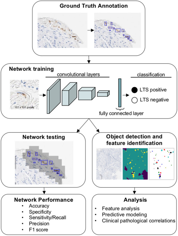
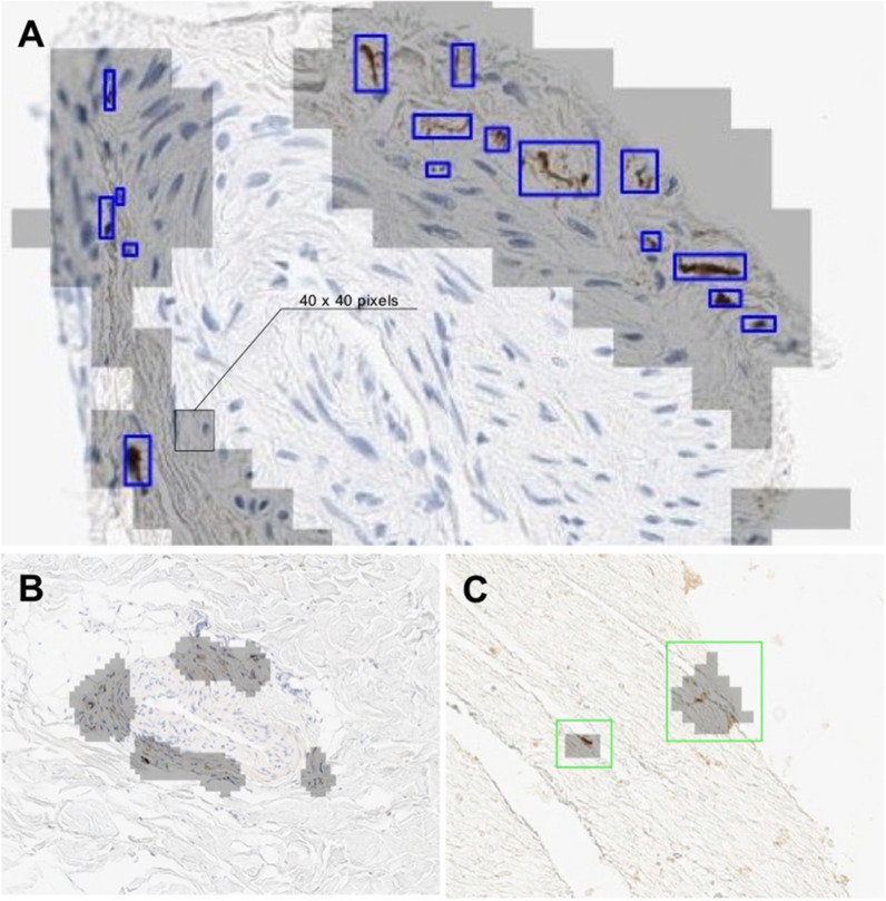
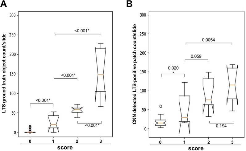
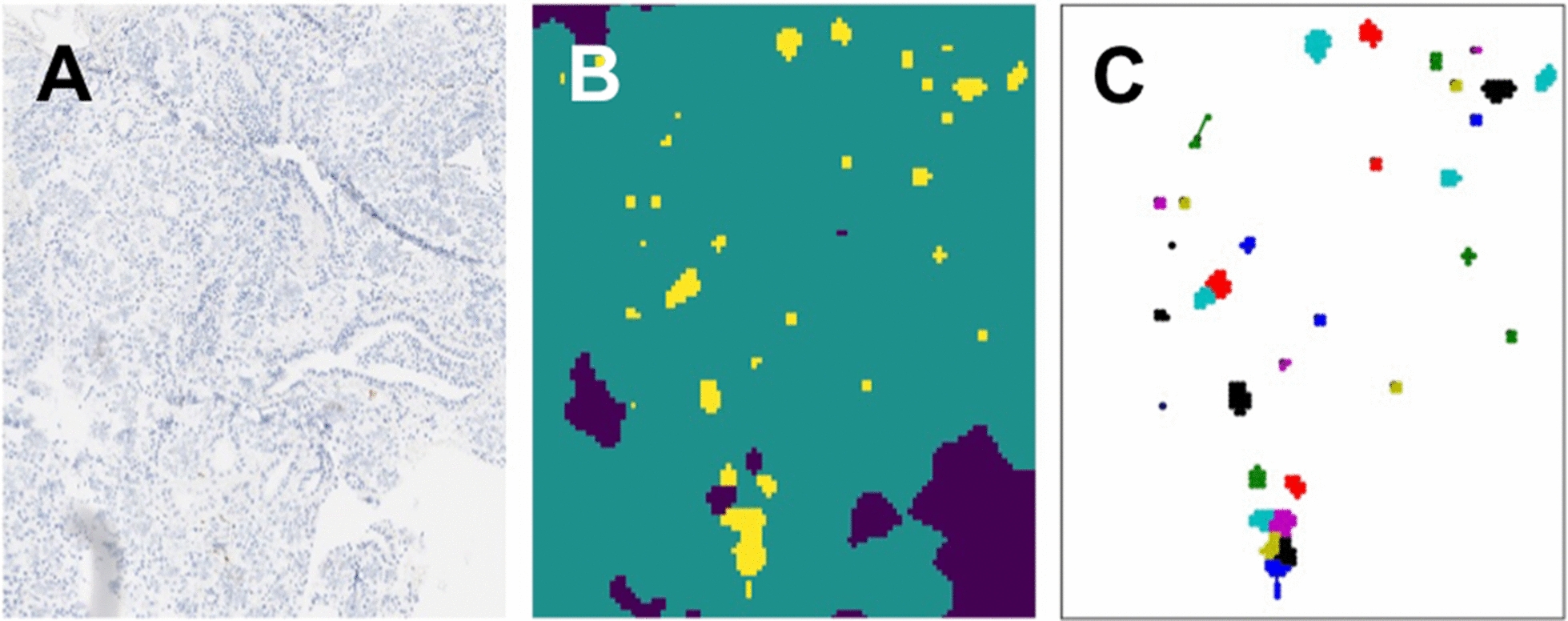
The diagnosis of Parkinson's disease (PD) is challenging at all stages due to variable symptomatology, comorbidities, and mimicking conditions. Postmortem assessment remains the gold standard for a definitive diagnosis. While it is well recognized that PD manifests pathologically in the central nervous system with aggregation of α-synuclein as Lewy bodies and neurites, similar Lewy-type synucleinopathy (LTS) is additionally found in the peripheral nervous system that may be useful as an antemortem biomarker. We have previously found that detection of LTS in submandibular gland (SMG) biopsies is sensitive and specific for advanced PD; however, the sensitivity is suboptimal especially for early-stage disease. Further, visual microscopic assessment of biopsies by a neuropathologist to identify LTS is impractical for large-scale adoption. Here, we trained and validated a convolutional neural network (CNN) for detection of LTS on 283 digital whole slide images (WSI) from 95 unique SMG biopsies. A total of 8,450 LTS and 35,066 background objects were annotated following an inter-rater reliability study with Fleiss Kappa = 0.72. We used transfer learning to train a CNN model to classify image patches (151 × 151 pixels at 20× magnification) with and without the presence of LTS objects. The trained CNN model showed the following performance on image patches: sensitivity: 0.99, specificity: 0.99, precision: 0.81, accuracy: 0.99, and F-1 score: 0.89. We further tested the trained network on 1230 naïve WSI from the same cohort of research subjects comprising 42 PD patients and 14 controls. Logistic regression models trained on features engineered from the CNN predictions on the WSI resulted in sensitivity: 0.71, specificity: 0.65, precision: 0.86, accuracy: 0.69, and F-1 score: 0.76 in predicting clinical PD status, and 0.64 accuracy in predicting PD stage, outperforming expert neuropathologist LTS density scoring in terms of sensitivity but not specificity. These findings demonstrate the practical utility of a CNN detector in screening for LTS, which can translate into a computational tool to facilitate the antemortem tissue-based diagnosis of PD in clinical settings.

Keywords: Artificial intelligence; Convolutional neural network; Deep learning; Machine learning; Parkinson’s disease; Peripheral biopsy; Submandibular gland; Synucleinopathy; Whole slide image.


Conflict of interest statement

Bahram Marami, Marcel Prastawa, Mary Sawyer, Israel Duran, John Koll, Gerardo Fernandez, Jack Zeineh, and Carlos Cordon-Cardo disclose their relationship with PreciseDx™, an artificial intelligence-guided pathology diagnostics company, which has developed proprietary platform and AI algorithms used in this study.

Figures

Fig. 1
Fig. 1
LTS object scoring and confidence ranking. Illustration of scoring density of sparse (A) moderate (B), and frequent (C). Submandibular gland biopsies immunohistochemically stained for α-synuclein with examples of sparse (D) moderate (E) and frequent (F). Examples of confidence ranking examples of definite (G), probable (H), and possible (I). LTS can be visualized and single or multiple immunopositive normal or misshapen axonal profiles in the submandibular gland parenchyma, nerve fascicles, or adjacent to blood vessels. LTS Lewy-type synucleinopathy
Fig. 2
Fig. 2
Schematic overview of data annotation and deep learning pipeline. LTS are annotated using WSI. The CNN was trained to classify image patches containing LTS from other image patches including tissue, artifacts and background. Different weights were used for the annotated objects while training using the cross entropy loss function for the final network. Image patches are extracted for network training that generates pixel-wise segmentations for LTS and background. Performance is determined using a separate novel set of images (test set) by comparing expert annotation with the trained network. The resulting trained network is further deployed on the naive WSI dataset for assessment of the predictive power of clinical outcomes. LTS Lewy-type synucleinopathy, WSI whole slide image, CNN convolutional neural network
Fig. 3
Fig. 3
Examples of CNN deployment on SMG biopsy WSI. A An example of annotated objects (blue) and CNN inference (grey shading). A 40 × 40 pixel inference patch is shown. B An example of true positive CNN classification. C An example of false positive CNN classification. CNN, convolutional neural network; SMG, submandibular gland; LTS, Lewy-type synucleinopathy, WSI, whole slide image
Fig. 4
Fig. 4
Comparison of ground truth annotations and CNN detection with expert scoring. A Expert annotation distribution boxplot in test WSI cohort (n = 56), Mann–Whitney two-tailed U test between score groups p values; Kruskal–Wallis H test of annotated LTS between expert score groups, and Spearman correlation between LTS burden and expert scores. B CNN, 40 × 40 patches positive for LTS distribution boxplot in test WSI cohort (n = 56), Mann–Whitney two-tailed U test between score groups p values; Kruskal–Wallis H test of 40 × 40 patches between expert score groups, and Spearman correlation between 40 × 40 patches positive for LTS burden and a score in test cohort. Scoring was performed as follows: each WSI was classified as positive or negative for α-synuclein pathology and assigned an LTS score ranging from 0 to 3 (Fig. 1), where 0 refers to being negative for α-synuclein, and scores 1–3 refer to scoring density of sparse (1) moderate (2), and frequent (3). CNN convolutional neural network, SMG submandibular gland, LTS Lewy-type synucleinopathy
Fig. 5
Fig. 5
Examples of CNN deployment on SMG biopsy WSI. A An Example of a WSI. B, C LTS patch detection and features generation (e.g., patches clustering, in color). CNN convolutional neural network, SMG submandibular gland, LTS Lewy-type synucleinopathy

Similar articles

Cited by

References

    1. (2016) Systemic synuclein sampling study s4: biospecimen collection, processing, and shipment manual. https://michaeljfox.Org/Files/S4_Biologics_Manual_Version_2.Pdf
    1. Adler CH, Beach TG, Hentz JG, Shill HA, Caviness JN, Driver-Dunckley E, Sabbagh MN, Sue LI, Jacobson SA, Belden CM, et al. Low clinical diagnostic accuracy of early vs advanced Parkinson disease: clinicopathologic study. Neurology. 2014;83:406–412. doi: 10.1212/Wnl.0000000000000641. - DOI - PMC - PubMed
    1. Adler CH, Dugger BN, Hentz JG, Hinni ML, Lott DG, Driver-Dunckley E, Mehta S, Serrano G, Sue LI, Duffy A. Peripheral synucleinopathy in early Parkinson's disease: submandibular gland needle biopsy findings. Mov Disord. 2016;31:250–256. doi: 10.1002/mds.26476. - DOI - PMC - PubMed
    1. Adler CH, Dugger BN, Hinni ML, Lott DG, Driver-Dunckley E, Hidalgo J, Henry-Watson J, Serrano G, Sue LI, Nagel T, et al. Submandibular gland needle biopsy for the diagnosis of Parkinson disease. Neurology. 2014;82:858–864. doi: 10.1212/Wnl.0000000000000204. - DOI - PMC - PubMed
    1. Adler CH, Serrano GE, Zhang N, Hinni ML, Lott DG, Mehta SH, Sue LI, Intorcia A, Beach TG. Feasibility of repeat and bilateral submandibular gland needle biopsies in Parkinson's disease. Parkinsonism Relat Disord. 2019;68:69–72. doi: 10.1016/J.Parkreldis.2019.10.006. - DOI - PMC - PubMed

Publication types