. 2022 Mar 24;8(2):a006193.
doi: 10.1101/mcs.a006193. Print 2022 Feb.

Ethylmalonic encephalopathy masquerading as meningococcemia

Affiliations

Ethylmalonic encephalopathy masquerading as meningococcemia

Ari Horton et al. Cold Spring Harb Mol Case Stud. .

Abstract

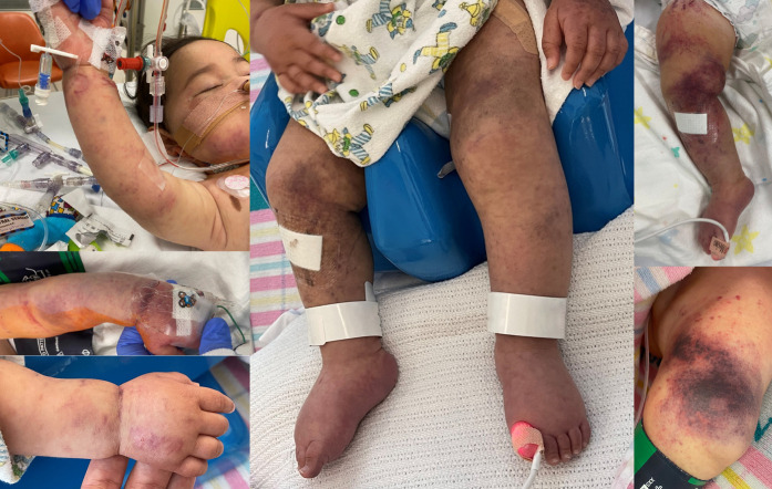
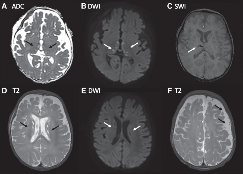
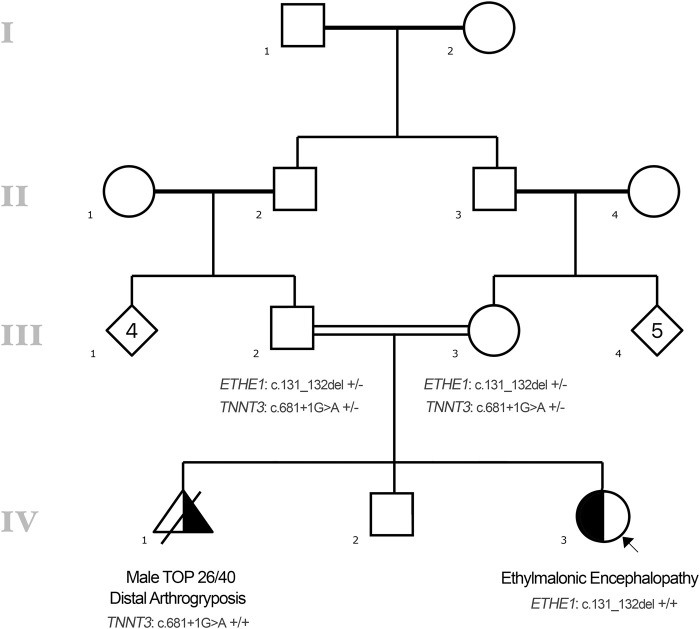
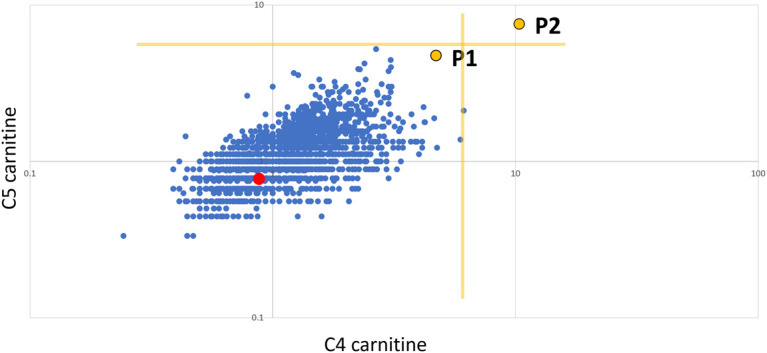
Ethylmalonic encephalopathy (MIM #602473) is a rare autosomal recessive metabolic condition caused by biallelic variants in ETHE1 (MIM #608451), characterized by global developmental delay, infantile hypotonia, seizures, and microvascular damage. The microvascular changes result in a pattern of relapsing spontaneous diffuse petechiae and purpura, positional acrocyanosis, and pedal edema, hemorrhagic suffusions of mucous membranes, and chronic diarrhea. Here, we describe an instructive case in which ethylmalonic encephalopathy masqueraded as meningococcal septicemia and shock. Ultrarapid whole-genome testing (time to result 60 h) and prompt biochemical analysis facilitated accurate diagnosis and counseling with rapid implementation of precision treatment for the metabolic crisis related to this condition. This case provides a timely reminder to consider rare genetic diagnoses when atypical features of more common conditions are present, with an early referral to ensure prompt biochemical and genomic diagnosis.

Keywords: acrocyanosis; ankle clonus; central hypotonia; cerebral ischemia; delayed fine motor development; delayed gross motor development; ethylmalonic aciduria; petechiae; progressive encephalopathy; recurrent cerebral hemorrhage.


Figures

Figure 1.
Figure 1.
Presenting features: cutaneous manifestations. Clinical photographs taken on day 4 of admission demonstrating widespread petechiae and purpura to the face, upper and lower limbs, and torso (not shown).
Figure 2.
Figure 2.
Presenting features: magnetic resonance imaging (MRI) brain findings. MRI day 2 postadmission: (A) Apparent diffusion coefficient (ADC) map and (B) corresponding diffusion-weighted image (DWI) show multiple punctate lesions in the thalami and globi pallidi that are associated with true diffusion restriction consistent with small acute (likely <7 d old) infarcts (arrows). (C) Susceptibility-weighted image (SWI) showing signal loss in a few of the right thalamic lesions consistent with microhemorrhage (arrow). (D) T2-weighted image demonstrating numerous T2 hyperintense periventricular punctate lesions that have no corresponding restricted diffusion (E) consistent with nonacute infarcts. (F) T2-weighted axial image demonstrating subtle widening of the left frontoparietal subarachnoid space (arrows) likely due to reduced left hemispheric volume consequent upon microscopic remote ischemic white matter/subplate injury postnatally or prenatally.
Figure 3.
Figure 3.
Pedigree.
Figure 4.
Figure 4.
Presenting features: positional acrocyanosis. Photographs taken by the family in the weeks prior to presentation showing positional acrocyanosis.
Figure 5.
Figure 5.
Retrospective reanalysis of newborn blood spot screening (NBS). C5 carnitine vs C4 carnitine expressed as multiples of median (log:log scale). P1 is the proband, P2 is an unrelated neonate with a confirmed ETHE1 defect, the red dot is the unaffected sibling of P1, the blue dots represent control neonates (n = 3215), and the yellow lines indicate decision limits for a repeat sample (∼99.95%ile).

References

    1. AlAbdi L, Alrashseed S, Alsulaiman A, Helaby R, Imtiaz F, Alhamed M, Alkuraya FS. 2021. Residual risk for additional recessive diseases in consanguineous couples. Genet Med 23: 2448–2454. 10.1038/s41436-021-01289-5 - DOI - PubMed
    1. Blau N, Duran M, Gibson KM, Dionisi-Vici C. 2014. Physician's guide to the diagnosis, treatment, and follow-up of inherited metabolic diseases. Springer-Verlag, Berlin.
    1. Boyer M, Sowa M, Di Meo I, Eftekharian S, Steenari MR, Tiranti V, Abdenur JE. 2018. Response to medical and a novel dietary treatment in newborn screen identified patients with ethylmalonic encephalopathy. Mol Genet Metab 124: 57–63. 10.1016/j.ymgme.2018.02.008 - DOI - PubMed
    1. Cardelo Autero N, Cordón Martínez AM, Ramos-Fernández JM. 2021. Ethylmalonic encephalopathy: phenotype-genotype description and review of its management. Neurologia (Engl Ed) 36: 729–731. 10.1016/j.nrl.2021.01.004 - DOI - PubMed
    1. den Dunnen JT, Dalgleish R, Maglott DR, Hart RK, Greenblatt MS, McGowan-Jordan J, Roux AF, Smith T, Antonarakis SE, Taschner PE. 2016. HGVS recommendations for the description of sequence variants: 2016 update. Hum Mutat 37: 564–569. 10.1002/humu.22981 - DOI - PubMed

Publication types

Substances

Supplementary concepts