. 2022 Feb 14;13(1):853.
doi: 10.1038/s41467-022-28510-6.

Structural basis of neuropeptide Y signaling through Y1 receptor

Affiliations

Structural basis of neuropeptide Y signaling through Y1 receptor

Chaehee Park et al. Nat Commun. .

Erratum in

Abstract

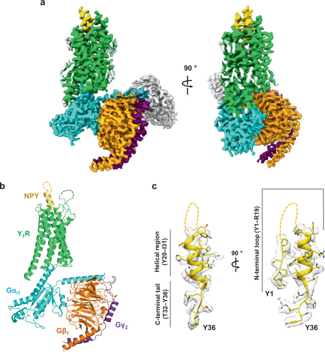
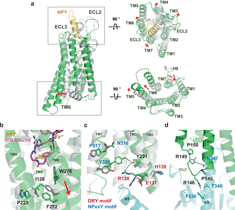
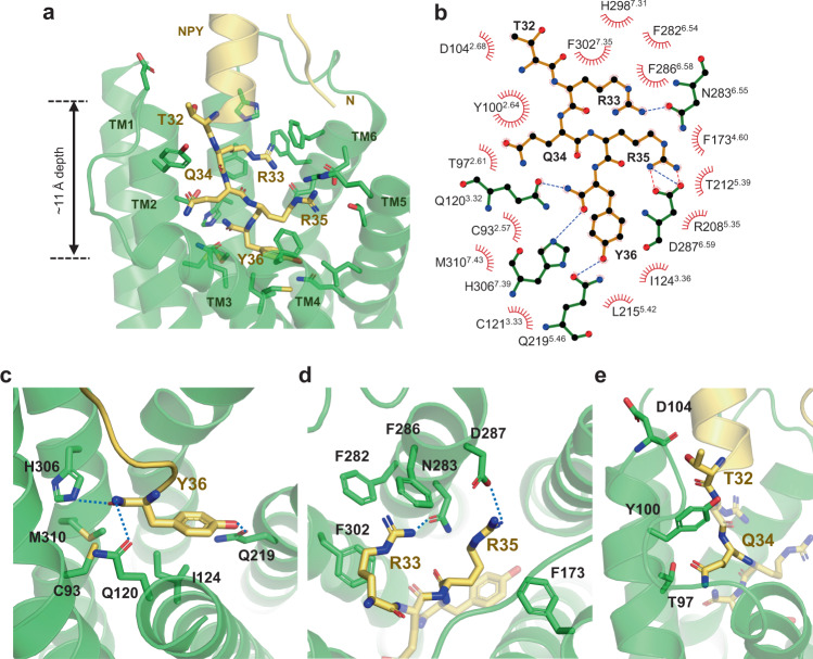
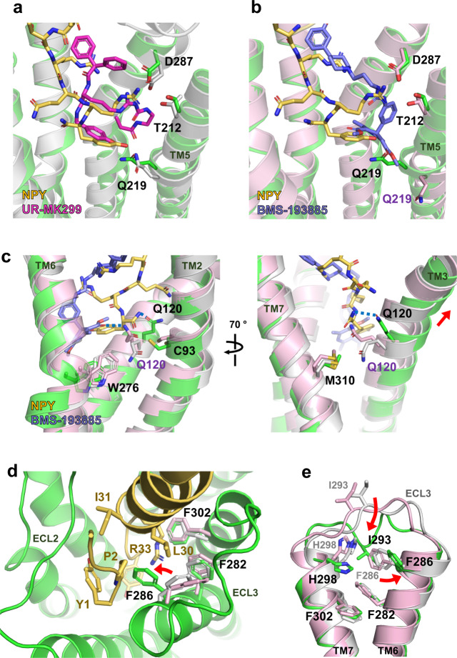
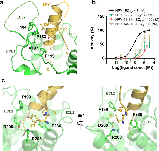
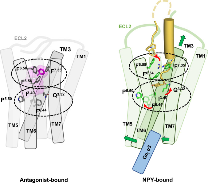
Neuropeptide Y (NPY) is highly abundant in the brain and involved in various physiological processes related to food intake and anxiety, as well as human diseases such as obesity and cancer. However, the molecular details of the interactions between NPY and its receptors are poorly understood. Here, we report a cryo-electron microscopy structure of the NPY-bound neuropeptide Y1 receptor (Y1R) in complex with Gi1 protein. The NPY C-terminal segment forming the extended conformation binds deep into the Y1R transmembrane core, where the amidated C-terminal residue Y36 of NPY is located at the base of the ligand-binding pocket. Furthermore, the helical region and two N-terminal residues of NPY interact with Y1R extracellular loops, contributing to the high affinity of NPY for Y1R. The structural analysis of NPY-bound Y1R and mutagenesis studies provide molecular insights into the activation mechanism of Y1R upon NPY binding.


Conflict of interest statement

The authors declare no competing interests.

Figures

Fig. 1
Fig. 1. Overall structure of the NPY–Y1R–Gi1–scFv16 complex.
a The cryo-EM map of the NPY–Y1R–Gi1–scFv16 complex is shown. Y1R, NPY, Gαi1, Gβ1, Gγ2, and scFv16 are colored green, yellow, cyan, orange, purple, and gray, respectively. The remaining micelle density is shown in light gray. Details on cryo-EM map generation are described in the “Methods” section. b The structure of the NPY–Y1R–Gi1 complex is shown. scFv16 is included in the final structure but omitted in this figure for clarity. c The sharpened cryo-EM map with the NPY peptide model is shown in two orientations. NPY residues 6–19 are not modeled in this structure and are shown as dashed lines. The three regions of NPY are marked as the N-terminal loop region (Y1–R19), the helical region (Y20–I31), and the C-terminal tail (T32–Y36).
Fig. 2
Fig. 2. Comparison between NPY-bound and antagonist-bound Y1R structures.
a Structural alignment between NPY (yellow)-bound Y1R (current structure, green) and antagonist-bound Y1R (PDB ID 5ZBQ, light gray, residues 18–32 of Y1R and antagonist are omitted for clarity) clearly shows characteristic TM6 movement as observed in the active structures of class A GPCRs. Views from extracellular (right-upper panel) and cytoplasmic (right-lower panel) sides are shown on the right. Each red arrow represents the movement of the TM helix. b Structural changes at the connector region (P5.50I3.40F6.44 motif) upon NPY binding are shown. The antagonist-bound Y1R (PDB ID 5ZBQ) is colored in light gray and an antagonist, UR-MK299, in magenta. c Structural changes at the D(E)/R/Y(H) (labeled in red) and NPxxY (labeled in blue) motifs are shown. d The binding interface between α5 of Gi1 (in cyan) and ICL2 is shown. Residues participating in the interactions and two Pro residues (P145ICL2 and P150ICL2) in ICL2 are shown as sticks. Dashed lines represent the polar interactions.
Fig. 3
Fig. 3. Binding of NPY C-terminal tail to Y1R.
a The C-terminal tail of NPY (yellow) and Y1R residues that participate in NPY binding are presented as sticks. b Schematic representation of NPY–Y1R interactions analyzed using the LigPlot+ program is shown. The hydrogen bonds and the salt bridges are shown in blue and red dashed lines, respectively. ce Detailed interactions between C-terminal residues of NPY and Y1R are shown. The polar contacts are shown as blue dashed lines.
Fig. 4
Fig. 4. Comparison of binding mode between NPY and antagonists.
a Ligand-binding site occupied by UR-MK299 (PDB ID 5ZBQ, magenta) and b BMS-193885 (PDB ID 5ZBH, slate) is compared with the binding site for NPY (yellow). The side chains of Q2195.46 are differently oriented in all three structures, whereas D2876.59 and T2125.39 maintain similar interactions. c Q1203.32-mediated interactions in the antagonist-bound and NPY-bound structures are compared. In the NPY-bound active structure of Y1R, TM3 including Q1203.32 shifts upward and Q1203.32 forms a polar interaction with the C-terminal amide of NPY through its upward-facing sidechain. d Upon NPY binding, F2866.58 rotamer is changed to form the interactions with NPY R33, L30, and Y1. e Upon NPY binding, I293ECL3 participates in a hydrophobic interaction network with F2866.58, H2987.31, and F3027.35. UR-MK299-bound Y1R is indicated in light gray, and BMS-193885-bound Y1R is in pink. Red arrows represent positional changes of I293ECL3 and F2866.58 upon NPY binding.
Fig. 5
Fig. 5. Binding of NPY N-terminus to Y1R.
a The sticks show the residues participating in van der Waals interactions between NPY helix and ECL2 of Y1R. Unresolved regions of NPY and ECL2 of Y1R are shown with dashed lines. b The results of Ca2+ assays performed with N-terminally truncated NPY peptides are shown. Removal of two N-terminal residues dramatically reduces NPY potency and efficacy. Symbol and error bar indicate the mean and S.E.M. (standard error of mean) of n = three (NPY(3–36), NPY(18–36), and NPY(AA-36)) or n = 17 (NPY) independent experiments, respectively. Calculated EC50 values (pEC50 ± SEM) are presented on each graph and are summarized in Supplementary Table 2. Source data are provided as a Source Data file. c Van der Waals interactions of Y1 and P2 of NPY with Y1R are shown in two different orientations.
Fig. 6
Fig. 6. Mechanism of Y1R activation by NPY binding.
Schematic figures representing antagonist-bound (left) and NPY-bound (right) Y1R are shown. Two key regions for Y1R activation are indicated by dashed ellipses. The three phenylalanine residues at TM6 and TM7, and Q1203.32, I1283.40, P2235.50, and F2726.44 in the connector region are shown in stick representation. The three phenylalanine residues participate in antagonist binding through π–π interactions, but these interactions are rearranged upon NPY binding. F2866.58 flips and forms a new interaction network with Y1 and R33 of NPY. Since F2866.58 is not conserved in Y2R, F2866.58-mediated interactions are Y1R-selective. Q1203.32, I1283.40 and F2726.44 show rotamer changes upon NPY binding. A series of these changes upon NPY binding pulls TM3 upward and causes outward movement of the cytoplasmic region of TM6.

References

    1. Michel MC, et al. XVI. International Union of Pharmacology recommendations for the nomenclature of neuropeptide Y, peptide YY, and pancreatic polypeptide receptors. Pharm. Rev. 1998;50:143–150. - PubMed
    1. Herzog H, et al. Cloned human neuropeptide Y receptor couples to two different second messenger systems. Proc. Natl Acad. Sci. USA. 1992;89:5794–5798. - PMC - PubMed
    1. Kohno D, Yada T. Arcuate NPY neurons sense and integrate peripheral metabolic signals to control feeding. Neuropeptides. 2012;46:315–319. - PubMed
    1. Tasan RO, et al. The central and basolateral amygdala are critical sites of neuropeptide Y/Y2 receptor-mediated regulation of anxiety and depression. J. Neurosci. 2010;30:6282–6290. - PMC - PubMed
    1. Gotzsche CR, Woldbye DP. The role of NPY in learning and memory. Neuropeptides. 2016;55:79–89. - PubMed

Publication types