. 2022 Apr;18(4):368-375.
doi: 10.1038/s41589-021-00966-5. Epub 2022 Feb 14.

Widespread occurrence of covalent lysine-cysteine redox switches in proteins

Affiliations

Widespread occurrence of covalent lysine-cysteine redox switches in proteins

Fabian Rabe von Pappenheim et al. Nat Chem Biol. 2022 Apr.

Abstract

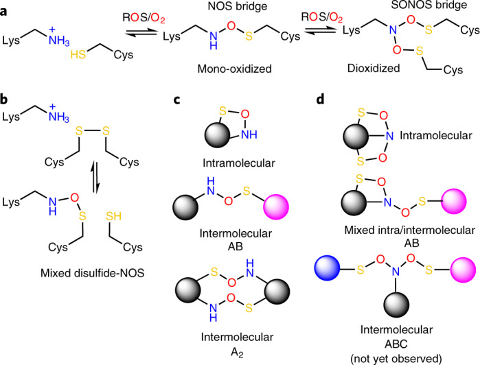
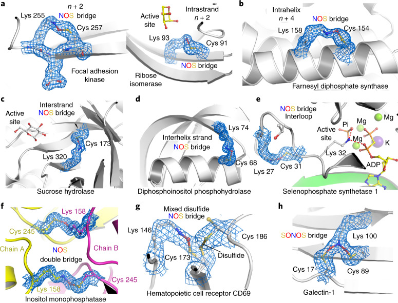
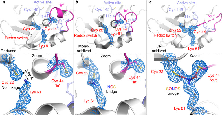
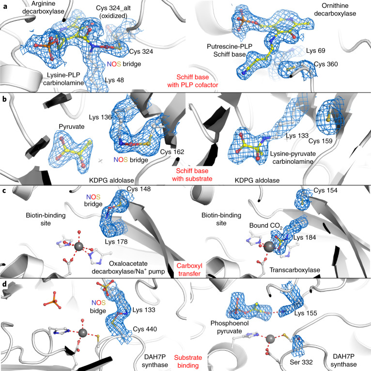
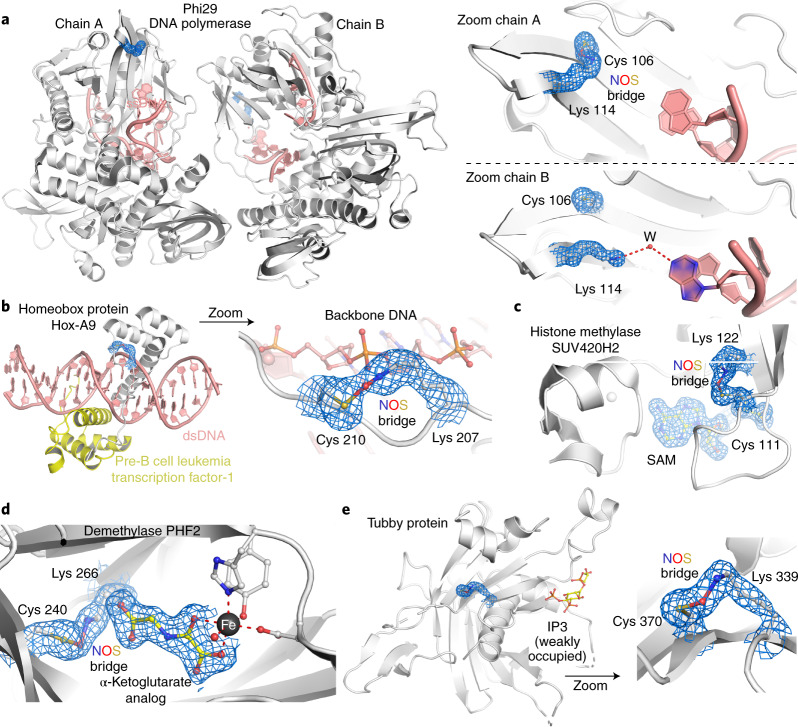
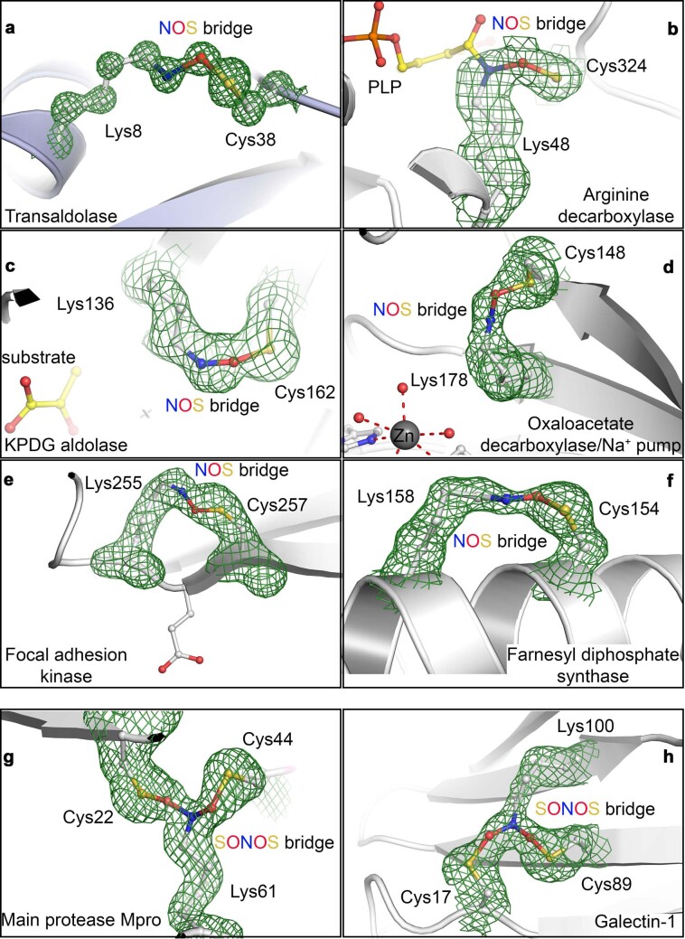
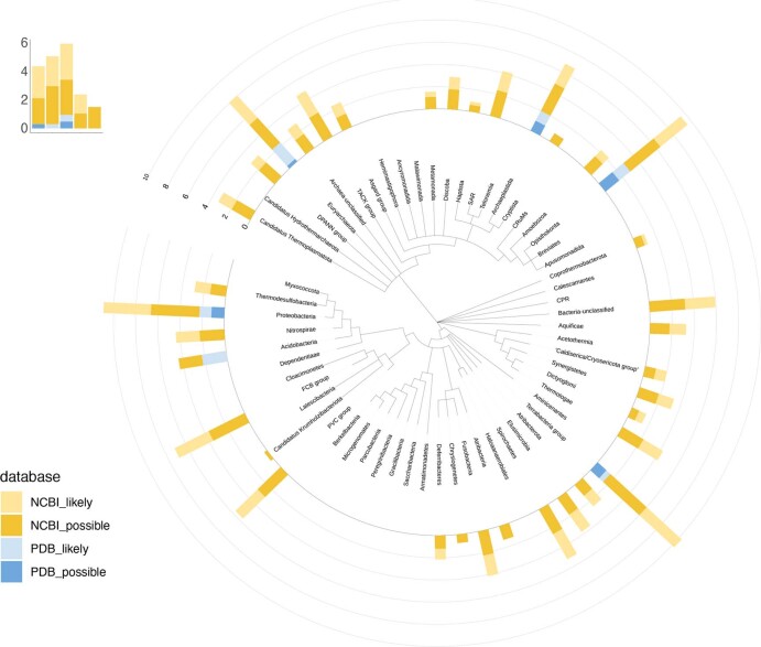
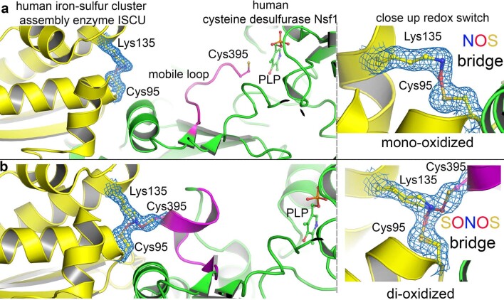
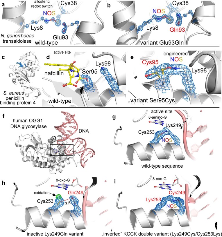
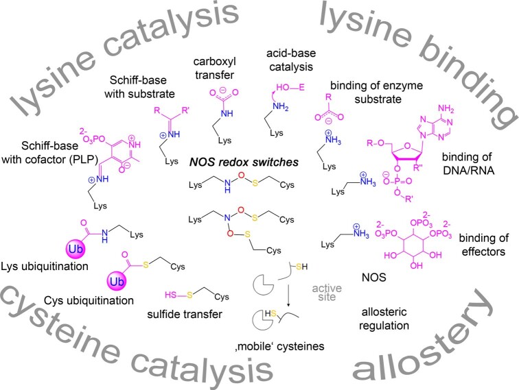
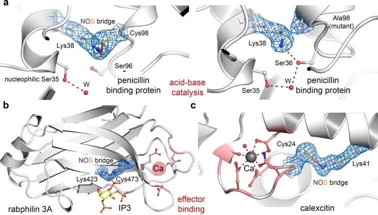
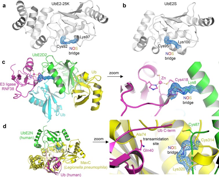
We recently reported the discovery of a lysine-cysteine redox switch in proteins with a covalent nitrogen-oxygen-sulfur (NOS) bridge. Here, a systematic survey of the whole protein structure database discloses that NOS bridges are ubiquitous redox switches in proteins of all domains of life and are found in diverse structural motifs and chemical variants. In several instances, lysines are observed in simultaneous linkage with two cysteines, forming a sulfur-oxygen-nitrogen-oxygen-sulfur (SONOS) bridge with a trivalent nitrogen, which constitutes an unusual native branching cross-link. In many proteins, the NOS switch contains a functionally essential lysine with direct roles in enzyme catalysis or binding of substrates, DNA or effectors, linking lysine chemistry and redox biology as a regulatory principle. NOS/SONOS switches are frequently found in proteins from human and plant pathogens, including severe acute respiratory syndrome coronavirus 2 (SARS-CoV-2), and also in many human proteins with established roles in gene expression, redox signaling and homeostasis in physiological and pathophysiological conditions.


Conflict of interest statement

F.R.v.P., M.W., L.-M.F., J.U., R.A.M. and K.T. have filed a European patent application (application number EP21164101.4) for regulating protein activities by targeting the NOS or SONOS redox switches. The remaining authors declare no competing interests.

Figures

Fig. 1
Fig. 1. Chemical structures and topologies of NOS and SONOS redox switches in proteins.
a, Structures and reaction scheme of NOS and SONOS redox bridge formation by ROS or oxygen in subsequent oxidation steps. b, Suggested structure of a ‘mixed’ NOS-disulfide redox switch, in which a disulfide is in equilibrium with an NOS bridge. c, Topologies of NOS bridges showing intramolecular and intermolecular cross-links as observed in experimentally determined protein structures. d, Topologies of SONOS bridges showing intramolecular and intermolecular cross-links as observed in experimentally determined protein structures. A SONOS bridge, where the lysine and two cysteines are contributed by three different proteins, has not been identified yet. The letters A, B and C indicate different proteins.
Fig. 2
Fig. 2. Structural and chemical motifs of NOS bridges in proteins.
The corresponding 2mFoDFc electron density maps are shown in blue at a contour level of 1σ. a, Examples for NOS bridges in intrastrand or strand-like motifs with a sequence distance of n + 2. Shown are focal adhesion kinase from Gallus gallus (PDB: 6CB0) and ribose isomerase from Acinetobacter sp. (PDB: 4Q0P). b, Example for an NOS bridge in intrahelix motifs with a sequence distance of n + 4. Shown is the farnesyl diphosphate synthase from Trypanosoma cruzi (PDB: 6SDP). c, Example for an NOS bridge in interstrand (cross-strand) motifs. Shown is the sucrose hydrolase from Xanthomonas axonopodis (PDB: 3CZG). d, Example for an NOS bridge connecting a helix and a neighboring strand showing human diphosphoinositol phosphohydrolase (PDB: 6PCK). e, Example for an intraloop NOS bridge showing human selenophosphate synthetase 1 (PDB: 3FD5). f, Example for an intermolecular NOS double bridge between two chains in a homodimeric assembly. Shown is the inositol monophosphatase from Medicago truncatula (PDB: 5EQA). The two chains are colored individually in yellow and magenta, respectively. g, Example for a ‘mixed’ NOS-disulfide switch showing the human hematopoietic cell receptor CD69 (PDB: 1E8I, chain A) with an NOS bridge between Lys 146 and Cys 173 (30% occupancy) and a disulfide bridge between Cys 173 and Cys 186 (70% occupancy). h, Example for a SONOS bridge linking a lysine and two cysteines at the same time showing galectin-1 from Rattus norvegicus (PDB: 4GA9).
Fig. 3
Fig. 3. NOS and SONOS bridges in Mpro from SARS-CoV-2.
The corresponding 2mFoDFc electron density maps are shown in blue at a contour level of 1σ. a, Structure of Mpro in the reduced state (PDB: 7JR3) showing the redox switch at the protein surface formed by residues Cys 22, Cys 44 and Lys 61 (highlighted in red) and the active site with residues Cys 145 (catalytic nucleophile), His 41 and Tyr 54 (highlighted in slate blue). A mobile loop bearing Cys 44 is indicated in magenta. Top, structural overview; bottom, close-up of the redox switch site. Note that residue Cys 44 is in the ‘in conformation’ and interacts with Tyr 54. b, Structure of Mpro in a mono-oxidized state with an NOS bridge formed between Cys 22 and Lys 61 (PDB: 6XMK). Top, structural overview; bottom, close-up of the redox switch site. Cys 44 is found in the ‘in conformation’. c, Structure of Mpro in a dioxidized state with a SONOS bridge formed between Cys 22, Lys 61 and Cys 44 (PDB: 7JR4). Top, structural overview; bottom, close-up of the redox switch site. Cys 44 is found in the ‘out conformation’. Competitive refinements (SONOS bridge only, two separate NOS bridges, mixture of SONOS and NOS) indicate full occupancy of the SONOS bridge.
Fig. 4
Fig. 4. Functional roles of NOS bridge lysines in enzyme catalysis and in binding of enzymatic substrates or effectors.
Left, proteins in the oxidized state with the NOS bridge present; right, same or closely related protein in the reduced state with the lysine exerting its function. The corresponding 2mFoDFc electron density maps are shown in blue at a contour level of 1σ. a, Catalytic lysines forming Schiff base intermediates in PLP-dependent enzymes; left, arginine decarboxylase from Paramecium bursaria chlorella virus (PDB: 2NV9), in which the catalytic Lys 48 forms an NOS bridge with Cys 324; right, ornithine decarboxylase from T. brucei in covalent complex with product putrescine (PDB: 1F3T). Note that in the presence of the NOS bridge, the reaction does not proceed beyond the carbinolamine. b, Catalytic lysines forming Schiff base intermediates with enzymatic substrates; left, KDPG aldolase from Oleispira antarctica in non-covalent complex with substrate pyruvate and with an NOS bridge between Lys 136 and Cys 162 (PDB: 3VCR); right, KDPG aldolase from Escherichia coli in covalent complex with substrate pyruvate (PDB: 1EUA). Note that in the presence of the NOS bridge, covalent catalysis is inhibited. c, Catalytic lysines in carboxyl transfer; left, oxaloacetate decarboxylase/Na+ pump from Vibrio cholerae with an NOS bridge between Lys 178 and Cys 148 (PDB: 2NX9); right, transcarboxylase 5S subunit from Propionibacterium freudenreichii with carboxylated Lys 184 (PDB: 1RQB) thought to be an intermediate in CO2 transfer to biotin. Note that in the presence of the NOS bridge, catalysis is inhibited. d, Lysines with roles in non-covalent binding of enzymatic substrates; left, DAH7P synthase from Mycobacterium tuberculosis (PDB: 3RZI) with an NOS bridge between Lys 133 and Cys 440; right, DAH7P synthase from Listeria monocytogenes in non-covalent complex with substrate phosphoenolpyruvate (PDB: 3TFC). This enzyme contains a serine (Ser 332) at the equivalent position of Cys 440 from M. tuberculosis DAH7P synthase and can therefore not form an NOS bridge. In the absence of the NOS bridge, the lysine forms a hydrogen bond with the carboxylate moiety of substrate phosphoenolpyruvate.
Fig. 5
Fig. 5. Proteins with NOS bridges inferred in DNA binding and transcription regulation.
The corresponding 2mFo–DFc electron density maps are shown in blue at a contour level of 1σ. a, DNA polymerase from Bacillus virus phi29 complexed with single-stranded DNA (PDB: 2PY5). Left, overall structure showing the two copies (chains A and B) in the asymmetric unit. The single-stranded DNA is highlighted in salmon red. Right, close-up of the NOS redox switch with residues Lys 114 and Cys 106. Note that in chain A, both residues form an NOS bridge (left), which is absent in chain B (right). In the absence of the NOS bridge, Lys 114 forms a hydrogen bond with a DNA base via a water molecule. b, Homeobox protein Hox-A9 from Mus musculus in complex with human pre-B cell leukemia transcription factor-1 and double-stranded DNA (dsDNA; PDB: 1PUF). Left, overall structure of the complex. The two proteins are colored individually, and the DNA is shown in salmon red. Right, close-up of the NOS bridge formed between Lys 207 and Cys 210 of Hox-A9. Note that the NOS bridge is at the binding interface with the backbone of the DNA, suggesting that Lys 207 in the reduced state interacts with the phosphate groups. c, Human histone-lysine N-methyltransferase SUV420H2 in complex with substrate S-adenosyl methionine (SAM) and with an NOS bridge formed between Lys 122 and Cys 111 (PDB: 3RQ4). Note the proximity of the NOS bridge with respect to the SAM-binding locale. d, Human demethylase PHF2 in complex with an analog of the α-ketoglutarate cofactor and with an NOS bridge formed between Lys 266 and Cys 240 (PDB: 3PU8). Note the proximity of the NOS bridge with respect to the cofactor-binding locale, suggesting a direct interaction of Lys 266 with the carboxylate moiety of the cofactor. e, Tubby protein from Mus musculus in complex with inositol trisphosphate (I3; PDB: 1I7E). Left, structural overview highlighting the ligand-binding site and the allosteric NOS switch. Right, close-up of the NOS bridge between Lys 339 and Cys 370.
Extended Data Fig. 1
Extended Data Fig. 1. Computed structural information for conformers of Lys and Cys residues with and without the NOS bond.
Computed structural information for conformers of Lys and Cys residues with and without the NOS bond, using different alpha-carbon distances (6 Å, 8 Å and 10 Å). The residues are computed as models, truncated at the alpha-carbons. The sampled structures at the semi-empirical level were refined at the B3LYP-D3(BJ)/def2-SVP level of theory (more details in the Supplementary Information), with the lowest conformers being reoptimised with a larger basis (def2-TZVPP). We have considered three different conformers: NOS - covalent bonding between the residues, NHS - hydrogen bond with the Lys deprotonated and NHS+ - hydrogen bond with the Lys protonated. (a) N-S distance (in Å) as a function of the relative conformer stability within each group computed with the SVP basis; (b) histogram for the SVP basis results; (c) overlap of SVP optimised structures for NHS and NOS; (d) histogram for all structures with the TZVPP basis. The images in c are obtained by an arbitrary alignment of the structures (given that only two points are fixed - the alpha-carbon positions). This can lead to somewhat different representations, or apparent ‘banding’ of the structures. This effect will depend on the alignment chosen and should not be misinterpreted. The figures are there to help illustrate the conformational freedom of these interactions..
Extended Data Fig. 2
Extended Data Fig. 2. Detectability of NOS bridges in protein crystal structures in dependence from resolution and deposited dose.
Detectability of NOS bridges in protein crystal structures in dependence from resolution and deposited dose. (a) Data truncation at different resolutions. A previously determined sub-ångstrøm resolution crystallographic dataset obtained for Neisseria gonorrheae transaldolase (pdb code 6XZ4), which forms an allosteric NOS bridge between Lys8 and Cys38, was truncated at different resolutions from 1.0-3.0 Å as indicated. For refinement, which included simulated annealing and B-factor blurring, the lysine and cysteine residues were modeled without the bridging oxygen atom. The calculated structural models and 2mFo-DFc electron density (blue: 3σ, grey: 1σ) and mFo-DFc difference electron density (green: 5σ) maps are shown. The respective intensities of the positive peaks in the difference electron density maps are indicated. Note the loss of structural information in both maps regarding the bridging oxygen atom with decreasing resolution. (b) Dose dependence. Crystallographic datasets for a single crystal of Neisseria gonorrheae transaldolase were collected at different doses 0.27, 2.7 MGy and 5.4 MGy at cryogenic temperature. The refined structural models of the NOS bridge formed between Lys8 and Cys38 and of a neighboring helix bearing residues Cys87 and Asp88 are shown for datasets obtained at doses of 2.7 MGy and 5.4 MGy. The respective 2mFo-DFc electron density maps shown in blue are contoured at 2σ. The calculated difference in intensity in the electron density maps relative to that obtained for a dose of 0.27 MGy is shown in red at a contour level of 3σ. Note the progressive loss in intensity for Cys87 and Asp88 with increasing dose, which indicates ‘radiation damage’. For the NOS bridge, radiation damage sets in at higher doses of 5.4 MGy.
Extended Data Fig. 3
Extended Data Fig. 3. Unbiased mFo-DFc omit maps for NOS and SONOS bridges in representative examples calculated by Phenix.polder.
Unbiased mFo-DFc omit maps for NOS and SONOS bridges in representative examples calculated by Phenix.polder. The lysine and cysteine residues incl. the NOS or SONOS bridges were excluded from the structural models prior to map calculation to eliminate model bias. The corresponding pdb codes and contour levels are indicated. (a) transaldolase from Neisseria gonorrhoeae (pdb code 6ZX4, 5σ), (b) arginine decarboxylase from Paramecium bursaria Chlorella virus (pdb code 2NV9, 3.5σ), (c) KPDG aldolase from Oleispira antarctica (pdb code 3VCR, 4σ), (d) oxaloacetate decarboxylase/Na+ pump from Vibrio cholerae (pdb code 2NX9, 3σ), (e) focal adhesion kinase from Gallus gallus (pdb code 6CB0, 5σ), (f) farnesyl diphoshate synthase from Trypanosoma cruzi (pdb code 6SDP, 3σ), (g) main protease from SARS-CoV-2 (pdb code 7JR4, 5σ) and (h) galectin-1 from Rattus norvegicus (pdb code 4GA9, 3σ).
Extended Data Fig. 4
Extended Data Fig. 4. Distribution of proteins containing NOS crosslinks across the Tree of Life.
Distribution of proteins containing NOS crosslinks across the Tree of Life. Shown are proteins likely/ probably containing NOS bridges (blue) as identified from PDB structures (Supplementary Data 1), as well as homologs of these proteins as identified in NCBI’s non-redundant (NR) database (yellow). Values are log10-transformed.
Extended Data Fig. 5
Extended Data Fig. 5. NOS and SONOS bridges in the human Fe/S cluster biosynthesis complex.
NOS and SONOS bridges in the human Fe/S cluster biosynthesis complex. The corresponding 2mFo-DFc electron density maps are shown in blue at a contour level of 1σ. (a) Structure of the complex consisting of iron-sulfur cluster assembly enzyme ISCU (highlighted in yellow) and cysteine desulfurase Nsf1 (highlighted in green) in the mono-oxidized state with an intramolecular NOS bridge between Cys95 and Lys131 of ISCU (pdb code 6UXE). A mobile loop from Nsf1 with catalytic residue Cys395 catalyzing sulfide transfer from the PLP cofactor at the active site of Nsf1 to ISCU is indicated in magenta. Left panel: structural overview. Right panel: close-up of the redox switch site. Note that Cys395 visits the active site of Nsf1. (b) Structure of the complex in the di-oxidized state with a SONOS bridge formed between Cys95 and Lys131 of ISCU and Cys395 of Nsf1 (pdb code 6WI2, also observed in 6WIH). Left panel: structural overview. Right panel: close-up of the redox switch site. Competitive refinements (SONOS bridge only, 2 separate NOS bridges, mixture of SONOS and NOS) indicate a mixture of the SONOS bridge (refined occupancy 60%) and the NOS bridge between Lys135 and Cys95 (refined occupancy 40%) for this dataset.
Extended Data Fig. 6
Extended Data Fig. 6. Structural requirements for formation and engineering of NOS bridges.
Structural requirements for formation and engineering of NOS bridges. (a, b) Structure of the allosteric NOS bridge in Neisseria gonorrhoeae transaldolase wild-type (a, pdb code 6XZ4) and variant Glu93Gln (b, this study) showing NOS residues Lys8 and Cys38 as well as neighboring residue 93. The structural models of Lys8, Cys38 and Glu93 or Gln93 are superposed with the corresponding 2mFo-DFc electron density maps at 3σ. Note that NOS bridge formation does not require the presence of a catalytic residue at position 93. (c, d, e) An unintentionally engineered lysine-cysteine crosslink in Staphylococcus aureus penicillin binding protein 4 showing the overall protein structure (c) and a close-up of the active site with catalytic residues Ser95 (nucleophile) in linkage with nafcillin and Lys98 (d, pdb code 5TY2). In variant Ser95Cys (e), where the catalytic Ser95 has been replaced by cysteine, an NOS bridge was formed between introduced Cys95 and Lys98. The 2mFo-DFc electron density map is contoured at 1σ. In variant Ser95Cys, residue 95 was modeled in two alternative conformations. Note that a sulfenamide linkage with an N-S bond could also explain the density. (f-i) NOS bridges in the human DNA glycosylase OGG1. (f) Overall structure of OGG1 in complex with DNA (pdb code 1M3Q). (g) Close-up of the active site showing catalytic residue Lys249 (Schiff-base with DNA) and neighboring residue Cys253 forming an NOS bridge. The 2mFo-DFc electron density map is contoured at 1.5σ⊡ (h) Active site structure of inactive OGG1 variant Lys249Gln (pdb code 1EBM), in which the catalytic lysine had been replaced by glutamine. The 2mFo-DFc electron density map is shown in blue at 1.5σ and the mFo-DFc difference electron density map in green at 3σ⊡ Note that Gln249 and Cys253 form a H-bond rather than an NOS bridge and that Cys253 is likely to be oxidized as indicated by the positve peak in the difference electron density map. (i) Active site structure of OGG1 KCCK double variant Lys249Cys/Cys253Lys (pdb code 2XHI), in which the sequence positions of the NOS-bridge-forming Lys and Cys residues are inverted. The 2mFo-DFc electron density map is shown at 1.5σ. Note the presence of the NOS bridge in this ‘inverted’ variant akin to the wild-type sequence argueing against a mechanism, where catalysis is provided by neighboring residues for the NOS bridge to form.
Extended Data Fig. 7
Extended Data Fig. 7. Chemical functions of lysine and cysteine residues forming NOS and SONOS bridges in proteins.
Chemical functions of lysine and cysteine residues forming NOS and SONOS bridges in proteins. Four major functional categories could be identified including i) lysines with catalytic roles in enzyme mechanisms, ii) lysines involved in binding of enzyme substrates, nucleic acids and effectors, iii) cysteines with catalytic roles in enzyme mechanisms and iv) allosteric bridges, which are located remotely relative to the active/functional site. Structures of key reaction intermediates and interaction partners are highlighted. Note that these functions are exerted under reducing conditions that is in the absence of NOS/SONOS bridges. Formation of the NOS or SONOS bridge under oxidizing conditions leads to either a loss-of-function (catalytic lysines, catalytic cysteines), diminished biologial activity (lysine with binding roles) or modulated function (allosteric switches). Specific information about all proteins regarding origin, biological function, type of NOS/SONOS redox switch, suggested mechanism of the redox switch and potential relevance in disease states is compiled in Supplementary Tables 2 and 3.
Extended Data Fig. 8
Extended Data Fig. 8. Functional roles of NOS bridge lysines in enzyme catalysis and in binding of enzymatic substrates or effectors (see also Fig. 4).
Functional roles of NOS bridge lysines in enzyme catalysis and in binding of enzymatic substrates or effectors (see also Fig. 4). The corresponding 2mFo-DFc electron density maps are shown in blue at a contour level of 1σ. (a) Catalytic lysines acting as acid-base catalysts. Left: Penicillin binding protein (transpeptidase) from Streptomyces sp. K15 with an NOS bridge between Lys38 and Cys98 (pdb code 1SKF). Right: Penicillin binding protein variant Cys98Ala from Streptomyces sp. K15 (pdb code 1ES3). As Lys38 is thought to activate the nucleopilic Ser35 by acid-base catalysis, formation of an NOS bridge under oxidizing conditions is likely to inhibit catalysis. (b,c) Lysines with roles in binding of effectors. (b) Rabphilin 3a from Rattus norvegicus in complex with effector inositol trisphosphate (IP3) and with an NOS bridge formed between Lys423 and Cys473 (pdb code 4NP9). Ligand IP3 is weakly occupied with traceable density for the phosphate portions only. Lys423 is directly located at the binding site of IP3. The proximal Ca2+ binding site is indicated. (c) Calexcitin from Loligo pealeii with an NOS bridge formed between Lys41 and Cys24 proximal to the Ca2+ binding site (pdb code 2CCM).
Extended Data Fig. 9
Extended Data Fig. 9. NOS bridges in proteins from the ubiquitin system.
NOS bridges in proteins from the ubiquitin system. The corresponding 2mFo-DFc electron density maps are shown in blue at a contour level of 1σ. (a) Human ubiquitin-conjugating enzyme E2-25K (UbE2-25K) with an NOS bridge formed between catalytic residue Cys92 and Lys97, a potential auto-ubiquitination site (pdb code 3E46). (b) Human ubiquitin-conjugating enzyme E2-S (UbE2S) with an NOS bridge formed between catalytic residue Cys95 and Lys100, a potential auto-ubiquitination site (pdb code 6QHK). (c) Complex between ubiquitin-conjugating enzyme E2 D2 (UbE2D2), E3 ring ligase RNF38 and two ubiquitin (Ub) molecules (pdb code 4V3L). Left: Overall structure of the complex showing the proteins in individual colors. Right: Close-up of the intermolecular NOS bridge formed between Lys8 of E2 and Cys418 of E3. Note the proximity of the NOS bridge with respect to the Zn2+-binding site of E3. (d) Structure of a complex between MavC from Legionella pneumophila in complex with human ubiquitin-conjugating enzyme E2 N (UbE2N) and human ubiquitin (Ub) (pdb code 6ULH). MavC is a bacterial effector that inactivates the human ubiquitin system by catalyzing a transamidation reaction between Lys92 of UbE2N and Gln40 of Ub forming a dead-end complex. Left: Overall structure of the complex showing the proteins in individual colors. Right: Close-up of the intramolecular NOS bridge in MavC formed between Lys320 and Cys314. Note the proximity of the NOS bridge with respect to the transamidation site.

References

    1. Schieber M, Chandel NS. ROS function in redox signaling and oxidative stress. Curr. Biol. 2014;24:R453–R462. - PMC - PubMed
    1. Sies H, Jones DP. Reactive oxygen species (ROS) as pleiotropic physiological signalling agents. Nat. Rev. Mol. Cell Biol. 2020;21:363–383. - PubMed
    1. Bazopoulou D, et al. Developmental ROS individualizes organismal stress resistance and lifespan. Nature. 2019;576:301–305. - PMC - PubMed
    1. Dickinson BC, Chang CJ. Chemistry and biology of reactive oxygen species in signaling or stress responses. Nat. Chem. Biol. 2011;7:504–511. - PMC - PubMed
    1. Sies H, Berndt C, Jones DP. Oxidative stress. Annu. Rev. Biochem. 2017;86:715–748. - PubMed

Publication types