

Internal Jugular Vein Fenestration: An Intraoperative Finding Without a Radiological Clue
- PMID: 35165616
- PMCID: PMC8831423
- DOI: 10.7759/cureus.21166
Internal Jugular Vein Fenestration: An Intraoperative Finding Without a Radiological Clue
Abstract
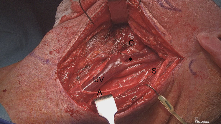
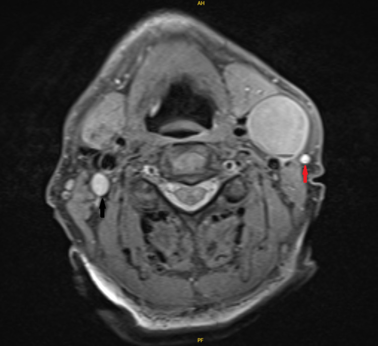
A comprehensive understanding of the anatomical variations of the internal jugular vein (IJV) is essential to prevent inadvertent injuries during neck procedures, particularly neck dissection. In addition, its relationship with the spinal accessory nerve in the upper part of the neck is relatively variable. IJV fenestration refers to bifurcation of the vein with reunion proximal to the subclavian vein, whereas IJV duplication refers to continued branching till joining the subclavian vein separately. We report a case of a fenestrated IJV identified intraoperatively with the spinal accessory nerve passing laterally to both divisions.
Keywords: anatomical anomalies; duplication; fenestration; internal jugular vein; neck dissection; split.
Copyright © 2022, Aladham et al.
Conflict of interest statement
The authors have declared that no competing interests exist.
Figures
Similar articles
-
Internal Jugular Vein Fenestration and Duplication: Anatomical Findings, Prevalence, and Literature Review.Front Surg. 2020 Nov 13;7:593367. doi: 10.3389/fsurg.2020.593367. eCollection 2020. Front Surg. 2020. PMID: 33282909 Free PMC article.
-
Fenestration and Bifurcation of the Internal Jugular Vein; Surprises During Head and Neck Surgery.Iran J Otorhinolaryngol. 2025;37(2):99-103. doi: 10.22038/ijorl.2025.83514.3810. Iran J Otorhinolaryngol. 2025. PMID: 40162379 Free PMC article.
-
Unilateral Fenestration of Internal Jugular Vein With a Radiological Clue: A Rare Case Report and Literature Review.Cureus. 2023 Jun 2;15(6):e39863. doi: 10.7759/cureus.39863. eCollection 2023 Jun. Cureus. 2023. PMID: 37404430 Free PMC article.
-
Internal jugular vein duplication and fenestration: Case series and literature review.Laryngoscope. 2016 Jul;126(7):1585-8. doi: 10.1002/lary.25743. Epub 2015 Oct 26. Laryngoscope. 2016. PMID: 26498831 Free PMC article. Review.
-
Bilateral duplicated internal jugular veins: case study and literature review.Clin Anat. 2007 Apr;20(3):260-6. doi: 10.1002/ca.20366. Clin Anat. 2007. PMID: 16838288 Review.
Cited by
-
Study of the Anatomical Variations of Spinal Accessory Nerve Seen During Neck Dissection.Indian J Otolaryngol Head Neck Surg. 2024 Jun;76(3):2295-2303. doi: 10.1007/s12070-023-04468-9. Epub 2024 Jan 18. Indian J Otolaryngol Head Neck Surg. 2024. PMID: 38883541 Free PMC article.
-
From incidental to intentional: ultrasound imaging in detecting internal jugular vein duplication/fenestration from a case report.Medicine (Baltimore). 2025 Jun 27;104(26):e42935. doi: 10.1097/MD.0000000000042935. Medicine (Baltimore). 2025. PMID: 40587714 Free PMC article.
References
-
- Anatomic relationship between the spinal accessory nerve and internal jugular vein in the upper neck. Hinsley ML, Hartig GK. Otolaryngol Head Neck Surg. 2010;143:239–241. - PubMed
-
- High duplication of the internal jugular vein: clinical incidence in the adult and surgical consequences, a report of three clinical cases. Prades JM, Timoshenko A, Dumollard JM, Durand M, Merzougui N, Martin C. Surg Radiol Anat. 2002;24:129–132. - PubMed
-
- Surgically important variations of the jugular veins. Nayak BS. Clin Anat. 2006;19:544–546. - PubMed
-
- A review of two cases of fenestrated internal jugular veins as seen by CT angiography. Towbin AJ, Kanal E. http://www.ajnr.org/content/25/8/1433.long. AJNR Am J Neuroradiol. 2004;25:1433–1434. - PMC - PubMed
Publication types
LinkOut - more resources
Full Text Sources