

Case Series of Neonatal Extravasation Injury: Importance of Early Identification and Management
- PMID: 35165627
- PMCID: PMC8837468
- DOI: 10.7759/cureus.21179
Case Series of Neonatal Extravasation Injury: Importance of Early Identification and Management
Abstract
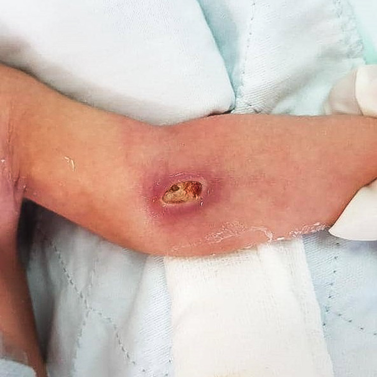
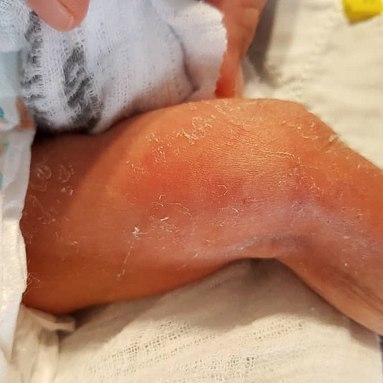
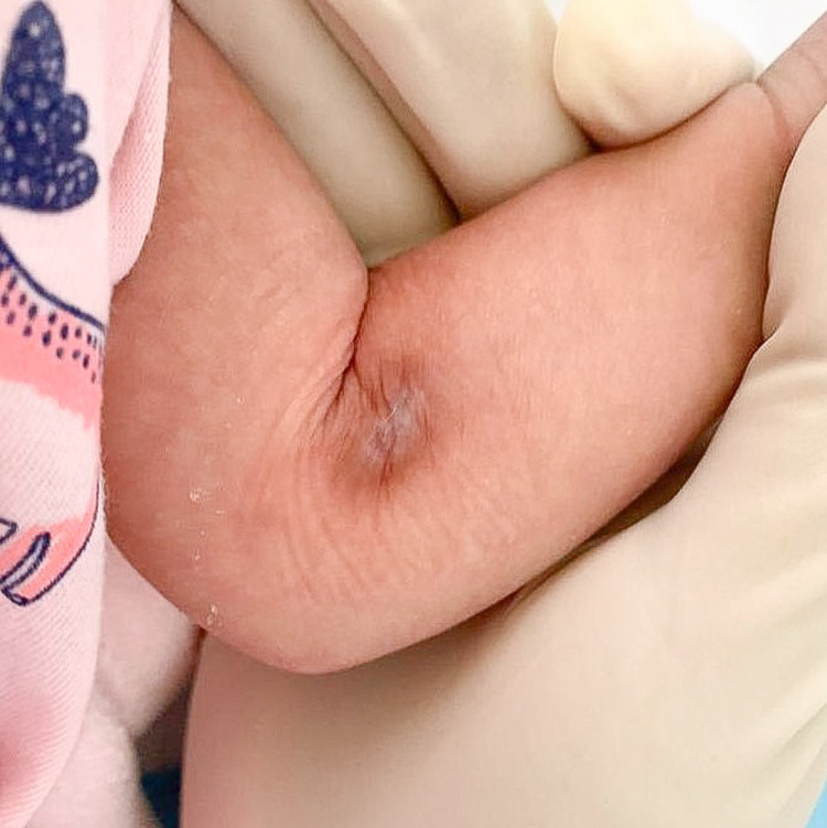
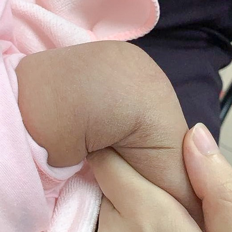
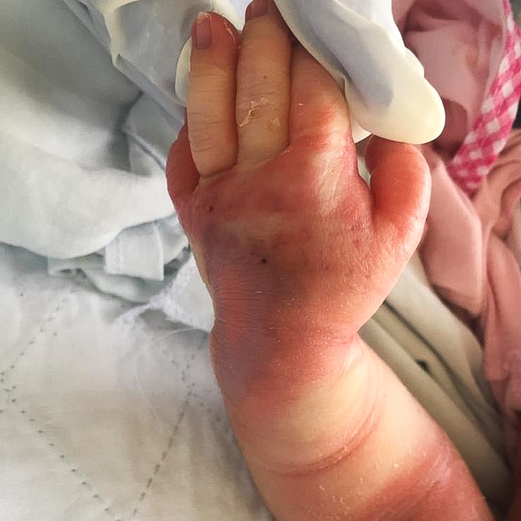
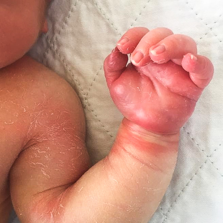
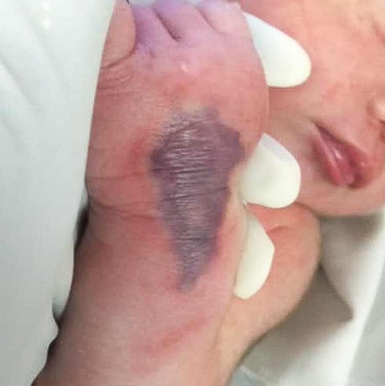
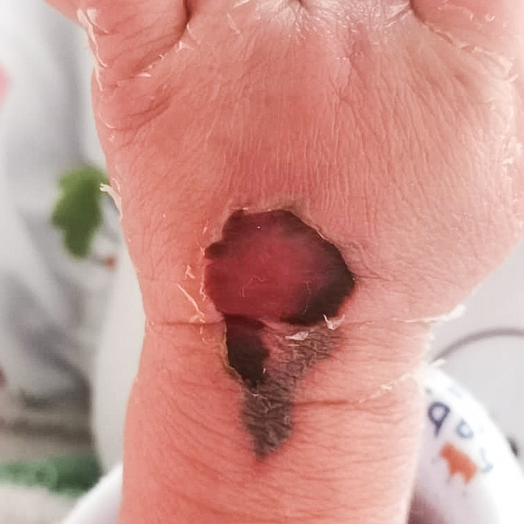
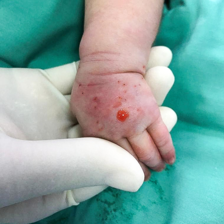
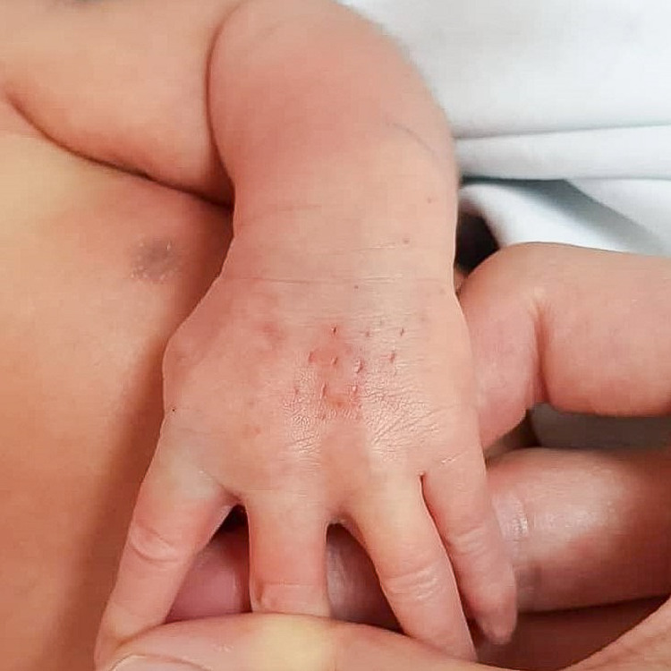
Extravasation injury is a common iatrogenic injury, especially in neonates. Intravenous access is essential in neonatal care, but neonatal extravasation injury is associated with severe morbidity. We present three cases of neonatal extravasation injuries with varying presentations, etiological agents, and timing of management. It shows that extravasation injuries treated with the saline flush-out technique and timely intervention have a superior outcome with almost immediate resolution and subsequent healing with no scars. This is in stark contrast with the lesions treated conservatively with dressings that took more time to heal. We are reminded to be vigilant with infusion therapies and the importance of early detection and prompt treatment in neonatal extravasation injuries.
Keywords: cannula; dressing; extravasation injury; extravasation of diagnostic and therapeutic materials; gault technique; intravenous fluid; necrosis; neonate; newborn; saline flush-out.
Copyright © 2022, Yew et al.
Conflict of interest statement
The authors have declared that no competing interests exist.
Figures
Similar articles
-
Severe Extravasation Injuries in Neonates: A Report of 34 Cases.Pediatr Dermatol. 2015 Nov-Dec;32(6):830-5. doi: 10.1111/pde.12664. Epub 2015 Sep 4. Pediatr Dermatol. 2015. PMID: 26337780
-
Treatment of a neonatal peripheral intravenous infiltration/extravasation (PIVIE) injury with hyaluronidase: a case report.Br J Nurs. 2022 Apr 21;31(8):S31-S36. doi: 10.12968/bjon.2022.31.8.S31. Br J Nurs. 2022. PMID: 35439074
-
Efficacy of Dehydrated Human Amniotic Membrane Allograft for the Treatment of Severe Extravasation Injuries in Preterm Neonates.Wounds. 2018 Aug;30(8):224-228. Wounds. 2018. PMID: 30212365
-
Management of extravasation injuries: a focused evaluation of noncytotoxic medications.Pharmacotherapy. 2014 Jun;34(6):617-32. doi: 10.1002/phar.1396. Epub 2014 Jan 13. Pharmacotherapy. 2014. PMID: 24420913 Review.
-
Management of large volume CT contrast medium extravasation injury: technical refinement and literature review.J Plast Reconstr Aesthet Surg. 2008;61(5):562-5; discussion 565. doi: 10.1016/j.bjps.2007.02.024. Epub 2007 Apr 25. J Plast Reconstr Aesthet Surg. 2008. PMID: 17459795 Review.
Cited by
-
Evaluation of optical sensor technology for the early detection of peripheral intravenous infiltration in neonates: a retrospective cohort study.BMJ Open. 2025 Jul 5;15(7):e094464. doi: 10.1136/bmjopen-2024-094464. BMJ Open. 2025. PMID: 40617616 Free PMC article.
References
-
- Extravasation injuries. Gault DT. Br J Plast Surg. 1993;46:91–96. - PubMed
-
- Skin necrosis from extravasation of intravenous fluids in children. Brown AS, Hoelzer DJ, Piercy SA. Plast Reconstr Surg. 1979;64:145–150. - PubMed
-
- Extravasation injuries: a review. Goutos I, Cogswell LK, Giele H. J Hand Surg Eur Vol. 2014;39:808–818. - PubMed
Publication types
LinkOut - more resources
Full Text Sources