Review
. 2022 Apr;19(3):931-941.
doi: 10.1007/s13311-022-01197-9. Epub 2022 Feb 14.

CRISPR-Cas9 Gene Therapy for Duchenne Muscular Dystrophy

Affiliations
Review

CRISPR-Cas9 Gene Therapy for Duchenne Muscular Dystrophy

Cedric Happi Mbakam et al. Neurotherapeutics. 2022 Apr.

Abstract

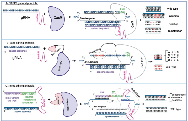
Discovery of the CRISPR-Cas (clustered regularly interspaced short palindromic repeat, CRISPR-associated) system a decade ago has opened new possibilities in the field of precision medicine. CRISPR-Cas was initially identified in bacteria and archaea to play a protective role against foreign genetic elements during viral infections. The application of this technique for the correction of different mutations found in the Duchenne muscular dystrophy (DMD) gene led to the development of several potential therapeutic approaches for DMD patients. The mutations responsible for Duchenne muscular dystrophy mainly include exon deletions (70% of patients) and point mutations (about 30% of patients). The CRISPR-Cas 9 technology is becoming increasingly precise and is acquiring diverse functions through novel innovations such as base editing and prime editing. However, questions remain about its translation to the clinic. Current research addressing off-target editing, efficient muscle-specific delivery, immune response to nucleases, and vector challenges may eventually lead to the clinical use of the CRISPR-Cas9 technology. In this review, we present recent CRISPR-Cas9 strategies to restore dystrophin expression in vitro and in animal models of DMD.

Keywords: CRISPR-Cas; DMD gene; Duchenne muscular dystrophy; Dystrophin; Gene therapy.


Figures

Fig. 1
Fig. 1
General CRISPR mechanism for genome modification. A The basic principle of CRISPR Cas9 gene modification which uses a guide RNA and a Cas9 to mediate the nuclease activity that could result in insertions, deletions, or INDELs. B The basic mechanism for base editing, which uses a sgRNA (spacer sequence + scaffold) and a Cas9 nickase fused with a cytosine or an adenine deaminase. Depending on the deaminase used, after binding with a specific DNA template, a cytosine is modified into a thymine or an adenosine into a guanine. C shows the two important components of prime editing: a PE2 protein and a pegRNA. The pegRNA is an extended sgRNA including in addition to the spacer sequence and the constant sequence (scaffold), a reverse transcriptase template (RTT), and a primer binding site (PBS). PE2 is a Cas9 nickase fused with an engineered reverse transcriptase enzyme from M-MLV. After the binding of the PE2 protein and pegRNA with a specific DNA target, the possible modification outcomes are an insertion, a deletion, or a substitution of a few nucleotides
Fig. 2
Fig. 2
Structure of dystrophin complex and dystrophin gene and regulator elements. A The approximate structure of the dystrophin complex in the normal context and in a DMD context. In the normal context, dystrophin is present under the sarcolemma and is properly attached to other components to form the dystrophin complex. In the DMD context, there is an absence of the dystrophin complex which results in muscle fiber degradation during muscle contraction. B The dystrophin gene (exons 1 to 79) with different functional domains. The N-terminal actin binding domain (ABD1 in green) with different dystrophin promoters (Dp427P, Dp427B, Dp427M, Dp260R, Dp140B3, Dp116S, and Dp71G, respectively, for cerebellar Purkinje cells, brain, muscle, retina, brain, Schwann cells, and general) and calponin-homology motifs (CH1 and CH2). The central rod domain (blue color) with spectrin-like repeats R1 to R24, the second actin-binding domain (ABD2), the nNos-binding site (nNos bs), and the spreading of proline rich hinge regions (H1, H2, H3, and H4 in red). The cysteine-rich domain contains the WW domain, EF hands, and ZZ domain. The C terminal domain serves as binding site for syntrophins and dystrobrevin glycoprotein
Fig. 3
Fig. 3
Different CRISPR approaches to mediate the dystrophin restoration in DMD patients. A The outcome of DMD exon 52 deletion and the two primary methods to restore dystrophin expression: (1) splice sites modifications: the donor site (gt) may be modified to ga or the acceptor site (ag) may be modified to gg. (2) The insertion in exon 51 of one nucleotide (A in red) to reframe exon 53 while conserving the same first amino acid codon for exon 53. B represents a strategy where a double-strand break can be made to generate an INDEL (micro-insertion or micro-deletion) in the exon preceding a stop codon. This stop codon was induced by a frameshift mutation which is compensated for by the engineered INDEL to restore the normal reading frame for the next exon. C The strategy where two single guide RNA can be used to completely delete one or several exons to restore the normal reading. D The strategy leading to the formation of a hybrid exon by inducing double-strand breaks. This hybrid exon not only restores the normal reading frame but also produces a dystrophin protein, which has a normal structure thus including a normal spectrin-like repeat structure at the junction site of the two exons. E The point mutation strategy where the abnormal nucleotide (G) in the figure forming a stop codon (TAG) is directly modified into another nucleotide (T in the figure) thus removing the stop codon

References

    1. Ishino Y, Shinagawa H, Makino K, et al. Nucleotide sequence of the iap gene, responsible for alkaline phosphatase isozyme conversion in Escherichia coli, and identification of the gene product. J Bacteriol. 1987;169:5429–5433. - PMC - PubMed
    1. Jansen R, van Embden JDA, Gaastra W, et al. Identification of genes that are associated with DNA repeats in prokaryotes. Mol Microbiol. 2002;43:1565–1575. - PubMed
    1. Barrangou R, Fremaux C, Deveau H, et al. CRISPR provides acquired resistance against viruses in prokaryotes. Science. 2007;315:1709–1712. - PubMed
    1. Bolotin A, Quinquis B, Sorokin A, et al. Clustered regularly interspaced short palindrome repeats (CRISPRs) have spacers of extrachromosomal origin. Microbiology. 2005;151:2551–2561. - PubMed
    1. Pourcel C, Salvignol G, Vergnaud GY. CRISPR elements in Yersinia pestis acquire new repeats by preferential uptake of bacteriophage DNA, and provide additional tools for evolutionary studies. Microbiology. 2005;151:653–663. - PubMed

Publication types

MeSH terms

Grants and funding