. 2022 Apr;26(8):2152-2162.
doi: 10.1111/jcmm.17237. Epub 2022 Feb 15.

CD93 in macrophages: A novel target for atherosclerotic plaque imaging?

Affiliations

CD93 in macrophages: A novel target for atherosclerotic plaque imaging?

Chen Su et al. J Cell Mol Med. 2022 Apr.

Abstract

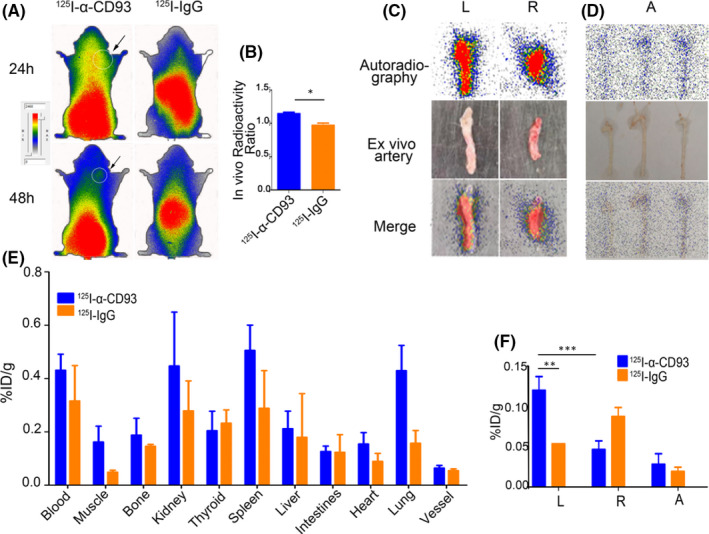
Noninvasive imaging atherosclerotic (AS) plaque is of great importance for early diagnosis. Recently, CD93 in MΦ was linked to atherosclerosis development. Herein, we have investigated whether CD93 in MΦ is a potential novel target for atherosclerotic plaque imaging. CD93hi and CD93lo MΦ were prepared with or without LPS stimulation, before biological activity was evaluated. A rat AS model was produced with left carotid artery clamped. Whole-body/ex vivo phosphor autoradiography of the artery and biodistribution were investigated after incorporation of 3 H-2-DG into CD93hi and CD93lo MΦ or after 125 I-α-CD93 (125 I-anti-CD93mAb) injection. The plaque tissue was subjected to CD93/CD68 immunofluorescence/immunohistochemistry staining. CD93hi and CD93lo MΦ cells were successfully prepared without significant effect on bioactivity after incorporative labelled with 3 H-2-DG. The AS model was successfully established. Biodistribution studies showed that adoptive transfer of 3 H-2-DG-CD93hi MΦ or 125 I- α-CD93 injection resulted in accumulation of radioactivity within the atherosclerotic plaque in the clamped left carotid artery. T/NT (target/non-target, left/right carotid artery) ratio was higher in the 3 H-2-DG-CD93hi MΦ adoptive transfer group than in the 3 H-2-DG-CD93lo MΦ group (p < .05). Plaque radioactivity in the 125 I-α-CD93 injection group was significantly higher than in the 125 I-IgG control group (p < .01). The higher radioactivity accumulated in the clamped left carotid artery was confirmed by phosphor autoradiography. More importantly, CD93/CD68 double-positive MΦ accumulated at the atherosclerotic plaque in 3 H-2-DG-CD93hi MΦ adoptive transfer group, which correlated with plaque radioactivity (r = .99, p < .01). In summary, both adoptive-transferred 3 H-2-DG-labelled CD93hi MΦ and 125 I-α-CD93 injection specifically targeted CD93 in atherosclerotic plaque. CD93 is a potential target in atherosclerotic plaque imaging.

Keywords: CD93; atherosclerosis; macrophage; molecular imaging; radionuclide.


Conflict of interest statement

The author declares that there is no conflict of interest.

Figures

FIGURE 1
FIGURE 1
H&E and Oil red staining for rat artery and aorta. (A) Left/right carotid artery and aorta arch from the rat AS model, with representative H&E staining images at 100× and 400× magnification. The black arrow points to the atherosclerotic plaque in the carotid artery (left) and the aorta. (B) Oil red O staining of aorta in the AS model, with black arrow pointing to lipid droplets. (C) Immunofluorescence staining of CD93hi and CD93lo MΦ and representative images (400×), CD93 (red), CD68 (green) and nucleus (blue). Data were obtained from three independent experiments
FIGURE 2
FIGURE 2
Effect of LPS stimulation and 3H‐2‐DG labelling on CD93 expression and MΦ function. (A) CD93 mRNA expression after LPS stimulation and 3H‐2‐DG labelling was detected by RT‐PCR. (B) CD93 protein expression after LPS stimulation and 3H‐2‐DG labelling was detected by Western Blot. (C) Phagocytosis of Dil‐Ox‐LDL by MΦ (400×), with Dil‐Ox‐LDL (red) and nuclear (blue). NT: non treated by LPS, no 3H‐2‐DG labelling, LPS: LPS‐stimulated, 3H‐2‐DG: labelled with 3H‐2‐DG. Data were analysed by Student's t‐test (n = 3), **p < .01
FIGURE 3
FIGURE 3
Effect of LPS stimulation and 3H‐2‐DG labelling on CD93 expression and MΦ function. (A) Morphology (400×) after LPS stimulation and 3H‐2‐DG labelling. (B) Apoptosis of MΦ detected by flow cytometry. (C) Cell cycle of MΦ detected by flow cytometry. G1: pre‐DNA synthesis stage, NT: non treated by LPS, no 3H‐2‐DG labelling, LPS: LPS‐stimulated, 3H‐2‐DG: labelled with 3H‐2‐DG. Data were obtained from three independent experiments
FIGURE 4
FIGURE 4
Evaluation of radiolabelled tracers. (A) Representative saturation binding curve and Scatchard plots with increasing 125I‐α‐CD93 binding to CD93hi MΦ; (B) the same for CD93lo MΦ. (C) Competition binding curve between 125I‐α‐CD93 and increasing unlabelled α‐CD93, n = 5
FIGURE 5
FIGURE 5
Tritium‐phosphor autoradiography and biodistribution of ex vivo aorta arch 72 h after 3H‐2‐DG‐labelled CD93hi and CD93lo MΦ adoptive transfer. (A) Representative ex vivo tritium‐phosphor autoradiography images of aortic arch in AS model after 72 h; (B/C) radioactivity biodistribution (%ID/g) of representative tissues in the 3H‐2‐DG‐labelled CD93hi and CD93lo MΦ adoptive transfer group after 72 h, with L (left carotid artery), R (right carotid artery) and A (aortic arch). n = 5, *** p < .001
FIGURE 6
FIGURE 6
Whole‐body and ex vivo artery phosphor autoradiography and biodistribution. (A) Representative images of 125I‐α‐CD93 and 125I‐IgG in AS model whole‐body phosphor autoradiography after 24 and 48 h, with black arrow pointing to the left carotid artery. (B) In vivo radioactivity ratio (left carotid artery/right carotid artery) between 125I‐α‐CD93 and 125I‐IgG; representative images of ex vivo (C) left/right carotid artery and (D) aorta artery after 48 h. (E) Biodistribution of 125I‐α‐CD93 and 125I‐IgG after 48 h. (F) Biodistribution of 125I‐α‐CD93 and 125I‐IgG in left/right carotid artery and aorta after 48 h. L (left carotid artery), R (right carotid artery) and A (aorta). Data were obtained from three independent experiments (n = 5), * p < .05, ** p < .01, *** p < .001
FIGURE 7
FIGURE 7
Immunofluorescence and immunohistochemistry imaging of diseased vessels. (A) Immunofluorescence staining of nucleus with DAPI (blue), CD68 (green) and CD93 (red). (B) Immunohistochemistry staining (400×) of CD93 and CD68, with positive staining shown as brown colour. (C) Correlation between IOD/Area of CD93 in immunohistochemistry staining and 125I‐α‐CD93 (DLU/mm2, digital light units) in ex vivo artery phosphor autoradiography. (D) Correlation between IOD/Area of CD68 in immunohistochemistry staining and 3H‐2‐DG‐CD93hi MΦ in ex vivo aorta tritum‐phosphor autoradiography. L (left carotid artery), R (right carotid artery) and A (aorta). n = 3

Similar articles

Cited by

References

    1. Finn AV, Nakano M, Narula J, Kolodgie FD, Virmani R. Concept of vulnerable/unstable plaque. Arterioscler Thromb Vasc Biol. 2010. Jul;30(7):1282‐1292. - PubMed
    1. Liu C, Zhang X, Song Y, et al. SPECT and fluorescence imaging of vulnerable atherosclerotic plaque with a vascular cell adhesion molecule 1 single‐chain antibody fragment. Atherosclerosis. 2016. Nov;254:263‐270. - PubMed
    1. Raggi P, Achenbach S. Computed tomography for atherosclerosis and coronary artery disease imaging. Discov Med. 2010. Feb;9(45):98‐104. - PubMed
    1. Jager NA, Westra J, Golestani R, et al. Folate receptor‐β imaging using 99mTc‐folate to explore distribution of polarized macrophage populations in human atherosclerotic plaque. J Nucl Med. 2014. Dec;55(12):1945‐1951. - PubMed
    1. van der Wall EE. Molecular imaging of coronary atherosclerosis; predictive of an acute myocardial infarction? Neth Heart J. 2014. Jan;22(1):1‐2. - PMC - PubMed

Publication types

MeSH terms