

A Bionic-Homodimerization Strategy for Optimizing Modulators of Protein-Protein Interactions: From Statistical Mechanics Theory to Potential Clinical Translation
- PMID: 35166067
- PMCID: PMC9008432
- DOI: 10.1002/advs.202105179
A Bionic-Homodimerization Strategy for Optimizing Modulators of Protein-Protein Interactions: From Statistical Mechanics Theory to Potential Clinical Translation
Abstract
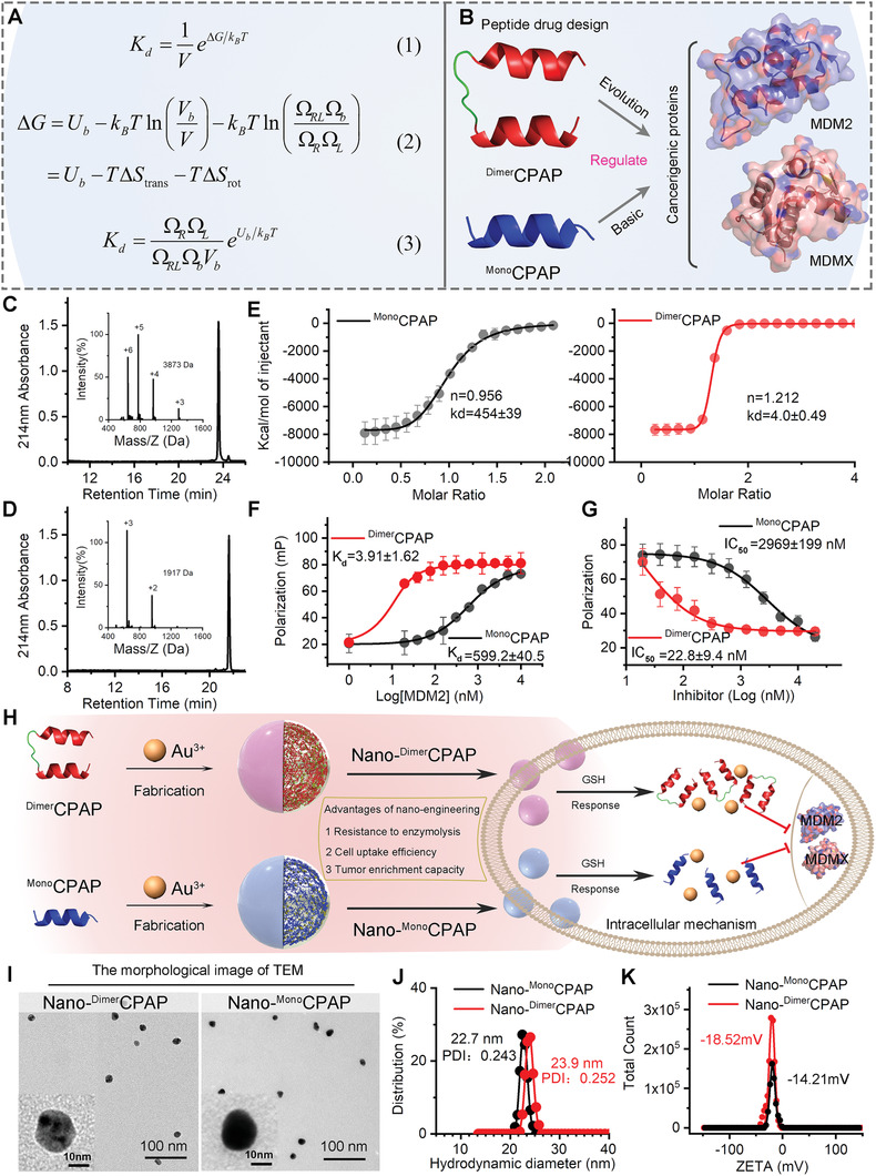
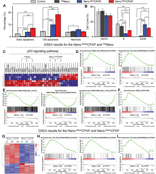
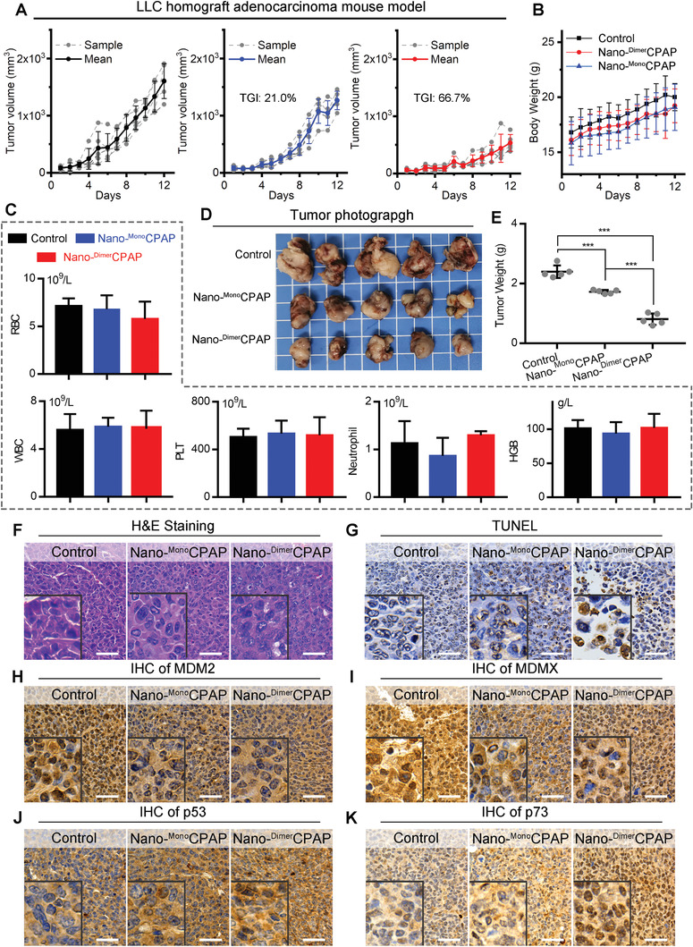
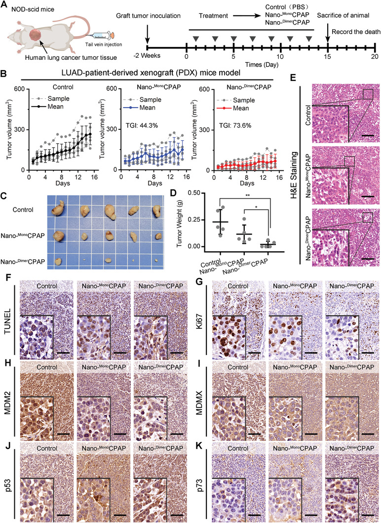
Emerging protein-protein interaction (PPI) modulators have brought out exciting ability as therapeutics in human diseases, but its clinical translation has been greatly hampered by the limited affinity. Inspired by the homodimerize structure of antibody, the homodimerization contributes hugely to generating the optimized affinity is conjectured. Herein, a statistical-mechanics-theory-guided method is established to quantize the affinity of ligands with different topologies through analyzing the change of enthalpy and the loss of translational and rotational entropies. A peptide modulator for p53-MDM2 termed CPAP is used to homodimerize connecting, and this simple homodimerization can significantly increase the affinity. To realize the cellular internalization and tumor accumulation, Dimer CPAP and Mono CPAP are nanoengineered into gold(I)-CPAP supermolecule by the aurophilic interaction-driven self-assembly. Nano-Dimer CPAP potently suppressed tumor growth in lung cancer allograft model and a patient-derived xenograft model in more action than Nano-Mono CPAP, while keeping a favorable drug safety profile. This work not only presents a physico-mechanical method for calculating the affinity of PPI modulators, but also provides a simple yet robust homodimerization strategy to optimize the affinity of PPI modulators.
Keywords: bionic-dimerization; nanomedicine; peptide; protein-protein interactions; statistical mechanics theory.
© 2022 The Authors. Advanced Science published by Wiley-VCH GmbH.
Conflict of interest statement
The authors declare no conflict of interest.
Figures
References
-
- a) Azzarito V., Long K., Murphy N. S., Wilson A. J., Nat. Chem. 2013, 5, 161; - PubMed
- b) Wilson A. J., Chem. Soc. Rev. 2009, 38, 3289; - PubMed
- c) Nim S., Jeon J., Corbi‐Verge C., Seo M.‐H., Ivarsson Y., Moffat J., Tarasova N., Kim P. M., Nat. Chem. Biol. 2016, 12, 275; - PMC - PubMed
- d) Liu Q., Zheng J., Sun W., Huo Y., Zhang L., Hao P., Wang H., Zhuang M., Nat. Methods 2018, 15, 715; - PubMed
- e) Luck K., Kim D.‐K., Lambourne L., Spirohn K., Begg B. E., Bian W., Brignall R., Cafarelli T., Campos‐Laborie F. J., Charloteaux B., Choi D., Coté A. G., Daley M., Deimling S., Desbuleux A., Dricot A., Gebbia M., Hardy M. F., Kishore N., Knapp J. J., Kovács I. A., Lemmens I., Mee M. W., Mellor J. C., Pollis C., Pons C., Richardson A. D., Schlabach S., Teeking B., Yadav A., et al., Nature 2020, 580, 402; - PMC - PubMed
- f) Lu H., Zhou Q., He J., Jiang Z., Peng C., Tong R., Shi J., Signal Transduction Targeted Ther. 2020, 5, 213. - PMC - PubMed
-
- a) White A. W., Westwell A. D., Brahemi G., Expert Rev. Mol. Med. 2008, 10, e8; - PubMed
- b) Rosell M., Fernandez‐Recio J., Expert Opin. Drug Discovery 2018, 13, 327; - PubMed
- c) Yan J., He W., Yan S., Niu F., Liu T., Ma B., Shao Y., Yan Y., Yang G., Lu W., Du Y., Lei B., Ma P. X., ACS Nano 2018, 12, 2017; - PubMed
- d) Yan J., Yan S., Hou P., Lu W., Ma P. X., He W., Lei B., Nano Lett.. 2019, 19, 7918. - PubMed
-
- a) Nevola L., Giralt E., Chem. Commun. 2015, 51, 3302; - PubMed
- b) Sheng C., Dong G., Miao Z., Zhang W., Wang W., Chem. Soc. Rev. 2015, 44, 8238; - PubMed
- c) Pelay‐Gimeno M., Glas A., Koch O., Grossmann T. N., Angew. Chem., Int. Ed. 2015, 54, 8896; - PMC - PubMed
- d) Chung C., Hann M., Structural Biology in Drug Discovery: Methods, Techniques, and Practices, John Wiley & Sons, Hoboken, NJ: 2020.
-
- a) Gide T. N., Quek C., Menzies A. M., Tasker A. T., Shang P., Holst J., Madore J., Lim S. Y., Velickovic R., Wongchenko M., Yan Y., Lo S., Carlino M. S., Guminski A., Saw R. P. M., Pang A., McGuire H. M., Palendira U., Thompson J. F., Rizos H., Silva I. P. d., Batten M., Scolyer R. A., Long G. V., Wilmott J. S., Cancer Cell 2019, 35, 238; - PubMed
- b) Boutros C., Tarhini A., Routier E., Lambotte O., Ladurie F. L., Carbonnel F., Izzeddine H., Marabelle A., Champiat S., Berdelou A., Lanoy E., Texier M., Libenciuc C., Eggermont A. M. M., Soria J.‐C., Mateus C., Robert C., Nat. Rev. Clin. Oncol. 2016, 13, 473; - PubMed
- c) De Castro L. N., Castro L. N., Timmis J., Artificial Immune Systems: A New Computational Intelligence Approach, Springer Science & Business Media, Secaucus, NJ: 2002;
- d) Borrebaeck C. A. K., Ohlin M., Nat. Biotechnol. 2002, 20, 1189; - PubMed
- e) Cooper M. D., Alder M. N., Cell 2006, 124, 815; - PubMed
- f) Feige M. J., Grawert M. A., Marcinowski M., Hennig J., Behnke J., Auslander D., Herold E. M., Peschek J., Castro C. D., Flajnik M., Hendershot L. M., Sattler M., Groll M., Buchner J., Proc. Natl. Acad. Sci. U. S. A. 2014, 111, 8155. - PMC - PubMed
-
- Woof J. M., Burton D. R., Nat. Rev. Immunol. 2004, 4, 89. - PubMed
Publication types
MeSH terms
Substances
Grants and funding
LinkOut - more resources
Full Text Sources
Medical
Research Materials
Miscellaneous