

Disrupted topological organization of resting-state functional brain networks in cerebral small vessel disease
- PMID: 35166416
- PMCID: PMC9057099
- DOI: 10.1002/hbm.25808
Disrupted topological organization of resting-state functional brain networks in cerebral small vessel disease
Abstract
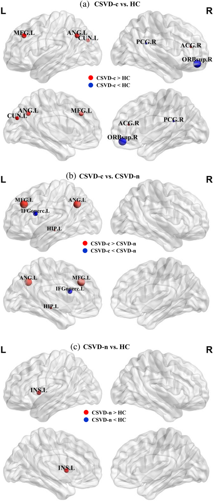
We aimed to investigate alterations in functional brain networks and assess the relationship between functional impairment and topological network changes in cerebral small vessel disease (CSVD) patients with and without cerebral microbleeds (CMBs). We constructed individual whole-brain, region of interest (ROI) level functional connectivity (FC) networks for 24 CSVD patients with CMBs (CSVD-c), 42 CSVD patients without CMBs (CSVD-n), and 36 healthy controls (HCs). Then, we used graph theory analysis to investigate the global and nodal topological disruptions between groups and relate network topological alterations to clinical parameters. We found that both the CSVD and control groups showed efficient small-world organization in FC networks. However, compared to CSVD-n patients and controls, CSVD-c patients exhibited a significantly decreased clustering coefficient, global efficiency, and local efficiency and an increased shortest path length, indicating a disrupted balance between local specialization and global integration in FC networks. Although both the CSVD and control groups showed highly similar hub distributions, the CSVD-c group exhibited significantly altered nodal betweenness centrality (BC), mainly distributed in the default mode network (DMN), attention, and visual functional areas. There were almost no global or regional alterations between CSVD-n patients and controls. Furthermore, the altered nodal BC of the right anterior/posterior cingulate gyrus and left cuneus were significantly correlated with cognitive parameters in CSVD patients. These results suggest that CSVD patients with and without CMBs had segregated disruptions in the topological organization of the intrinsic functional brain network. This study advances our current understanding of the pathophysiological mechanisms underlying CSVD.
Keywords: cerebral microbleeds; cerebral small vessel disease; functional connectivity; graph theory; topological organization.
© 2022 The Authors. Human Brain Mapping published by Wiley Periodicals LLC.
Conflict of interest statement
The authors declare that they have no competing interests.
Figures


Similar articles
-
Disrupted brain structural networks associated with depression and cognitive dysfunction in cerebral small vessel disease with microbleeds.Prog Neuropsychopharmacol Biol Psychiatry. 2024 Apr 20;131:110944. doi: 10.1016/j.pnpbp.2024.110944. Epub 2024 Jan 20. Prog Neuropsychopharmacol Biol Psychiatry. 2024. PMID: 38246218
-
Disrupted Gray Matter Networks Associated with Cognitive Dysfunction in Cerebral Small Vessel Disease.Brain Sci. 2023 Sep 22;13(10):1359. doi: 10.3390/brainsci13101359. Brain Sci. 2023. PMID: 37891728 Free PMC article.
-
Decreased Local Specialization of Brain Structural Networks Associated with Cognitive Dysfuntion Revealed by Probabilistic Diffusion Tractography for Different Cerebral Small Vessel Disease Burdens.Mol Neurobiol. 2024 Jan;61(1):326-339. doi: 10.1007/s12035-023-03597-0. Epub 2023 Aug 22. Mol Neurobiol. 2024. PMID: 37606718 Free PMC article.
-
Functional connectivity changes in cerebral small vessel disease - a systematic review of the resting-state MRI literature.BMC Med. 2021 May 5;19(1):103. doi: 10.1186/s12916-021-01962-1. BMC Med. 2021. PMID: 33947394 Free PMC article.
-
Neuroimaging studies on cognitive impairment due to cerebral small vessel disease.Stroke Vasc Neurol. 2019 Apr 5;4(2):99-101. doi: 10.1136/svn-2018-000209. eCollection 2019 Jul. Stroke Vasc Neurol. 2019. PMID: 31338220 Free PMC article. Review.
Cited by
-
Alterations in regional homogeneity and functional connectivity associated with cognitive impairment in patients with hypertension: a resting-state functional magnetic resonance imaging study.Hypertens Res. 2023 May;46(5):1311-1325. doi: 10.1038/s41440-023-01168-3. Epub 2023 Jan 23. Hypertens Res. 2023. PMID: 36690806
-
Association between functional network connectivity, retina structure and microvasculature, and visual performance in patients after thalamic stroke: An exploratory multi-modality study.Brain Behav. 2024 Jan;14(1):e3385. doi: 10.1002/brb3.3385. Brain Behav. 2024. PMID: 38376035 Free PMC article.
-
Structural network disruption of corticothalamic pathways in cerebral small vessel disease.Brain Imaging Behav. 2024 Oct;18(5):979-988. doi: 10.1007/s11682-024-00889-4. Epub 2024 May 8. Brain Imaging Behav. 2024. PMID: 38717572 Free PMC article.
-
Graph Metrics Reveal Brain Network Topological Property in Neuropathic Pain Patients: A Systematic Review.J Pain Res. 2024 Oct 9;17:3277-3286. doi: 10.2147/JPR.S483466. eCollection 2024. J Pain Res. 2024. PMID: 39411193 Free PMC article. Review.
-
Long-range connections damage in white matter hyperintensities affects information processing speed.Brain Commun. 2024 Feb 21;6(1):fcae042. doi: 10.1093/braincomms/fcae042. eCollection 2024. Brain Commun. 2024. PMID: 38410619 Free PMC article.
References
-
- Ashburner, J. (2007). A fast diffeomorphic image registration algorithm. NeuroImage, 38, 95–113. - PubMed
-
- Bergeron, D. , Flynn, K. , Verret, L. , Poulin, S. , Bouchard, R. W. , Bocti, C. , … Laforce, R., Jr. (2017). Multicenter validation of an MMSE‐MoCA conversion table. Journal of the American Geriatrics Society, 65, 1067–1072. - PubMed
Publication types
MeSH terms
LinkOut - more resources
Full Text Sources
Medical
