. 2022 Mar 23;14(637):eabn8057.
doi: 10.1126/scitranslmed.abn8057. Epub 2022 Mar 23.

Neutralizing antibody responses elicited by SARS-CoV-2 mRNA vaccination wane over time and are boosted by breakthrough infection

Affiliations

Neutralizing antibody responses elicited by SARS-CoV-2 mRNA vaccination wane over time and are boosted by breakthrough infection

John P Evans et al. Sci Transl Med. .

Abstract

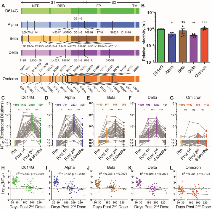
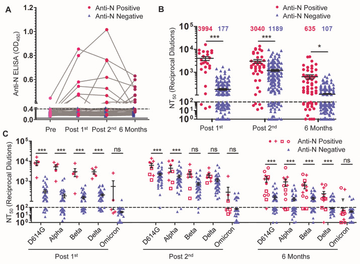
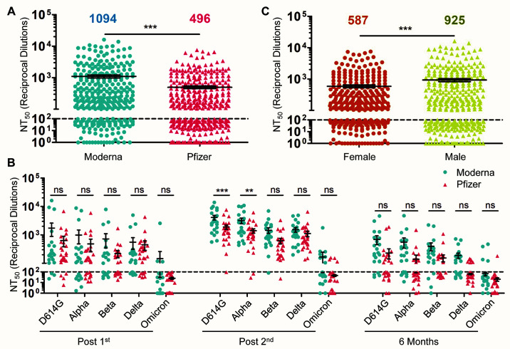
The waning efficacy of SARS-CoV-2 vaccines, combined with the continued emergence of variants resistant to vaccine-induced immunity, has reignited debate over the need for booster vaccine doses. To address this, we examined the neutralizing antibody response against the spike protein of five major SARS-CoV-2 variants, D614G, Alpha (B.1.1.7), Beta (B.1.351), Delta (B.1.617.2), and Omicron (B.1.1.529), in health care workers (HCWs) vaccinated with SARS-CoV-2 mRNA vaccines. Serum samples were collected before vaccination, 3 weeks after first vaccination, 1 month after second vaccination, and 6 months after second vaccination. Minimal neutralizing antibody titers were detected against Omicron pseudovirus at all four time points, including for most patients who had SARS-CoV-2 breakthrough infections. Neutralizing antibody titers against all other variant spike protein-bearing pseudoviruses declined markedly from 1 to 6 months after the second mRNA vaccine dose, although SARS-CoV-2 infection boosted vaccine responses. In addition, mRNA-1273-vaccinated HCWs exhibited about twofold higher neutralizing antibody titers than BNT162b2-vaccinated HCWs. Together, these results demonstrate possible waning of antibody-mediated protection against SARS-CoV-2 variants that is dependent on prior infection status and the mRNA vaccine received. They also show that the Omicron variant spike protein can almost completely escape from neutralizing antibodies elicited in recipients of only two mRNA vaccine doses.


Figures

Fig. 1.
Fig. 1.. The durability of vaccine-induced immunity wanes over time.
Gaussia luciferase reporter gene-containing pseudotyped lentiviruses were produced bearing the spike protein from SARS-CoV-2 variants. (A) Schematic representations of the SARS-CoV-2 variant spike proteins tested are shown, including D614G, Alpha (B.1.1.7), Beta (B.1.351), Delta (B.1.617.2), and Omicron (B.1.1.529). The schematics highlight the location of the S1 and S2 subunits of the spike protein as well as the N-terminal domain (NTD), receptor-binding domain (RBD), fusion peptide (FP), transmembrane region (TM), and the indicated mutations. (B) Pseudotyped lentiviruses were used to infect HEK293T-ACE2 cells. Media was harvested from infected cells 48 hours after infection and assayed for Gaussia luciferase activity to determine the relative infectivity of each variant pseudotyped virus; infectivity was measured as luminescence signal relative to D614G signal, relative units (ru). Significance relative to D614G was determined by one-way ANOVA with Bonferroni’s correction (n ≥ 3 biological replicates); error bars represent means ± standard error. (C to G) Lentivirus pseudotyped with SARS-CoV-2 spike protein from D614G (C), Alpha (D), Beta (E), Delta (F), and Omicron (G) were neutralized with serum samples from health care workers (HCWs) (n = 48 biological replicates) collected pre-vaccination (Pre), post-vaccination with a first mRNA vaccine dose (Post 1st), post-vaccination with a second mRNA vaccine dose (Post 2nd), and six months post second dose (Six Months). Neutralization titers 50% (NT50) values were determined by least-squares fit non-linear regression. Mean NT50 values are shown at the top of the plots, and NT50 values below 100 were considered background, as indicated by a dashed line; significance was determined by one-way repeated measures ANOVA with Bonferroni’s correction. (H to L) Log10-transformed NT50 values against D614G (H), Alpha (I), Beta (J), Delta (K), and Omicron (L) variants were plotted against days post-second vaccine dose of sample collection (n = 96 biological replicates). The goodness of fit (R2) and p-values are displayed on each plot as determined by least-squares fit linear regression. The dotted lines correspond to the background (NT50 < 100). In all cases, *p < 0.05; **p < 0.01; ***p < 0.001; ns: not significant.
Fig. 2.
Fig. 2.. The durability of the nAb response is influenced by prior COVID-19 infection and mRNA vaccine type.
COVID-19 status was determined by anti-N protein ELISA. Individuals were characterized as anti-N protein positive (optical density [OD]450 > 0.4 at any time point; n = 12) or anti-N protein negative (OD450 < 0.4 for all time points; n = 36). (A) ELISA optical density at 450nm (OD450) are shown for anti-N protein positive and anti-N protein negative HCWs. The dashed line indicates the cut-off of 0.4. (B) Comparisons of NT50 values between anti-N protein positive and anti-N protein negative HCWs are shown for the indicated time points. NT50 values against all variants were combined and plotted at post-first vaccine dose (n = 25 anti-N protein positive; n = 215 anti-N protein negative), post-second vaccine dose (n = 40 anti-N protein positive; n = 200 anti-N protein negative), and six months post-second vaccine dose (n = 60 anti-N protein positive; n = 180 anti-N protein negative). Significant differences were determined by two-way ANOVA with Bonferroni’s correction. (C) Comparisons of NT50 values against different variants between anti-N protein positive and anti-N protein negative HCWs are shown for the indicated time points. For anti-N protein positive HCWs, timing of first anti-N protein positive sample is distinguished as: pre-vaccination (solid magenta circle ●, n = 1); post-first dose (magenta cross +, n = 4); post-second dose (magenta open square □, n = 3); and six months-post second (magenta open circle ○, n =4). These are plotted alongside anti-N protein negative HCWs (all shown in green blue ▲) for three time points: post-first vaccine dose (n = 5 anti-N protein positive; n = 43 anti-N protein negative), post-second vaccine dose (n = 8 anti-N protein positive; n = 40 anti-N protein negative), and six months post-second vaccine dose (n = 12 anti-N protein positive; n = 36 anti-N protein negative). Significant differences were determined by two-way repeated-measures ANOVA with Bonferroni’s correction. For panels B and C, error bars indicate means ± standard errors, and the dashed horizontal line indicates the limit of detection (NT50 < 100). In all cases, *p < 0.05; **p < 0.01; ***p < 0.001; ns: not significant.
Fig. 3.
Fig. 3.. nAb response is influenced by vaccine type and sex.
(A) HCWs were divided by types of mRNA vaccine received, either Moderna mRNA-1273 (n = 22) or Pfizer/BioNTech BNT162b2 (n = 26), and NT50 values were combined for all variants at all time points. Significance between vaccine type was determined by unpaired two-tailed t test with Welch’s correction and means are indicated at the top of the plot. (B) NT50 values of serum samples from individuals vaccinated with Moderna mRNA-1273 (n = 22) or Pfizer/BioNTech BNT162b2 (n = 26) were grouped by variant and time point, with significance determined by two-way repeated-measures ANOVA with Bonferroni’s correction. (C) NT50 values against all variants at post-first dose, post-second dose, and six months post-second dose combined were compared for male (n = 390) and female (n = 330) participants; mean NT50 values are displayed at the top of the plot, with significance determined by unpaired, two-tailed t test with Welch’s correction. For all panels, error bars indicate means ± standard errors, and the dashed horizontal line indicates the limit of detection (NT50 < 100). In all cases, *p < 0.05; **p < 0.01; ***p < 0.001; ns: not significant.

Similar articles

Cited by

References

    1. World Health Organization. COVID-19 weekly epidemiological update, 18 January 2022. (2021).
    1. Plante J. A., Liu Y., Liu J., Xia H., Johnson B. A., Lokugamage K. G., Zhang X., Muruato A. E., Zou J., Fontes-Garfias C. R., Mirchandani D., Scharton D., Bilello J. P., Ku Z., An Z., Kalveram B., Freiberg A. N., Menachery V. D., Xie X., Plante K. S., Weaver S. C., Shi P.-Y., Spike mutation D614G alters SARS-CoV-2 fitness. Nature 592, 116–121 (2021). 10.1038/s41586-020-2895-3 - DOI - PMC - PubMed
    1. Korber B., Fischer W. M., Gnanakaran S., Yoon H., Theiler J., Abfalterer W., Hengartner N., Giorgi E. E., Bhattacharya T., Foley B., Hastie K. M., Parker M. D., Partridge D. G., Evans C. M., Freeman T. M., de Silva T. I., McDanal C., Perez L. G., Tang H., Moon-Walker A., Whelan S. P., LaBranche C. C., Saphire E. O., Montefiori D. C.; Sheffield COVID-19 Genomics Group , Tracking changes in SARS-CoV-2 Spike: Evidence that D614G increases infectivity of the COVID-19 virus. Cell 182, 812–827.e19 (2020). 10.1016/j.cell.2020.06.043 - DOI - PMC - PubMed
    1. Abdool Karim S. S., de Oliveira T., New SARS-CoV-2 variants—Clinical, public health, and vaccine implications. N. Engl. J. Med. 384, 1866–1868 (2021). 10.1056/NEJMc2100362 - DOI - PMC - PubMed
    1. Singh J., Rahman S. A., Ehtesham N. Z., Hira S., Hasnain S. E., SARS-CoV-2 variants of concern are emerging in India. Nat. Med. 27, 1131–1133 (2021). 10.1038/s41591-021-01397-4 - DOI - PubMed

Publication types

Supplementary concepts