. 2022 Feb 28;50(4):1864-1874.
doi: 10.1093/nar/gkac097.

DNA methylation cues in nucleosome geometry, stability and unwrapping

Affiliations

DNA methylation cues in nucleosome geometry, stability and unwrapping

Shuxiang Li et al. Nucleic Acids Res. .

Abstract

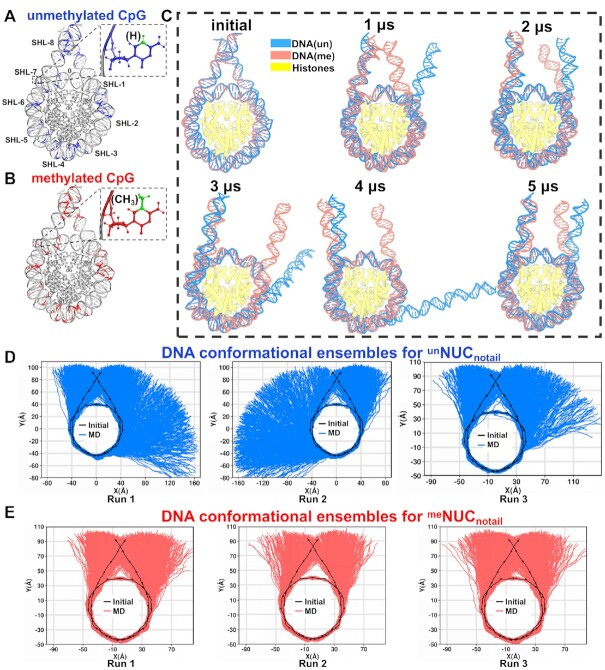
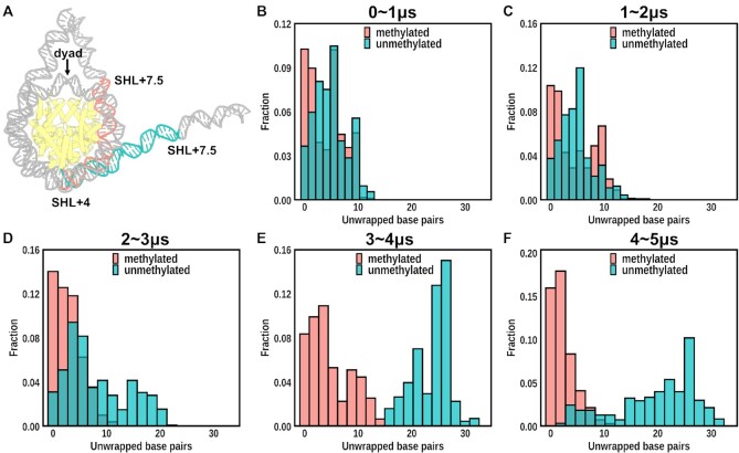
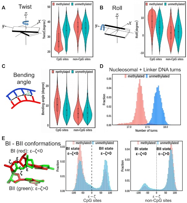
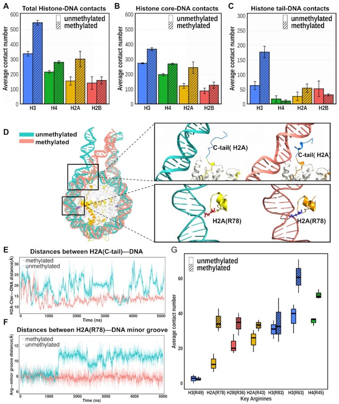
Cytosine methylation at the 5-carbon position is an essential DNA epigenetic mark in many eukaryotic organisms. Although countless structural and functional studies of cytosine methylation have been reported, our understanding of how it influences the nucleosome assembly, structure, and dynamics remains obscure. Here, we investigate the effects of cytosine methylation at CpG sites on nucleosome dynamics and stability. By applying long molecular dynamics simulations on several microsecond time scale, we generate extensive atomistic conformational ensembles of full nucleosomes. Our results reveal that methylation induces pronounced changes in geometry for both linker and nucleosomal DNA, leading to a more curved, under-twisted DNA, narrowing the adjacent minor grooves, and shifting the population equilibrium of sugar-phosphate backbone geometry. These DNA conformational changes are associated with a considerable enhancement of interactions between methylated DNA and the histone octamer, doubling the number of contacts at some key arginines. H2A and H3 tails play important roles in these interactions, especially for DNA methylated nucleosomes. This, in turn, prevents a spontaneous DNA unwrapping of 3-4 helical turns for the methylated nucleosome with truncated histone tails, otherwise observed in the unmethylated system on several microseconds time scale.


Figures

Figure 1.
Figure 1.
The dynamics of nucleosomal DNA in systems without histone tails. (A, B) Illustrations of nucleosome systems with unmethylated (blue) and methylated (red) cytosines of the KRAS gene sequence (see Supplementary Figure S1 for the full DNA sequence and methylated CpG sites in detail). (C) Nucleosome conformations of unNUCnotail (blue) and meNUCnotail (red) at 0, 1, 2, 3, 4 and 5 μs simulation time points for two representative simulations. Two Run #1 simulations from (D) and (E) are used for illustrating the representative conformations of unNUCnotail (blue) and meNUCnotail (red), respectively. (D) The DNA conformational ensembles in three independent unNUCnotail simulation run replicas, five microsecond each. (E) The DNA conformational ensembles in three independent meNUCnotail simulation runs, five microsecond each. The black line and dots indicate the integer and half-integer SHL values of the initial conformation.
Figure 2.
Figure 2.
Time evolution of the number of unwrapped DNA base pairs during simulations for nucleosome systems without tails. (A) The number of unwrapped DNA base pairs were ranged from SHL + 4 to SHL + 7.5 (outer DNA region, see Supplementary Figure S3A for definitions of DNA regions). (B–F) The distributions of the number of unwrapped DNA base pairs for unNUCnotail (green) and meNUCnotail (red) in different time intervals. The y-axis shows the fraction of frames with a certain number of unwrapped base pairs. Out of three unNUCnotail simulation runs, one run with the most extensive DNA unwrapping from the exit side is shown. The simulation runs with the most extensive DNA unwrapping from the entry side are illustrated in Supplementary Figure S3.
Figure 3.
Figure 3.
Effect of CpG methylation on DNA structural parameters. (A) Left: A schematic diagram of the twist parameter values to describe the relative orientation of two successive base pair planes. Right: Methylation reduces twist values at CpG sites where cytosines are methylated in the meNUCtail systems. Distributions corresponding to twist parameters from unNUCtail and meNUCtail systems are colored in green and red, respectively. No significant changes of twist were observed at non-CpG sites between unNUCtail and meNUCtail systems. The positions of CpG sites and non-CpG sites are shown in Supplementary Figure S1. (B) Same as (A) but for the roll parameter with a schematic diagram of roll shown on the left. The right figure shows that methylation increases roll values at CpG sites. (C) Same as (A) but for the bending angle. The figure shows that methylation induces a more curved DNA with the increased bending angles at CpG sites. (D) Methylation leads to undertwisted DNA and a decreased number of helical turns. The results for unNUCtail and meNUCtail systems are shown in blue and red, respectively. (E) Left: CpG dinucleotide structures illustrate BI (red) and BII (green) conformations based on the sugar-phosphate backbone angles ϵ and ζ. Middle: at CpG sites, DNA methylation (red) shifts the DNA backbone BI/BII equilibrium toward the BI conformations compared to unmethylated CpG sites (blue). Right: No significant changes are observed at non-CpG sites between unmethylated (blue) and methylated (red) systems.
Figure 4.
Figure 4.
Atomic-level interactions between DNA and histones. (A-C) An average number of contacts of histone–DNA interactions in unNUCtail (without stripes) and meNUCtail (with stripes) systems. (A) Interactions for (histone core + histone tails)-DNA. (B) Interactions for histone core–DNA. (C) Interactions for histone tails–DNA. (D) Representative snapshots from MD simulation trajectories showing the interactions between histones and DNA in meNUCtail (red) and unNUCtail (green) nucleosomes. The upper inset shows the interactions between H2A C-terminal tail with DNA and the lower inset shows the interactions between H2A R78 and the DNA minor groove. (E) Time evolution of the distances between the H2A C-terminal tail (the geometric center of amino acid residues from 119 to 128) and the geometric center of DNA segment (base pair positions from –65 to –73 relative to dyad) during the MD simulations. The lines were smoothed with Savitzky-Golay filter using a ten ns window and first-degree polynomial. (F) Time evolution of the distances between the key arginine H2A(R78) and the DNA minor grooves (base pair positions from –58 to –54 relative to dyad) during MD simulations. (G) An average contact number between DNA and the key arginines in the unNUCtail (without stripes) and meNUCtail (with stripes) systems. Locations of each key arginine and their SHL positions relative to the dyad are shown in Supplementary Figure S22. Error bars represent the standard errors calculated from two copies of each type of histone from three independent simulation runs.

Similar articles

Cited by

References

    1. Jones P.A. Functions of DNA methylation: islands, start sites, gene bodies and beyond. Nat. Rev. Genet. 2012; 13:484–492. - PubMed
    1. Bergman Y., Cedar H.. DNA methylation dynamics in health and disease. Nat. Struct. Mol. Biol. 2013; 20:274–281. - PubMed
    1. Carmona F.J., Davalos V., Vidal E., Gomez A., Heyn H., Hashimoto Y., Vizoso M., Martinez-Cardus A., Sayols S., Ferreira H.J.et al. .. A comprehensive DNA methylation profile of epithelial-to-mesenchymal transition. Cancer Res. 2014; 74:5608–5619. - PubMed
    1. Cedar H., Bergman Y.. Linking DNA methylation and histone modification: patterns and paradigms. Nat. Rev. Genet. 2009; 10:295–304. - PubMed
    1. Hu S., Wan J., Su Y., Song Q., Zeng Y., Nguyen H.N., Shin J., Cox E., Rho H.S., Woodard C.et al. .. DNA methylation presents distinct binding sites for human transcription factors. Elife. 2013; 2:e00726. - PMC - PubMed

Publication types