. 2022 Apr 26;34(5):1980-2000.
doi: 10.1093/plcell/koac049.

Phosphorylation of MdERF17 by MdMPK4 promotes apple fruit peel degreening during light/dark transitions

Affiliations

Phosphorylation of MdERF17 by MdMPK4 promotes apple fruit peel degreening during light/dark transitions

Shuai Wang et al. Plant Cell. .

Abstract

As apple fruits (Malus domestica) mature, they accumulate anthocyanins concomitantly with losing chlorophyll (Chl); however, the molecular pathways and events that coordinate Chl degradation and fruit coloration have not been elucidated. We showed previously that the transcription factor ETHYLENE RESPONSE FACTOR17 (MdERF17) modulates Chl degradation in apple fruit peels and that variation in the pattern of MdERF17 serine (Ser) residues is responsible for differences in its transcriptional regulatory activity. Here, we report that MdERF17 interacts with and is phosphorylated by MAP KINASE4 (MdMPK4-14G). Phosphorylation of MdERF17 at residue Thr67 by MdMPK4-14G is necessary for its transcriptional regulatory activity and its regulation of Chl degradation. We also show that MdERF17 mutants with different numbers of Ser repeat insertions exhibit altered phosphorylation profiles, with more repeats increasing its interaction with MdMPK4. MdMPK4-14G can be activated by exposure to darkness and is involved in the dark-induced degreening of fruit peels. We also demonstrate that greater phosphorylation of MdERF17 by MdMPK4-14G is responsible for the regulation of Chl degradation during light/dark transitions. Overall, our findings reveal the mechanism by which MdMPK4 controls fruit peel coloration.


Figures

Figure 1
Figure 1
Interaction assays between MdERF17 and MdMPK4. A, BiFC assays to assess the interaction between MdERF17 and MdMPK4 in N. benthamiana leaves. The indicated constructs were transiently expressed in N. benthamiana leaves, using CA forms of the kinases. Co-expression of MdERF17-3S-cYFP, MdERF17-8S-cYFP, and nYFP or CA-MdMPK4-06G-nYFP, CA-MdMPK4-14G-nYFP, and cYFP were used as negative controls. Yellow fluorescence indicates a positive interaction, and DAPI staining was used to visualize the nuclei. Scale bars 20 µm. B, SPR binding profiles of MdERF17-3S and MdERF17-8S onto CA-MdMPK4-06G and CA-MdMPK4-14G. Various concentrations of recombinant MdERF17 protein were injected over the MdMPK4-immobilized sensor chip. The curves represent the concentrations of the injected MdERF17. From bottom to top: 0.094, 0.187, 0.375, 0.75, 1.5, and 3 µM were used for MdERF17-8S and 0.0625, 0.125, 0.25, 0.5, 1, and 2 µM were used for MdERF17-3S. RU, resonance units.
Figure 2
Figure 2
MdMPK4-14G directly phosphorylates MdERF17 in vitro and in vivo. A, In vitro phosphorylation analysis of MdERF17 by CA-MdMPK4-06G and CA-MdMPK4-14G. Upper panel, phosphorylated MdERF17, and autophosphorylation of MdMPK4 were detected using anti-phosphoserine/threonine. Bottom panel, recombinant MdMPK4 and MdERF17 proteins in CBB-stained gel. The MdMPK4 and MdERF17 proteins are indicated by arrows, and two intense MdERF17 protein bands are indicated with a red asterisk. B, Identification of the MdERF17 site phosphorylated by CA-MdMPK4-14G in vitro. One phosphopeptide peak (m/z 803.38) matching LGSYDpT67PEKAARAF originating from the AP2/ERF domain was detected from phosphorylated MdERF17, and Thr67 was identified as the phosphorylated residue by LC–MS/MS. C, In vitro analysis phosphorylation of wild-type and mutated MdERF17 by MdMPK4-14G. Recombinant MdERF17 and MdERF17T67A were incubated with CA-MdMPK4-14G. Upper, immunoblot with anti-phosphoserine/threonine antibody; bottom panel, CBB-stained gel. The MdMPK4 and MdERF17 proteins are indicated by arrows. D, The Thr67 residue in the AP2/ERF domain of MdERF17 is conserved in other plant species, but the number of Ser repeat insertions varies. E, Phosphorylation of MdERF17 at Thr67 by MdMPK4-14G in vivo. The indicated constructs were transiently infiltrated in N. benthamiana leaves. Phosphorylated MdERF17 was detected using anti-Phos-MdERF17 antibodies. MdERF17, MdERF17T67A, and MdMPK4 loading control were detected by immunoblot analysis using anti-Myc and anti-GFP antibodies. Wild-type plants were used as the negative controls. F and G, MdERF17 is phosphorylated by MdMPK4-14G in apple fruit calli. Apple calli transformation with OE-MdMPK4-14G and OE-CA-MdMPK4-14G was confirmed by RT-qPCR (F) and immunoblotting with anti-GFP antibodies (G). The levels of phosphorylated MdERF17 in apple calli were determined by immunoblotting with anti-Phos-MdERF17 antibodies. In (F), the expression levels in WT were set to 1; error bars indicate the standard deviation (sd) of three biological replicates. Asterisks indicate statistically significant differences (**P < 0.01) according to one-way ANOVA, followed by Tukey’s honestly significant difference test.
Figure 3
Figure 3
MdMPK4-14G enhances the DNA-binding activity of MdERF17 by Thr67 phosphorylation. A, Schematic diagram of the effector constructs expressing MdMPK4-14G, MdERF17, and MdERF17T67A and the reporter vectors containing the MdPPH and MdNYC promoters. B and C, LUC/REN ratios after co-infiltration into N. benthamiana leaves with different reporter and effector construct combinations, calculated using a dual-LUC reporter assay system. LUC activity was normalized to REN activity, with LUC/REN ratios from the control without effector set to 1 (B and C, upper). Protein levels of MdERF17, MdERF17T67A and MdMPK4 in different combinations (B and C, lower). In (B) and (C), error bars indicate the SD of three biological replicates. Asterisks indicate statistically significant differences (**P  < 0.01) according to Student’s t test.
Figure 4
Figure 4
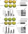
MdMPK4-14G-mediated MdERF17 phosphorylation promotes Chl degradation. A, Phenotypes of vacuum-infiltrated apple fruit transformed with pSuper1300-MdERF17 or pSuper1300-MdERF17T67A were visualized 3 days after infiltration. Fruits infiltrated with an empty pSuper1300 vector were used as a control. Scale bar, 1 cm. B, Chl contents in the infiltrated apple fruits shown in (A). C, Relative expression levels of MdERF17 and the Chl degradation genes MdPPH and MdNYC in infiltrated apple fruit, as determined by RT-qPCR. The expression levels in the control were set to 1. D, Immunoblot analysis showing phosphorylated MdERF17 in infiltrated apple fruit. MdERF17 and MdERF17T67A were detected by immunoblotting with anti-Myc antibodies. Actin was used as a protein loading control. In (B) and (C), error bars indicate the SD of three biological replicates. Asterisks indicate statistically significant differences (**P < 0.01) according to one-way ANOVA, followed by Tukey’s honestly significant difference test.
Figure 5
Figure 5
Differences in the levels of phosphorylated MdERF17-8S and MdERF17-3S caused by Ser repeat number are associated with Chl degradation. A, Kinetic parameters for the interaction between MdERF17 and MdMPK4, as determined by SPR assays. ka: association rate. kd: dissociation rate. KD: kd/ka, dissociation equilibrium constant. B, Phenotypes and Chl degradation rate of “Zisai Pearl” and “Red Fuji” fruits at different developmental stages. Scale bar, 1 cm. C, Detection of MdERF17 and MdMPK4 phosphorylation and MdERF17 and MdMPK4 protein levels in the peels of “Zisai Pearl” and “Red Fuji” fruits from 110 to 170 DAFB using anti-Phos-MdERF17, anti-Phos-MdMPK4, anti-MdERF17, and anti-MdMPK4 antibodies. Actin was used as a protein loading control. In (B), error bars indicate the sd of three biological replicates.
Figure 6
Figure 6
MdMPK4-14G transcript and phosphorylation levels are higher in the dark during fruit development. Phenotypes (A) and Chl contents (B) of “Gala” fruits at different developmental stages. Scale bar, 1 cm. Asterisks indicate significant differences compared with Chl contents at 20 DAFB to show the Chl degradation period. C, Schematic illustration showing the distribution of putative circadian and light-responsive cis-elements present in the promoter regions of MdMPK4-14G and MdMPK4-06G, as predicted by the PLACE database. D–F, Time course analyses of endogenous MdMPK4-14G expression in apple peels, by RT-qPCR. Apple fruits at 60, 80, and 100 DAFB from plants grown under natural conditions were harvested in 3-h intervals throughout a 24-h period. The light period is indicated according to sunrise (represented by the white areas); darkness is represented by the dark areas. Asterisks indicate significant differences compared to the MdMPK4-14G expression levels at 9 am. G, MdMPK4-14G transcription levels change between 9 am and 9 pm from 60 to 100 DAFB. H, MdMPK4 and MdERF17 phosphorylation levels change between 9 am and 9  pm from 60  to 100 DAFB. Anti-MdMPK4 and anti-MdERF17 antibodies were used to detect endogenous MdMPK4 and MdERF17 proteins. Actin was used as a protein loading control. In (B) and (D–G), the expression levels at 9 am were set to 1; error bars indicate the SD of three biological replicates. Asterisks indicate statistically significant differences (*P<0.05; **P<0.01) according to Student’s t test.
Figure 7
Figure 7
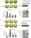
Silencing or overexpression of MdMPK4-14G in “Gala” apple under 14-h light/10-h dark conditions. A, Phenotypes of TRV control and MdMPK4-14G-silenced (TRV-MdMPK4-14G) fruits. Scale bar, 1 cm. B, Chl contents in the peels of the TRV control and MdMPK4-14G-silenced fruits. C, Relative expression levels of MdMPK4-14G, MdERF17, and the Chl degradation genes MdPPH and MdNYC sampled at 9 pm in infiltrated apple fruits, as detected by RT-qPCR. D, Detection of MdERF17 and MdMPK4 phosphorylation and protein levels in infiltrated apple fruits sampled at 9 pm by immunoblot analysis with anti-MdERF17, anti-MdMPK4, anti-Phos-MdERF17 and anti-Phos-MdMPK4 antibodies. Actin was used as a protein loading control. E, Phenotypes of the pRI101 control and CA-MdMPK4-14G-overexpressing (pRI101-CA-MdMPK4-14G) fruits. Scale bar, 1 cm. F, Chl contents in the peels of the pRI101 control and CA-MdMPK4-14G-overexpressing fruits. G, Relative expression levels of MdMPK4-14G, MdERF17 and the Chl degradation genes MdPPH and MdNYC in infiltrated apple fruits sampled at 9 am, as detected by RT-qPCR. H, Detection of MdERF17 and MdMPK4 phosphorylation and protein levels in infiltrated apple fruits sampled at 9 am by immunoblot analysis with anti-MdERF17, anti-MdMPK4, anti-Phos-MdERF17, and anti-Phos-MdMPK4 antibodies. Actin was used as a protein loading control. In (B), (C), (F), and (G), the expression levels in the control were set to 1; error bars indicate the SD of three biological replicates. Asterisks indicate statistically significant differences (*P < 0.05, **P  < 0.01) according to Student’s t test.
Figure 8
Figure 8
Overexpression or silencing of MdMPK4-14G in “Gala” apple under continuous dark conditions. A, Phenotypes of the pRI101 control and CA-MdMPK4-14G-overexpressing (pRI101-CA-MdMPK4-14G) fruits. Scale bar, 1 cm. B, Chl contents in the peels of the pRI101 control and CA-MdMPK4-14G-overexpressing fruits. C, Relative expression levels of MdMPK4-14G, MdERF17, and the Chl degradation genes MdPPH and MdNYC in infiltrated apple fruits, as detected by RT-qPCR. D, Detection of MdERF17 and MdMPK4 phosphorylation and protein levels in infiltrated apple fruits. Samples were collected from the peels of the pRI101 control and CA-MdMPK4-14G-overexpressing fruits and then were subjected to immunoblot analysis with anti-MdERF17, anti-MdMPK4, anti-Phos-MdERF17, and anti-Phos-MdMPK4 antibodies. Actin was used as a protein loading control. E, Phenotypes of the TRV control and MdMPK4-14G-silenced (TRV-MdMPK4-14G) fruits. Scale bar, 1 cm. F, Chl contents in the peels of the TRV control and MdMPK4-14G-silenced fruits. G, Relative expression levels of MdMPK4-14G, MdERF17, and the Chl degradation genes MdPPH and MdNYC in infiltrated apple fruits, as detected by RT-qPCR. H, Detection of MdERF17 and MdMPK4 phosphorylation and protein levels in infiltrated apple fruits. Samples were collected from the peels of the TRV control and MdMPK4-14G-silenced fruits and then were subjected to immunoblot analysis with anti-MdERF17, anti-MdMPK4, anti-Phos-MdERF17, and anti-Phos-MdMPK4 antibodies. Actin was used as a protein loading control. In (B), (C), (F), and (G), the expression levels in the control were set to 1; error bars indicate the SD of three biological replicates. Asterisks indicate statistically significant differences (*P<0.05; **P<0.01) according to Student’s t test.
Figure 9
Figure 9
Model of Chl degradation controlled by MdMPK4-14G-mediated phosphorylation of MdERF17 in apple fruits. In the dark, MdMYB1 is ubiquitinated and degraded by the 26S proteasome pathway. In contrast, MdERF17 is phosphorylated by dark-activated MdMPK4-14G, which results in high MdERF17 transcriptional activity and the subsequent induction of the expression of target genes, such as MdPPH and MdNYC, thus promoting Chl degradation. Upon light exposure, MdMYB1 is stabilized by MdMPK4-06G-mediated phosphorylation, and activated MdMYB1 binds to its target gene promoters to promote anthocyanin accumulation. MdERF17 phosphorylation is reduced by lower MdMPK4-14G expression and prevented from binding to its target genes for Chl degradation.

Similar articles

Cited by

References

    1. Albert NW, Davies KM, Lewis DH, Zhang H, Montefiori M, Brendolise C, Boase MR, Ngo H, Jameson PE, Schwinn KE (2014) A conserved network of transcriptional activators and repressors regulates anthocyanin pigmentation in Eudicots. Plant Cell 26: 962–980 - PMC - PubMed
    1. Allan AC, Hellens RP, Laing WA (2008) MYB transcription factors that colour our fruit. Trends Plant Sci 13: 99–102 - PubMed
    1. An X, Tian Y, Chen K, Liu X, Liu D, Xie X, Cheng C, Cong P, Hao Y (2015) MdMYB9 and MdMYB11 are involved in the regulation of the JA-induced biosynthesis of anthocyanin and proanthocyanidin in apples. Plant Cell Physiol 56: 650–662 - PubMed
    1. An X, Tian Y, Chen K, Wang X, Hao Y (2012) The apple WD40 protein MdTTG1 interacts with bHLH but not MYB proteins to regulate anthocyanin accumulation. J Plant Physiol 169: 710–717 - PubMed
    1. Andreasson E, Ellis B (2010) Convergence and specificity in the Arabidopsis MAPK nexus. Trends Plant Sci 15: 106–113 - PubMed

Publication types