

CDK6 increases glycolysis and suppresses autophagy by mTORC1-HK2 pathway activation in cervical cancer cells
- PMID: 35167417
- PMCID: PMC9037534
- DOI: 10.1080/15384101.2022.2039981
CDK6 increases glycolysis and suppresses autophagy by mTORC1-HK2 pathway activation in cervical cancer cells
Abstract
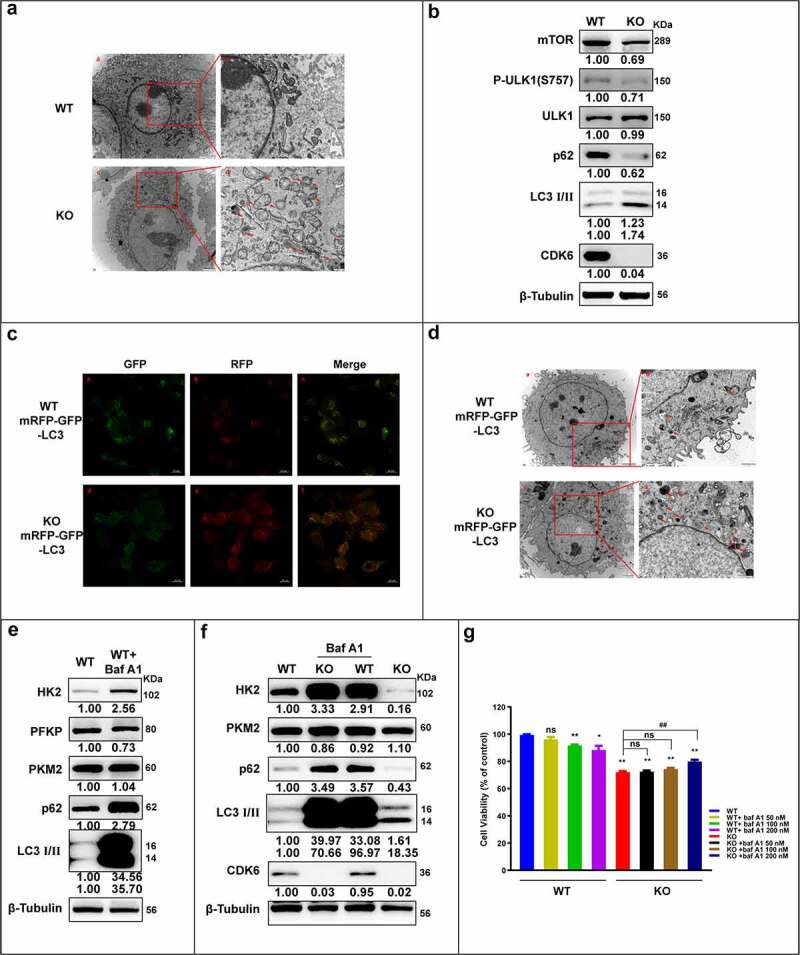
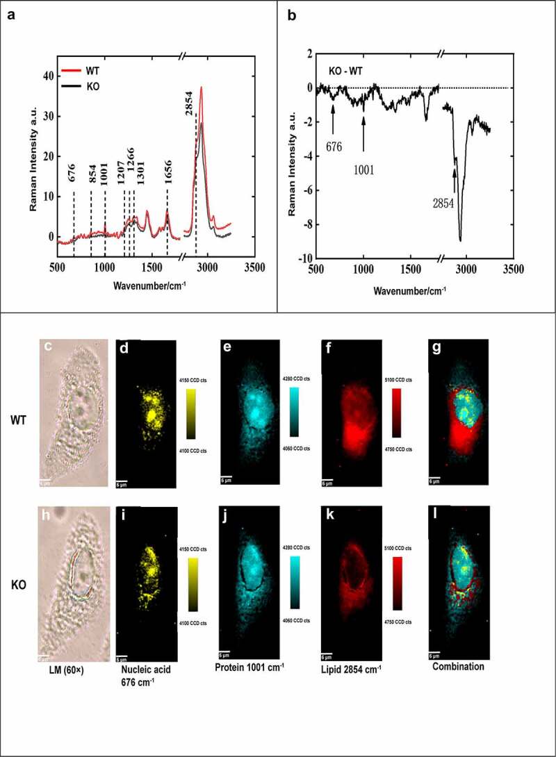
Cervical carcinoma is a leading malignant tumor among women worldwide, characterized by the dysregulation of cell cycle. Cyclin-dependent kinase 6 (CDK6) plays important roles in the cell cycle progression, cell differentiation, and tumorigenesis. However, the role of CDK6 in cervical cancer remains controversial. Here, we found that loss of CDK6 in cervical adenocarcinoma HeLa cell line inhibited cell proliferation but induced apoptosis as well as autophagy, accompanied by attenuated expression of mammalian target of rapamycin complex 1 (mTORC1) and hexokinase 2 (HK2), reduced glycolysis, and production of protein, nucleotide, and lipid. Similarly, we showed that CDK6 knockout inhibited the survival of CDK6-high CaSki but not CDK6-low SiHa cervical cancer cells by regulation of glycolysis and autophagy process. Collectively, our studies indicate that CDK6 is a critical regulator of human cervical cancer cells, especially with high CDK6 level, through its ability to regulate cellular apoptosis and metabolism. Thus, inhibition of CDK6 kinase activity could be a powerful therapeutic avenue used to treat cervical cancers.
Keywords: CDK6; Cervical carcinoma; apoptosis; autophagy; glycolysis; mTOR.
Conflict of interest statement
No potential conflict of interest was reported by the author(s).
Figures


Similar articles
-
CircCDKN2B-AS1 interacts with IMP3 to stabilize hexokinase 2 mRNA and facilitate cervical squamous cell carcinoma aerobic glycolysis progression.J Exp Clin Cancer Res. 2020 Dec 11;39(1):281. doi: 10.1186/s13046-020-01793-7. J Exp Clin Cancer Res. 2020. PMID: 33308298 Free PMC article.
-
Targeting hexokinase 2 inhibition promotes radiosensitization in HPV16 E7-induced cervical cancer and suppresses tumor growth.Int J Oncol. 2017 Jun;50(6):2011-2023. doi: 10.3892/ijo.2017.3979. Epub 2017 May 2. Int J Oncol. 2017. PMID: 28498475 Free PMC article.
-
Ribociclib, a selective cyclin D kinase 4/6 inhibitor, inhibits proliferation and induces apoptosis of human cervical cancer in vitro and in vivo.Biomed Pharmacother. 2019 Apr;112:108602. doi: 10.1016/j.biopha.2019.108602. Epub 2019 Feb 18. Biomed Pharmacother. 2019. PMID: 30784916
-
HK2/hexokinase-II integrates glycolysis and autophagy to confer cellular protection.Autophagy. 2015;11(6):963-4. doi: 10.1080/15548627.2015.1042195. Autophagy. 2015. PMID: 26075878 Free PMC article. Review.
-
How does hypoxia inducible factor-1α participate in enhancing the glycolysis activity in cervical cancer?Ann Diagn Pathol. 2013 Jun;17(3):305-11. doi: 10.1016/j.anndiagpath.2012.12.002. Epub 2013 Feb 1. Ann Diagn Pathol. 2013. PMID: 23375385 Review.
Cited by
-
CDK6 inhibits de novo lipogenesis in white adipose tissues but not in the liver.Nat Commun. 2024 Feb 5;15(1):1091. doi: 10.1038/s41467-024-45294-z. Nat Commun. 2024. PMID: 38316780 Free PMC article.
-
CDK6 is essential for mesenchymal stem cell proliferation and adipocyte differentiation.Front Mol Biosci. 2023 Aug 16;10:1146047. doi: 10.3389/fmolb.2023.1146047. eCollection 2023. Front Mol Biosci. 2023. PMID: 37664186 Free PMC article.
-
Comprehensive analysis of autophagy associated genes and immune infiltrates in cervical cancer.Iran J Basic Med Sci. 2024;27(7):813-824. doi: 10.22038/IJBMS.2024.74431.16168. Iran J Basic Med Sci. 2024. PMID: 38800011 Free PMC article.
-
Upregulation of FAM83F by c-Myc promotes cervical cancer growth and aerobic glycolysis via Wnt/β-catenin signaling activation.Cell Death Dis. 2023 Dec 16;14(12):837. doi: 10.1038/s41419-023-06377-9. Cell Death Dis. 2023. PMID: 38104106 Free PMC article.
-
Potential Target of CDK6 Signaling Pathway for Cancer Treatment.Curr Drug Targets. 2024;25(11):724-739. doi: 10.2174/0113894501313781240627062206. Curr Drug Targets. 2024. PMID: 39039674 Review.
References
-
- Cohen PA, Jhingran A, Oaknin A, et al. Cervical cancer. Lancet. 2019;393(10167):169–182. - PubMed
-
- Arafah M, Rashid S, Tulbah A, et al. Carcinomas of the uterine cervix: comprehensive review with an update on pathogenesis, nomenclature of precursor and invasive lesions, and differential diagnostic considerations. Adv Anat Pathol. 2021;28(3):150–170. - PubMed
-
- Meijer CJLM, Steenbergen RDM.. Gynaecological cancer: novel molecular subtypes of cervical cancer - potential clinical consequences. Nat Rev Clin Oncol. 2017;14(7):397–398. - PubMed
-
- Pirog EC, Lloveras B, Molijn A, et al. HPV prevalence and genotypes in different histological subtypes of cervical adenocarcinoma, a worldwide analysis of 760 cases. Mod Pathol. 2014;27(12):1559–1567. - PubMed
Publication types
MeSH terms
Substances
LinkOut - more resources
Full Text Sources
Other Literature Sources
Medical
Research Materials
Miscellaneous