. 2022 May;66(5):497-509.
doi: 10.1165/rcmb.2021-0357OC.

eNAMPT Is a Novel Damage-associated Molecular Pattern Protein That Contributes to the Severity of Radiation-induced Lung Fibrosis

Affiliations

eNAMPT Is a Novel Damage-associated Molecular Pattern Protein That Contributes to the Severity of Radiation-induced Lung Fibrosis

Alexander N Garcia et al. Am J Respir Cell Mol Biol. 2022 May.

Abstract

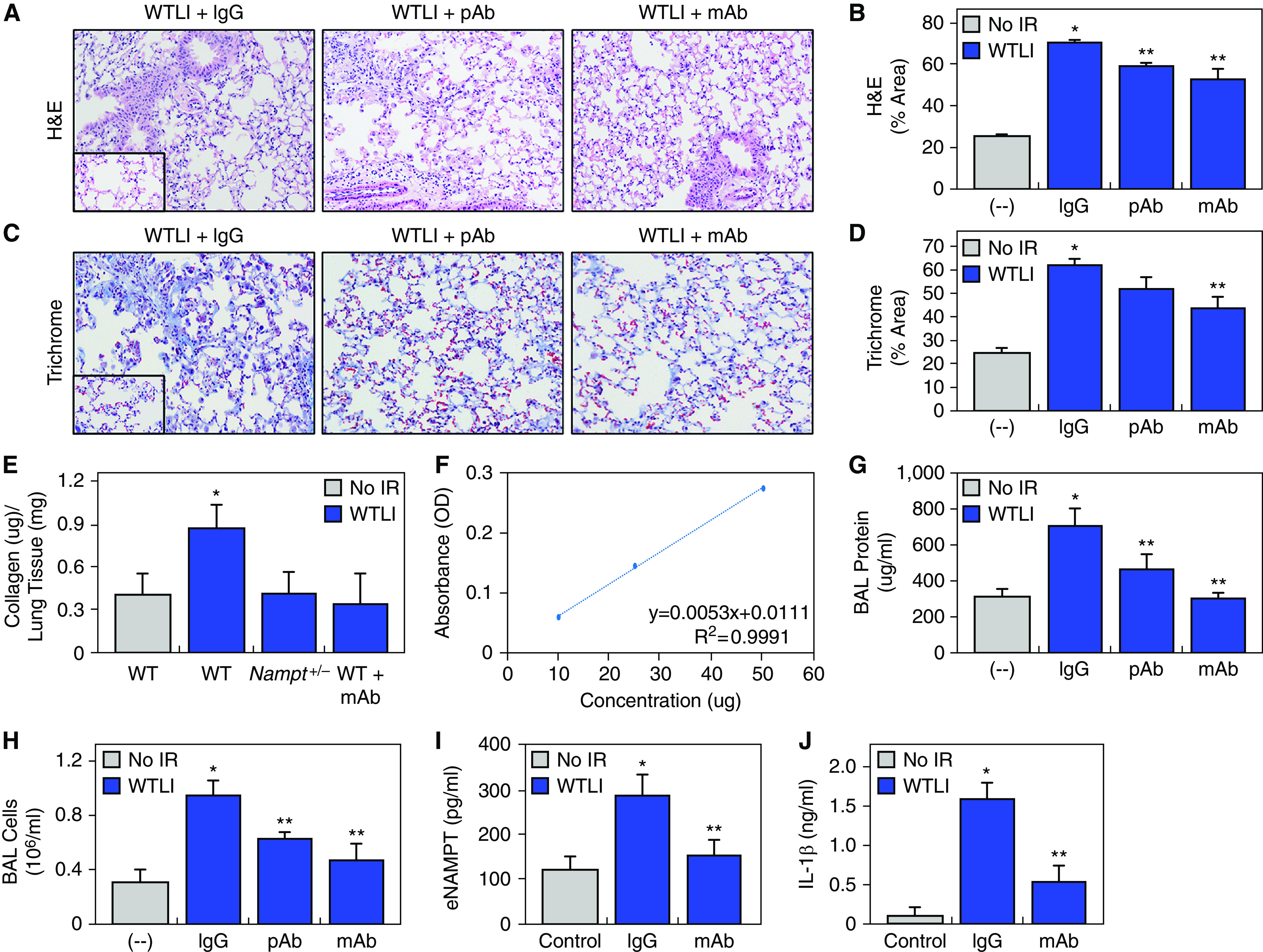
The paucity of therapeutic strategies to reduce the severity of radiation-induced lung fibrosis (RILF), a life-threatening complication of intended or accidental ionizing radiation exposure, is a serious unmet need. We evaluated the contribution of eNAMPT (extracellular nicotinamide phosphoribosyltransferase), a damage-associated molecular pattern (DAMP) protein and TLR4 (Toll-like receptor 4) ligand, to the severity of whole-thorax lung irradiation (WTLI)-induced RILF. Wild-type (WT) and Nampt+/- heterozygous C57BL6 mice and nonhuman primates (NHPs, Macaca mulatta) were exposed to a single WTLI dose (9.8 or 10.7 Gy for NHPs, 20 Gy for mice). WT mice received IgG1 (control) or an eNAMPT-neutralizing polyclonal or monoclonal antibody (mAb) intraperitoneally 4 hours after WTLI and weekly thereafter. At 8-12 weeks after WTLI, NAMPT expression was assessed by immunohistochemistry, biochemistry, and plasma biomarker studies. RILF severity was determined by BAL protein/cells, hematoxylin and eosin, and trichrome blue staining and soluble collagen assays. RNA sequencing and bioinformatic analyses identified differentially expressed lung tissue genes/pathways. NAMPT lung tissue expression was increased in both WTLI-exposed WT mice and NHPs. Nampt+/- mice and eNAMPT polyclonal antibody/mAb-treated mice exhibited significantly attenuated WTLI-mediated lung fibrosis with reduced: 1) NAMPT and trichrome blue staining; 2) dysregulated lung tissue expression of smooth muscle actin, p-SMAD2/p-SMAD1/5/9, TGF-β, TSP1 (thrombospondin-1), NOX4, IL-1β, and NRF2; 3) plasma eNAMPT and IL-1β concentrations; and 4) soluble collagen. Multiple WTLI-induced dysregulated differentially expressed lung tissue genes/pathways with known tissue fibrosis involvement were each rectified in mice receiving eNAMPT mAbs.The eNAMPT/TLR4 inflammatory network is essentially involved in radiation pathobiology, with eNAMPT neutralization an effective therapeutic strategy to reduce RILF severity.

Keywords: DAMP; TLR4; eNAMPT; nicotinamide phosphoribosyltransferase; whole-lung thoracic irradiation.


Figures

Figure 1.
Figure 1.
Thoracic irradiation exposure increases nicotinamide phosphoribosyltransferase (NAMPT) lung tissue expression and lung fibrosis in preclinical murine and nonhuman primate (NHP) models. Initial studies were designed to assess murine (C57Bl6) and Macaca mulatta (NHP) responses to thoracic irradiation. (A) Shown is representative hematoxylin and eosin (H&E) lung tissue staining (20× magnification) in an established 20-Gy whole-thorax lung irradiation (WTLI) murine model of radiation fibrosis at 12 weeks after ionizing radiation (IR) exposure (compared with sham-IR–exposed control mice, inset) showing alveolar inflammation and areas of fibrosis. ImageJ software was used to quantify the extent of hematoxylin and eosin (H&E) staining (three images from each animal, n = 5/group) as previously described (25). (B) Representative immunohistochemical (IHC) lung tissue staining of NAMPT (BLR058F, Bethyl) in C57BL/6J mice exposed to 20 Gy WTLI with ImageJ quantification. Significantly increased NAMPT expression in murine lung tissues was noted at 12 weeks after a single 20-Gy exposure (compared with control mice, inset) consistent with IR-induced NAMPT expression. (C) Representative trichrome blue staining image (Gomori’s trichrome stain, Newcomer Supply) performed in lung tissues from 20-Gy WTLI-exposed mice with ImageJ quantification. Significantly increased trichrome blue staining was noted at 12 weeks after a single 20-Gy exposure (compared with control mice, inset). (D) Representative H&E lung tissue staining in NHPs (Macaca mulatta) 9 weeks after exposure to a single 10.7-Gy IR exposure (compared with non-IR–exposed control animals, inset). ImageJ software was used to quantify the extent of H&E staining in NHPs that exhibited alveolar inflammation, alveolar wall thickening, and areas of fibrosis. (E) Similar to B, the representative IHC NAMPT lung tissue staining image shows significantly increased NAMPT protein expression in NHPs at 10 weeks after exposure (compared with control animals, inset) with ImageJ quantification. (F) Similar to C, IHC trichrome blue staining in WTLI-exposed NHP lung tissues with ImageJ quantification showed significantly increased trichrome blue staining at 6–12 weeks after exposure (representative image shown, compared with control animals, inset) consistent with the presence of lung fibrosis. For DF, three histologic or IHC images were selected for each animal and used for ImageJ quantification, with four animals/group, 8–11 weeks after WTLI. *P < 0.05.
Figure 2.
Figure 2.
Extracellular NAMPT (eNAMPT) neutralization reduces the severity of preclinical WTLI-mediated murine lung fibrosis. (A and B) Similar to results described in Figure 1A, compared with sham-IR–exposed C57BL/6J mice (inset), H&E staining revealed marked increases in inflammatory lung injury and areas of fibrosis in wild-type (WT) mice exposed to 20 Gy radiation at 12 weeks (quantified by ImageJ analysis). WTLI 20 Gy–exposed WT mice receiving either the eNAMPT-neutralizing polyclonal antibody (pAb) (4 mg/kg, weekly i.p.) or the humanized monoclonal antibody (mAb) (0.4 mg/kg, weekly i.p.) demonstrated significant reductions in WTLI-mediated histologic lung injury compared with the PBS/IgG1 control. (C and D) Similar to results described in Figure 1C, IHC studies in lung tissue from 20-Gy WTLI-exposed mice shows marked increases in trichrome blue staining with ImageJ quantification at 12 weeks (compared with control mice, inset) consistent with the presence of lung fibrosis. WTLI-exposed mice receiving the eNAMPT-neutralizing mAb (0.4 mg/kg, weekly i.p.) demonstrated significant reductions in trichrome blue staining. Mice receiving the eNAMPT pAb (4 mg/kg, weekly i.p.) showed reduced staining, which trended to significance (P = 0.08). (E and F) Sircol detection of murine lung tissue collagen in 12-week WTLI-exposed mice. A portion of the right lung retrieved from no-WTLI control mice and mice exposed to WTLI (20 Gy, 12 wk) was homogenized in 0.5 M acetic acid with 0.1 mg/kg of pepsin and total soluble collagen quantification performed using the Sircol kit (Biocolor Ltd.). WTLI groups included WT IgG-treated mice, Nampt heterozygous mice, and WT mice treated weekly with the ALT-100 mAb (0.4 mg/kg). These measurements of soluble collagen concentrations (μg), expressed per milligram lung tissue, show significantly increased soluble collagen concentrations in WT WTLI-exposed mice compared with sham-IR–exposed WT control mice (*P < 0.05). WTLI soluble collagen concentrations were reduced in Nampt heterozygous mice (P = 0.07) and in eNAMPT mAb-treated mice (P = 0.09). The standard curve for the Sircol detection of murine lung tissue collagen is displayed in F. (G and H) Both WTLI-mediated BAL protein concentrations and number of BAL cells were increased at 12 weeks. Mice treated with either the eNAMPT-neutralizing pAb or mAb exhibited marked reductions in BAL protein concentrations and BAL cells 12 weeks after WTLI exposure. (I and J) The Meso Scale ELISA-based Discovery platform was used to assess eNAMPT mAb effects on plasma cytokine concentrations of eNAMPT (I), and the inflammatory cytokine, IL-1β (J) in control sham-IR–exposed mice (n = 5), untreated 20-Gy WTLI-exposed mice receiving weekly intraperitoneal PBS/IgG1 (n = 7, 12 wk), and WTLI-exposed mice receiving the eNAMPT-neutralizing mAb weekly intraperitoneally (n = 5, 12 wk). Exposure to 20 Gy WTLI increased plasma concentrations of eNAMPT and IL-1β, which were significantly reduced in mice receiving the eNAMPT-neutralizing mAb. *P < 0.05 compared with no IR and **P < 0.05 compared to IgG control. IgG = immunoglobulin G; OD = optical density.
Figure 3.
Figure 3.
eNAMPT neutralization rectifies dysregulated expression of proteins involved in fibrosis/matrix-remodeling signaling pathways. Biochemical studies were next conducted in lung tissue homogenates to confirm eNAMPT/Toll-like receptor (TLR) influence on WTLI-induced dysregulated gene/protein signaling. Western blot studies of lung tissue homogenates from WTLI-exposed, PBS/IgG-, and eNAMPT mAb-treated mice (n = 5/group, 12 wk) were compared with sham-IR–exposed mice with representative blots (n = 3) shown. (A and C) Marked increases in immunoreactivity of smooth muscle actin (SMA), a hallmark of tissue fibrosis, and (A and D) the inflammatory mediator IL-1β, was noted at 12 weeks after WTLI exposure in IgG control mice. (A and E) Also noted were increased expression of TGF-β signaling pathway proteins, phosphorylated SMAD2, (A and F) phospho-SMAD1/5/9, and (A and G) TGF-β expression. WTLI-exposed mice also exhibited increased expression of the extracellular matrix protein TSP1 (thrombospondin-1) (B and H) and the reactive oxygen species (ROS)-generating NOX4 (B and I), well-known contributors to fibrosis (–43), while demonstrating reduced expression of the ROS-sensing and transcriptional signaling molecule NRF2 (B and J). Each dysregulated protein expressed in WTLI-challenged, IgG control–treated mice was markedly rectified in mice treated weekly with the eNAMPT-neutralizing mAb (0.4 mg/kg). *P < 0.05 compared with no IR and **P < 0.05 compared to IgG control. Ch. = change; Ctrl = control; NOX4 = NADPH oxidase 4; TGFβ = transforming growth factor β.
Figure 4.
Figure 4.
Nampt+/ heterozygous mice are protected from preclinical WTLI-induced lung fibrosis. As Nampt/ homozygous knockout mice are embryonically lethal, we used Nampt+/− heterozygous mice to assess the role of NAMPT in WTLI responses as we have previously described (23, 24, 26, 30). (A and B) Histologic evidence (H&E staining) of inflammatory lung injury in WTLI (20 Gy)-exposed C57BL/6J WT mice at 12 weeks similar to that described in Figures 1A and 2A (compared with sham-IR–exposed mice, inset). WTLI-exposed Nampt+/− heterozygous mice, in contrast, exhibited significantly reduced inflammation and fibrosis compared with WT mice (ImageJ software quantification). (C and D) Similar to results described in Figures 1C and 2B, IHC studies in lung tissue from 20-Gy WTLI-exposed mice show marked increases in trichrome blue staining with ImageJ quantification at 12 weeks after 20-Gy exposure, which was significantly reduced in Nampt+/− heterozygous mice, consistent with reduced lung fibrosis. (E and F) Compared with WTLI-exposed WT mice, WTLI-exposed Nampt+/− heterozygous mice (12 wk) demonstrated significant reductions in total BAL protein/BAL cells (*P < 0.05 and **P < 0.05 compared to WT mice). (G) Representative IHC lung tissue staining of NAMPT exactly as described in Figure 1B. Compared with WTLI-exposed WT C57BL/6J mice, Nampt+/− heterozygous mice show significantly reduced NAMPT lung tissue expression at 12 weeks.
Figure 5.
Figure 5.
eNAMPT/TLR4 signaling dysregulates gene/protein signaling pathways involved in WTLI-induced lung fibrosis. RNA sequencing was used to assess eNAMPT-mediated dysregulation of lung inflammatory signaling and injury/repair processes in WTLI-exposed lung tissues from untreated mice (12 wk) and mice receiving weekly eNAMPT mAb treatment. (A) A total of 561 differentially expressed genes (DEGs) with a false discovery rate (FDR) less than 0.05 were identified comparing post-WTLI samples (radiated versus nonradiated) (Table E1). Further stringency-based transcriptome filtering yielded 50 DEGs comparing WTLI-exposed untreated mice to mAb-treated WTLI mice. These DEGs produced the biological protein interactome network (STRING) depicted in A (adjusted P < 0.05, fold change [FC]  ± 1). Enrichment was prioritized by strength (ratio of number of proteins annotated between proteins expected annotated with this term) and FDR P values corrected by Benjamini-Hochberg (BH) procedure (<0.01). Network nodes represent genes, the edges indicate the functional association, and the line thickness indicates the strength of the data support. These analyses highlighted a very strong signal for matrix metalloproteinase 9 (MMP9) as a hub of eNAMPT mAb-influenced interacting genes with direct interactions with gene/proteins known to be associated with regulation of tissue fibrosis (Il6ra, Nfatc1, Eln, Thsp1). (B and C) Shown are the top Gene Ontology and Reactome pathways and biologic processes identified via analysis of the 50 DEGs. Highlighted are lung fibrosis, IL-1 signaling, Fra1/Fra2 transcription regulation, immune development, and histone deacetylase (HDAC) signaling. (D) Shown is the rectifying effect of the eNAMPT-neutralizing mAb on expression of 50 WTLI DEGs, which conform to pathways known to be associated with lung fibrosis, endothelial proliferation, TGF-β regulation, and angiogenesis. Arrows identify genes that are highlighted in A as MMP9 and MMP9-interacting proteins. Ap-1 = activator protein 1; Fra = Fos-related antigen.

Comment in

  • Targeting Danger Signals to Rescue Fibrosis.
    Tsoyi K, Rosas IO. Tsoyi K, et al. Am J Respir Cell Mol Biol. 2022 May;66(5):468-470. doi: 10.1165/rcmb.2022-0022ED. Am J Respir Cell Mol Biol. 2022. PMID: 35271415 Free PMC article. No abstract available.

Similar articles

Cited by

References

    1. Cytlak UM, Dyer DP, Honeychurch J, Williams KJ, Travis MA, Illidge TM. Immunomodulation by radiotherapy in tumour control and normal tissue toxicity. Nat Rev Immunol . 2021 doi: 10.1038/s41577-021-00568-1. - DOI - PubMed
    1. Giuranno L, Ient J, De Ruysscher D, Vooijs MA. Radiation-induced lung injury (RILI) Front Oncol . 2019;9:877. - PMC - PubMed
    1. Hanania AN, Mainwaring W, Ghebre YT, Hanania NA, Ludwig M. Radiation-induced lung injury: assessment and management. Chest . 2019;156:150–162. - PMC - PubMed
    1. Keffer S, Guy CL, Weiss E. Fatal radiation pneumonitis: literature review and case series. Adv Radiat Oncol . 2019;5:238–249. - PMC - PubMed
    1. Rodrigues G, Lock M, D’Souza D, Yu E, Van Dyk J. Prediction of radiation pneumonitis by dose - volume histogram parameters in lung cancer: a systematic review. Radiother Oncol . 2004;71:127–138. - PubMed

Publication types