. 2022 Mar 22;7(6):e153672.
doi: 10.1172/jci.insight.153672.

Transcriptional analysis of lung fibroblasts identifies PIM1 signaling as a driver of aging-associated persistent fibrosis

Affiliations

Transcriptional analysis of lung fibroblasts identifies PIM1 signaling as a driver of aging-associated persistent fibrosis

Tho X Pham et al. JCI Insight. .

Abstract

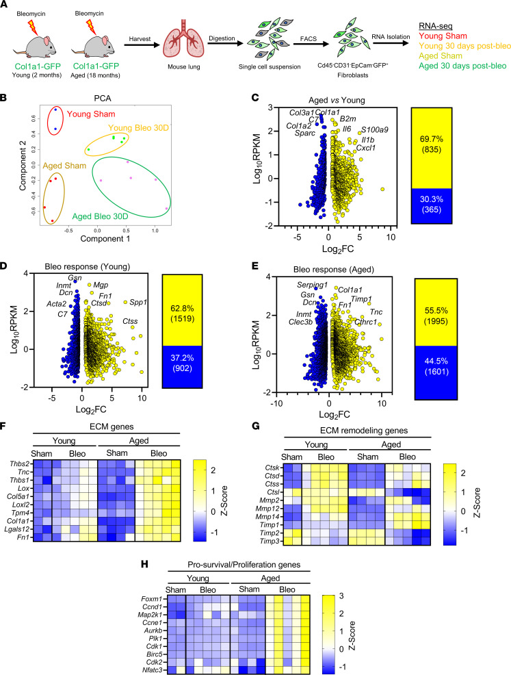
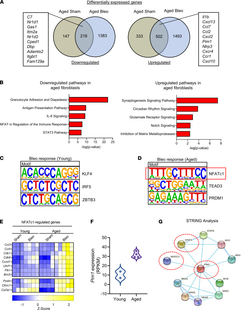
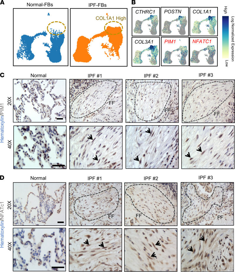
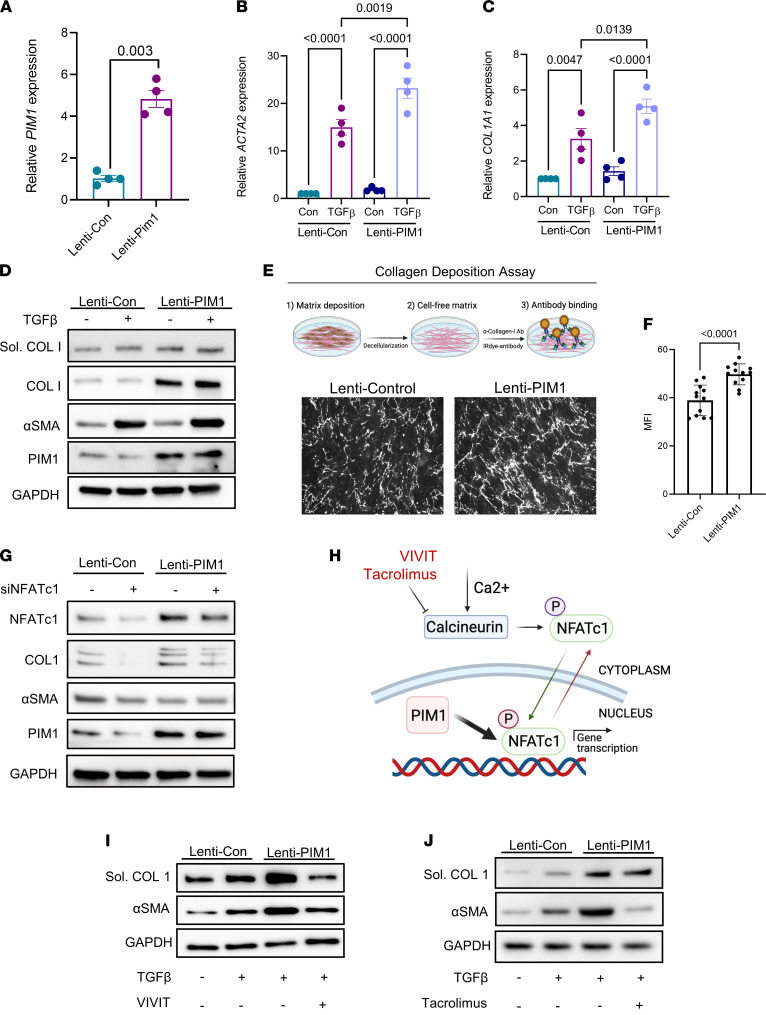
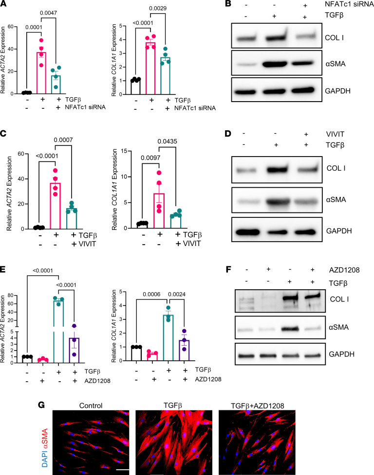
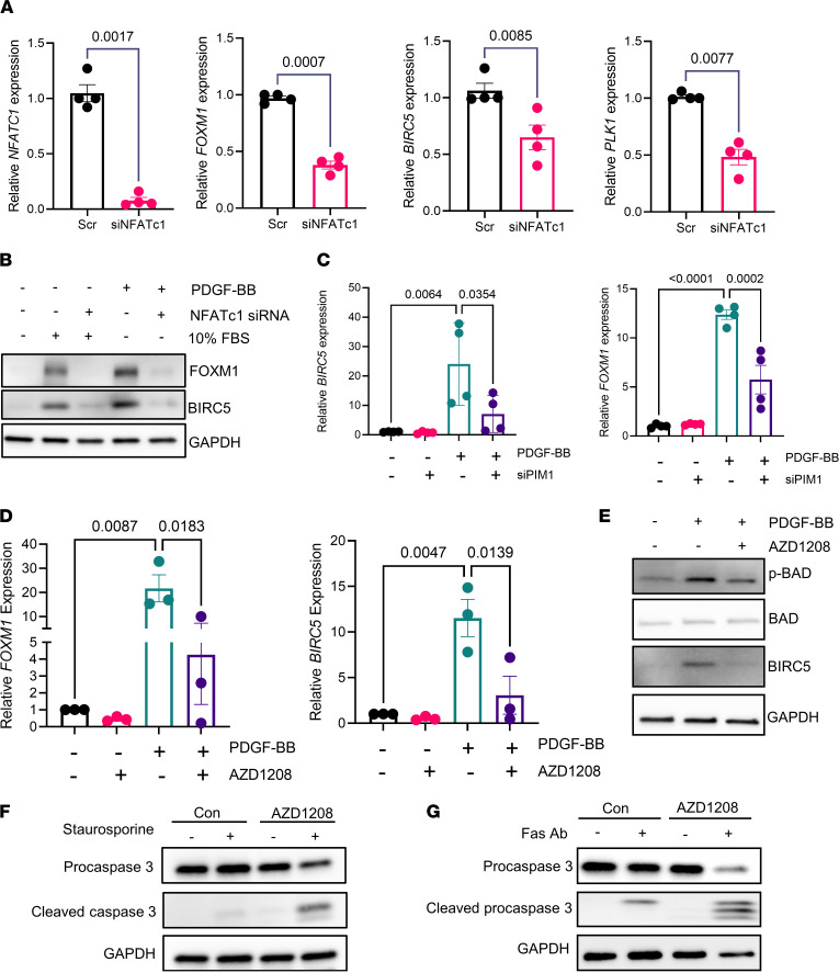
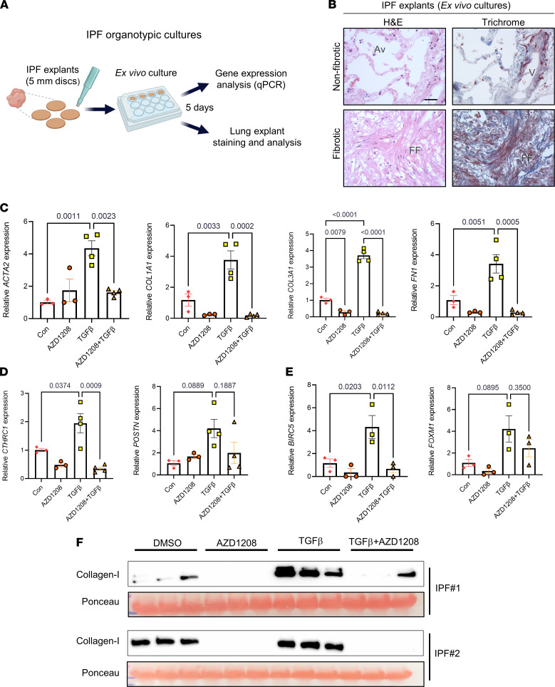
Idiopathic pulmonary fibrosis (IPF) is an aging-associated disease characterized by myofibroblast accumulation and progressive lung scarring. To identify transcriptional gene programs driving persistent lung fibrosis in aging, we performed RNA-Seq on lung fibroblasts isolated from young and aged mice during the early resolution phase after bleomycin injury. We discovered that, relative to injured young fibroblasts, injured aged fibroblasts exhibited a profibrotic state characterized by elevated expression of genes implicated in inflammation, matrix remodeling, and cell survival. We identified the proviral integration site for Moloney murine leukemia virus 1 (PIM1) and its target nuclear factor of activated T cells-1 (NFATc1) as putative drivers of the sustained profibrotic gene signatures in injured aged fibroblasts. PIM1 and NFATc1 transcripts were enriched in a pathogenic fibroblast population recently discovered in IPF lungs, and their protein expression was abundant in fibroblastic foci. Overexpression of PIM1 in normal human lung fibroblasts potentiated their fibrogenic activation, and this effect was attenuated by NFATc1 inhibition. Pharmacological inhibition of PIM1 attenuated IPF fibroblast activation and sensitized them to apoptotic stimuli. Interruption of PIM1 signaling in IPF lung explants ex vivo inhibited prosurvival gene expression and collagen secretion, suggesting that targeting this pathway may represent a therapeutic strategy to block IPF progression.

Keywords: Aging; Fibrosis; Pulmonology.


Conflict of interest statement

Conflict of interest: The authors have declared that no conflict of interest exists.

Figures

Figure 1
Figure 1. Impaired lung fibrosis resolution in aged mice is associated with increased inflammatory, ECM remodeling, and survival gene signatures in lung fibroblasts.
(A) Young and aged Col1a1-GFP mice were exposed to saline or bleomycin and sacrificed after 30 days during early fibrosis resolution. Lungs were harvested, digested, and sorted by FACS for CD31CD45EpCAMGFP+ lung fibroblasts and used for RNA-Seq analysis (young sham, n = 2; young bleo 30 days, n = 5; aged sham, n = 4; aged bleo 30 days, n = 5). (B) Principal components analysis (PCA) displaying clusters of samples from experimental groups and the similarity of their transcriptomes. (CE) (Left) Plots of log10 (avg.RPKM) versus log2 fold change of significantly upregulated or downregulated genes relative to young sham; fold-change ≥ 1.5, FDR ≤ 0.1. (Right) Percentage and total numbers of genes significantly upregulated (yellow) or downregulated (blue) relative to young sham. (FH) Heatmaps of differentially regulated gene signatures showing extracellular matrix (ECM) genes (F), extracellular matrix remodeling genes (G), and prosurvival/proliferation genes (H), displayed as Z scores of RPKM.
Figure 2
Figure 2. PIM1 and NFATc1 signaling pathways are associated with impaired fibrosis resolution in aged mice after bleomycin injury.
(A) Venn diagrams of differentially regulated genes from uninjured aged and injured aged fibroblasts relative to uninjured young fibroblasts. Intersection genes with the highest average expression are displayed. (B) Ingenuity pathway analysis of the intersection genes identifies differentially regulated canonical pathways. (C) De novo transcription factor binding motif analysis of differentially expressed genes in young fibroblasts after injury showing the top 3 binding motif and their associated transcription factors. (D) De novo transcription factor binding motif analysis of differentially expressed genes in aged lung fibroblasts after injury showing the top 3 binding motif and their associated transcription factor. Red box indicates highest ranked transcription factor binding motif and associated transcription factor in aged lung fibroblasts. (E) Heatmap of genes with NFATc1 binding motifs displayed as Z scores of RPKM (young sham, n = 2; young bleo 30 days, n = 5; aged sham, n = 4; aged bleo 30 days, n = 5). (F) Expression of PIM1 gene in young and aged lung fibroblasts (young sham, n = 2; aged sham, n = 4). (G) STRING functional protein association networks showing an interconnection between PIM1, STAT3, and NFATc1. Each node represents a protein, and each line represents a curated interaction.
Figure 3
Figure 3. NFATc1 and PIM1 expression is enriched in pathogenic fibroblasts in IPF.
(A) UMAP plots derived from publicly available scRNA-Seq data set (GSE132771) of mesenchymal cells sorted by FACS (CD31CD45CD235aEpCAM) isolated from normal and IPF lungs. (B) Normalized expression of CTHRC1, POSTN, COL1A1, COL3A1, PIM1, and NFATc1 in normal and pathogenic lung fibroblast populations. (C) IHC of human normal and IPF lung sections showing PIM1 nuclear expression. Dotted lines indicate fibrotic foci (FF). Arrows show PIM1-positive nuclei. Scale bar: 50 μm. (D) IHC of human normal and IPF lung sections showing NFATc1 nuclear expression. Dotted lines indicate fibrotic foci (FF). Arrows show NFATc1-positive nuclei. Scale bar: 50 μm.
Figure 4
Figure 4. NFATc1 inhibition attenuates PIM1-promoted lung fibroblast activation.
(A) Normal human lung fibroblasts transduced with control lentivirus (pLV-EGFP-CMV-mCherry) or lentivirus carrying the human PIM1 gene (pLV-EGFP-CMV-hPIM1) for 48 hours, followed by FACS to collect GFP+ cells. Shown is the mRNA expression of PIM1 evaluated by qPCR. Data are shown as mean ± SEM of n = 4 independent experiments. P values were calculated using 2-tailed, paired Student’s t test. (B and C) mRNA expression of ACTA2 (B) and COL1A1 (C) in control and PIM1 overexpressing cells treated with 2 ng/mL of TGF-β for 24 hours. Data are shown as mean ± SEM of n = 4 independent experiments. P values were calculated using 1-way ANOVA with Holm-Šidák post hoc test. (D) Normal human lung fibroblasts transduced with control lentivirus or PIM1 lentivirus were treated with 2 ng/mL of TGF-β for 24 and then analyzed by Western blotting. Shown is a representative blot of gels run in parallel. (E) Representative images of collagen-I fibers from a collagen deposition assay carried out in control and PIM1-overexpressing cells. (F) Quantification of collagen-I secretion measured as mean fluorescence intensity. n = 12. Data are shown as mean ± SEM. P values were calculated using 2-tailed Student’s t test. (G) Control and PIM1-overexpressing cells were transfected with scrambled or NFATc1 siRNAs for 48 hours, followed by Western blotting analysis. Shown is a representative blot of 3 independent experiments. (H) Schematic showing mechanisms of NFATc1 inhibition by VIVIT and tacrolimus. Following phosphorylation, NFATc1 is sequestered in the cytoplasm. Upon dephosphorylation by calcineurin, NFATc1 translocates into the nucleus, where it becomes transcriptionally active. In the nucleus, PIM1 can phosphorylate NFATc1 to enhance its transcription. (I and J) Western blotting analysis of control and PIM1 overexpressing lung fibroblasts cotreated with 2 ng/mL of TGF-β and the NFATc1 inhibitors VIVIT (5 μM) and tacrolimus (1 μM). Shown is a representative blot of 3 independent experiments.
Figure 5
Figure 5. Inhibition of PIM1 signaling pathway reduces profibrotic gene expression in IPF-derived lung fibroblasts.
(A) qPCR analysis of profibrotic gene expression in IPF-derived lung fibroblasts transfected with scrambled siRNA or siRNA for NFATc1 for 48 hours, followed by treatment with 2 ng/mL of TGF-β for an additional 24 hours. Data are shown as mean ± SEM of n = 4 independent experiments. P values were calculated using 1-way ANOVA with Holm-Šidák post hoc test. (B) IPF-derived lung fibroblasts were transfected with scrambled or NFATc1 siRNAs for 48 hours and then treated with 2 ng/mL of TGF-β for an additional 24 hours, followed by Western blotting analysis. Shown is a representative blot of 3 independent experiments. (C) qPCR analysis of profibrotic gene expression in IPF-derived lung fibroblasts treated with VIVIT (5 μM) and 2 ng/mL of TGF-β for 24 hours. Data are shown as mean ± SEM of n = 4 independent experiments. P values were calculated using 1-way ANOVA with Holm-Šidák post hoc test. (D) IPF-derived lung fibroblasts were treated with VIVIT (5 μM) and 2 ng/mL of TGF-β for 24 hours and analyzed via Western blot. Shown is a representative blot of n = 2 independent experiments. (E) qPCR analysis of profibrotic genes in IPF-derived lung fibroblasts cotreated with 10 μM of AZD1208 and 2 ng/mL of TGF-β for 24 hours. Data are shown as mean ± SEM of n = 3 independent experiments. P values were calculated using 1-way ANOVA with Holm-Šidák post hoc test. (F) IPF-derived lung fibroblasts were cotreated with 10 μM of AZD1208 and 2 ng/mL of TGF-β for 24 hours and analyzed by Western blotting. Shown is a representative blot of 3 independent experiments. (G) IHC of IPF-derived fibroblasts cotreated with 10 μM of AZD1208 and 2 ng/mL of TGF-β for 24 hours. Scale bar: 50 μm. Representative images of 3 independent experiments are shown.
Figure 6
Figure 6. Inhibition of PIM1 signaling pathway reduces prosurvival gene expression and sensitizes IPF-derived lung fibroblasts to apoptotic cues.
(A) qPCR analysis of aging-associated prosurvival genes and those with NFATc1 binding sites in IPF-derived fibroblasts transfected with scrambled siRNA or siRNA for NFATc1 for 48 hours. Data are shown as mean ± SEM of n = 4 independent experiments. P values were calculated using 2-tailed, paired Student’s t test. (B) Western blot analysis of FOXM1 and BIRC5 in IPF-derived fibroblasts transfected with scrambled siRNA or siNFATc1 for 48 hours and then stimulated with 50 ng/mL of PDGF-BB or 10% FBS for an additional 24 hours. Shown is a representative blot of 2 independent experiments. (C) qPCR analysis of prosurvival genes in IPF-derived fibroblasts transfected with scrambled siRNA or siRNA for PIM1 for 48 hours. Data are shown as mean ± SEM of n = 4 independent experiments. P values were calculated using 1-way ANOVA with Holm-Šidák post hoc test. (D) qPCR analysis of prosurvival gene expression in IPF-derived lung fibroblasts cotreated with 10 μM of AZD1208 and 50 ng/mL of PDGF-BB for 24 hours. Data are shown as mean ± SEM of n = 3 independent experiments. P values were calculated using 1-way ANOVA with Holm-Šidák post hoc test. (E) IPF-derived lung fibroblasts were cotreated with 10 μM of AZD1208 and 50 ng/mL of PDGF-BB for 24 hours and analyzed by Western blotting using antibodies against phospho-BAD, total-BAD, BIRC5, and GAPDH. (F) IPF-derived lung fibroblasts were pretreated for 24 hours with 10 μM of AZD1208 and with 300 nM of staurosporine for an additional 2 hours, followed by Western blotting analysis using antibodies against procaspase-3, cleaved caspase-3, and GAPDH. (G) IPF-derived lung fibroblasts were preincubated for 24 hours with 10 μM of AZD1208, followed by treatment with 0.5 μg/mL of FAS-activating antibody for an additional 24 hours. Procaspase-3 and cleaved caspase-3 were detected by Western blotting. Shown is a representative blot of 3 independent experiments.
Figure 7
Figure 7. PIM1 inhibition inhibits profibrotic gene expression and collagen secretion in organotypic IPF lung cultures ex vivo.
(A) Schematic of organotypic culture of IPF lungs. (B) H&E and trichrome staining of IPF lung tissue explants after 5 days in culture showing intact tissue architecture of nonfibrotic area (top panels) and distorted architecture of fibrotic area (bottom panels). Scale bar: 50 μm. FF, fibrotic foci; V, vein; Av, alveoli. (C) qPCR analysis of ECM gene expression in IPF lung explants treated with 10 μM of AZD1208 or DMSO control in combination with or without 10 ng/mL of TGF-β for 5 days. n ≥ 3 IPF lung explants. Data are shown as mean ± SEM. P values were calculated using 1-way ANOVA with Holm-Šidák post hoc test. (D) qPCR analysis of pathogenic lung fibroblasts gene markers in IPF lung explants treated with 10 μM of AZD1208 or DMSO in the presence or absence of 10 ng/mL of TGF-β for 5 days. n ≥ 3 IPF lung explants. Data are shown as mean ± SEM. P values were calculated using 1-way ANOVA with Holm-Šidák post hoc test. (E) qPCR analysis of prosurvival genes in IPF lung explants treated with 10 μM of AZD1208 or DMSO in the presence or absence of 10 ng/mL of TGF-β for 5 days. n = 3 IPF lung explants. Data are shown as mean ± SEM. P values were calculated using 1-way ANOVA with Holm-Šidák post hoc test. (F) Soluble collagen-I secreted from IPF lung explants into the media was evaluated by Western blot analysis. Each lane contained equal volume of conditioned medium of different lung sections obtained from single IPF lung explants. Ponceau staining was used as loading control for secreted collagen-I.

References

    1. Lederer DJ, Martinez FJ. Idiopathic pulmonary fibrosis. N Engl J Med. 2018;378(19):1811–1823. doi: 10.1056/NEJMra1705751. - DOI - PubMed
    1. Selman M, et al. Aging and pulmonary fibrosis. Rev Invest Clin. 2016;68(2):75–83. - PubMed
    1. El Agha E, et al. Mesenchymal stem cells in fibrotic disease. Cell Stem Cell. 2017;21(2):166–177. doi: 10.1016/j.stem.2017.07.011. - DOI - PubMed
    1. Glasser SW, et al. Mechanisms of lung fibrosis resolution. Am J Pathol. 2016;186(5):1066–1077. doi: 10.1016/j.ajpath.2016.01.018. - DOI - PMC - PubMed
    1. Jun JI, Lau LF. Resolution of organ fibrosis. J Clin Invest. 2018;128(1):97–107. doi: 10.1172/JCI93563. - DOI - PMC - PubMed

Publication types