

Association between the saphenous vein diameter and venous reflux on computed tomography venography in patients with varicose veins
- PMID: 35167584
- PMCID: PMC8846520
- DOI: 10.1371/journal.pone.0263513
Association between the saphenous vein diameter and venous reflux on computed tomography venography in patients with varicose veins
Abstract
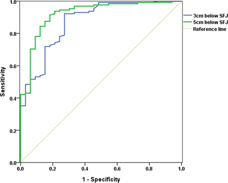
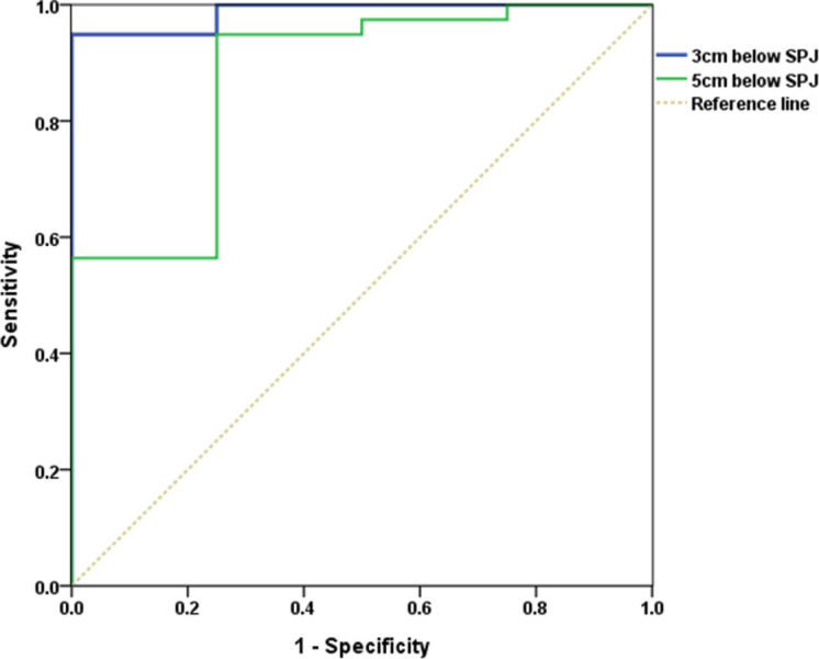
Three-dimensional computed tomography venography is a useful tool to identify increased saphenous vein diameter and provides a complementary road map for surgery in patients with varicose veins. In this study, we investigated the correlation between saphenous vein diameter on computed tomography venography and venous reflux detected on duplex ultraonography. We enrolled 152 patients (213 extremities) who underwent endovenous laser ablation therapy, following high ligation of the saphenofemoral junction between January 2014 and December 2019. All patients underwent preoperative computed tomography venography evaluation. The saphenous vein diameter was measured on computed tomography venography, and venous reflux was evaluated in the operating room using Doppler ultrasonography. Among the 152 patients included in the study, 61 showed varicose veins affecting the bilateral extremities. Among the 213 extremities investigated, 165 (77.5%) and 48 (22.5%) extremities showed varicosities involving the greater and lesser saphenous veins, respectively. Among all extremities, venous reflux was detected in 172 (80.8%). The mean diameter of the greater saphenous vein measured 5 cm distal to the saphenofemoral junction was 8.07±1.82 mm in patients with reflux and 5.11±1.20 mm in patients without reflux (p < .05). The small saphenous vein diameter measured 5 cm distal to the saphenopopliteal junction was 7.65±1.74 mm in patients with reflux and 5.04±1.80 mm in patients without reflux (p < .05). Based on the receiver operating characteristic curve, the greater saphenous vein threshold diameter of 5.880 mm measured 5 cm distal to the saphenofemoral junction was the optimal cut-off value to predict reflux (sensitivity 91.4%, specificity 81.8%). The lesser saphenous vein diameter of 5.285 mm measured 5 cm distal to the saphenopopliteal junction was the optimal cut-off value to predict reflux (sensitivity 94.9%, specificity 75.0%). Vein diameter cannot be used as an absolute reference for venous reflux; however, it may have predictive value in patients with varicose veins. Computed tomography venography based measurements of vein diameter may serve as a useful diagnostic tool to predict venous reflux and recommend treatment.
Conflict of interest statement
The authors have declared that no competing interests exist.
Figures
Similar articles
-
Anatomic variations of lower extremity venous system in varicose vein patients: demonstration by three-dimensional CT venography.Acta Radiol. 2017 May;58(5):542-549. doi: 10.1177/0284185116665420. Epub 2016 Sep 30. Acta Radiol. 2017. PMID: 27565630
-
Ultrasound-based topographic analysis of tributary vein connection with the saphenous vein during ambulatory conservative hemodynamic correction of chronic venous insufficiency.J Vasc Surg Venous Lymphat Disord. 2019 May;7(3):356-363. doi: 10.1016/j.jvsv.2018.09.011. Epub 2019 Feb 15. J Vasc Surg Venous Lymphat Disord. 2019. PMID: 30777672
-
Efficiency of Prophylactic Ablation of the Tributary Venous Pathways Draining Around the Saphenofemoral Junction to Decrease the Rate of Future Varicose Vein and Symptoms Occurence.Ann Vasc Surg. 2021 Aug;75:267-274. doi: 10.1016/j.avsg.2021.02.047. Epub 2021 Apr 3. Ann Vasc Surg. 2021. PMID: 33823264
-
Unusual causes of varicose veins in the lower extremities: CT venographic and Doppler US findings.Radiographics. 2009 Mar-Apr;29(2):525-36. doi: 10.1148/rg.292085154. Radiographics. 2009. PMID: 19325063 Review.
-
Endovenous laser ablation and sclerotherapy for treatment of varicose veins.Semin Cutan Med Surg. 2008 Dec;27(4):264-75. doi: 10.1016/j.sder.2008.08.001. Semin Cutan Med Surg. 2008. PMID: 19150298 Review.
References
Publication types
MeSH terms
LinkOut - more resources
Full Text Sources
Medical
