. 2022 Aug 8;107(Suppl_1):S1-S12.
doi: 10.1210/clinem/dgac045.

It Takes Two to Tango: IGF-I and TSH Receptors in Thyroid Eye Disease

Affiliations

It Takes Two to Tango: IGF-I and TSH Receptors in Thyroid Eye Disease

Leonard Girnita et al. J Clin Endocrinol Metab. .

Abstract

Context: Thyroid eye disease (TED) is a complex autoimmune disease process. Orbital fibroblasts represent the central orbital immune target. Involvement of the TSH receptor (TSHR) in TED is not fully understood. IGF-I receptor (IGF-IR) is overexpressed in several cell types in TED, including fibrocytes and orbital fibroblasts. IGF-IR may form a physical and functional complex with TSHR.

Objective: Review literature relevant to autoantibody generation in TED and whether these induce orbital fibroblast responses directly through TSHR, IGF-IR, or both.

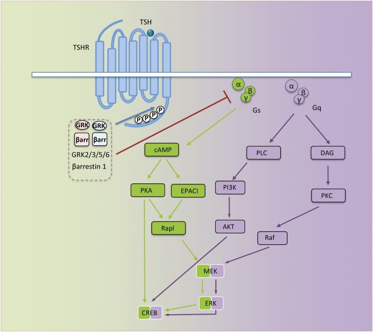
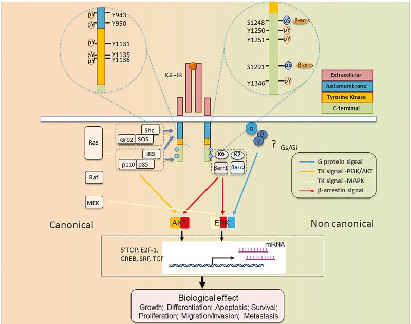
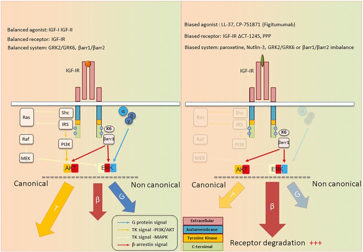
Evidence: IGF-IR has traditionally been considered a typical tyrosine kinase receptor in which tyrosine residues become phosphorylated following IGF-I binding. Evidence has emerged that IGF-IR possesses kinase-independent activities and can be considered a functional receptor tyrosine kinase/G-protein-coupled receptor hybrid, using the G-protein receptor kinase/β-arrestin system. Teprotumumab, a monoclonal IGF-IR antibody, effectively reduces TED disease activity, proptosis, and diplopia. In addition, the drug attenuates in vitro actions of both IGF-I and TSH in fibrocytes and orbital fibroblasts, including induction of proinflammatory cytokines by TSH and TED IgGs.

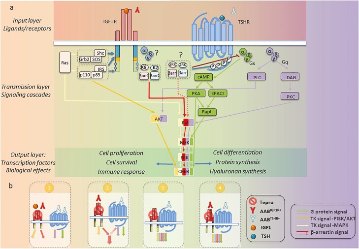
Conclusions: Although teprotumumab has been proven effective and relatively safe in the treatment of TED, many questions remain pertaining to IGF-IR, its relationship with TSHR, and how the drug might be disrupting these receptor protein/protein interactions. Here, we propose 4 possible IGF-IR activation models that could underlie clinical responses to teprotumumab observed in patients with TED. Teprotumumab is associated with several adverse events, including hyperglycemia and hearing abnormalities. Underpinning mechanisms of these are being investigated. Patients undergoing treatment with drug must be monitored for these and managed with best medical practices.

Keywords: GPCR; IGF-I; IGF-IR; IGF1; IGF1R; RTK; TSHR; beta-arrestins; biased signaling.


Figures

Figure 1.
Figure 1.
TSH receptor (TSHR) signaling. Schematic representation of major signaling pathways downstream TSHR. When not stimulated by activated receptor, Gα (α) is bound to guanosine diphosphate (GDP) and Gβγ (βγ), forming inactive Gs or Gq containing trimer, αβγ. Ligand-activated receptor leads to GDP/GTP exchange on the α subunit, resulting in dissociation into α and βγ subunits. α and βγ protein subunits subsequently interact with second effector proteins, promoting multiple downstream pathway activation; TSHR/Gs activates adenylyl cyclase (AC), resulting in increased cAMP, which in turn regulates multiple signaling pathways, including ion channels, protein kinase A and EPAC. TSHR/Gq initiates phospholipase C (PLC)/phosphoinositide 3-kinase (PI3K) cascade activation, promoting cell proliferation and survival. Likewise, TSHR/Gq enhances diacyl glycerol (DAG) generation and protein kinase C (PKC) activation. Both Gs and Gq, eventually activate Raf/MEK/ERK, also known as mitogen-activated protein kinase pathways. βγ subunits initiate receptor-distinct signaling processes including G protein-coupled receptor kinase (GRK) recruitment. GRKs phosphorylate serine/threonine residues within the TSHR C-terminus, creating binding sites for β-arrestins (βarr). βarr recruitment prevents further G protein activation (desensitization). All signaling pathways downstream from TSHR culminate in biological effects mediated through transcription factors such as ERK and cAMP response element binding protein.
Figure 2.
Figure 2.
Structure-function relationship of IGF-I receptor (IGF-IR) informing RTK/GPCR dualism. IGF-IR is annotated with numbered aa residues. Known key residues/sites of posttranslational modifications (PTMs) as determinant substrates/adaptor protein binding within β-subunit, thus controlling both canonical RTK signaling (left) and noncanonical GPCR-like signaling. IGF-IR kinase-dependent signaling pathways: IGF-I (or IGF-II) binding to IGF-IR promotes intrinsic tyrosine kinase activity and autophosphorylation. Activated receptor can recruit and phosphorylate substrates such as IRS and Shc. Phosphorylated tyrosine 950 within Asn-Pro-X-Tyr juxta membrane motif is essential for insulin receptor substrate-1 (IRS1)/IRS-2 and SHC-transforming protein (Shc) recruitment. Tyrosine phosphorylation of IRS and Shc proteins leads to as Grb2 and PI 3-kinase (p110/p85) binding. These protein associations induce downstream signaling activation, primarily through the rat sarcoma virus/Raf/MAPK and PI3K/Akt pathways, which in turn activate transcription factors coordinating downstream IGF biological effects. In addition to kinase signaling, agonist-induced IGF-IR stimulation results in noncanonical GPCR signaling through heterotrimeric G proteins (α, β, γ) (? = not yet fully understood pathways), followed by rapid IGF-IR phosphorylation by G protein-coupled receptor kinases (GRKs, K2, K6) at serine residues within the C-terminus. Serine-phosphorylated receptors present high-affinity binding sites for multifunctional adaptor protein β-arrestin 1/2 (βarr1/2). βarr recruitment: βarr acquires an active conformation following IGF-IR binding with MAPK pathway scaffold components. These events result in second wave IGF-IR kinase-dependent MAPK/ERK signaling activation. Abbreviations: Akt, protein kinase B; ERK, extracellular-signal-regulated kinase; IGF-IR, IGF receptor; P = major phosphorylation sites; IRS, insulin receptor substrate; MEK, mitogen-activated protein kinase/Erk kinase; Shc, Src homology and collagen domain protein.
Figure 3.
Figure 3.
Balanced vs biased IGF-IR signaling. IGF-I (balanced agonist) binding to IGF-IR within an unbiased system (GRKs and β-arrestin [βarr] balance) exhibits equivalent potencies for all signaling pathways, tyrosine kinase (T), βarr signaling (β), and G-protein signaling (G). Cell surface IGF-IR levels are preserved by GRK2/βarr2 (receptor recycling) and balanced by GRK6/βarr 1 (receptor degradation). Functional selectivity (biased signaling toward specific pathways) could be achieved by biased ligands (eg, anti-IGF-IR antibodies) or biased receptors (eg, TK inhibitors). System bias may be achieved through differential expression/inhibition of signaling effectors or cofactors, such as GRK and/or βarr isoforms. Demonstrated here is a model based on βarr biased signaling activation by anti-IGF-IR antibodies (eg, figitumumab), resulting in IGF-IR downregulation with cancer-protective βarr-ERK biased signaling activation.
Figure 4.
Figure 4.
Modeling proposed for teprotumumab mechanism of action in TED. (A) IGF-IR/TSHR system can be dissected into 3 distinct layers. The input layer comprises ligands (canonical or autoantibodies) and surface receptors. Following stimulation, signal transmission within second layer is controlled by a variety of messenger proteins. Confluence of signaling cascade components results in transcription factor activation within output layer. These factors control site-specific transcription and generate distinct biological effects. (B) Potential alternative signaling in TED, based on different mechanisms of IGF-IR activation. (1) Independent model: TSHR and IGF-IR function as separate entities. IGF-IR and TSHR activated as indicated with independent downstream signaling. Teprotumumab effects result entirely from IGF-IR inhibition. (2) Codependent IGF-IR signaling: IGF-IR activated by AABIGF-IR ligation. Biological effects are codependent on crosstalk between IGF-IR and TSHR downstream signaling pathways. Teprotumumab effects result from both IGF-IR and codependent TSHR inhibition. (3) Codependent TSHR signaling: AABTSHR activates TSHR, which in turn transactivates IGF-IR. Downstream IGF-IR/TSHR signaling pathways overlap extensively and are simultaneously triggered by activation of both receptor proteins. In this scenario, teprotumumab-engaged IGF-IR fails to become transactivated, whereas TSHR signaling remains unaffected. (4) Codependent IGF-IR/TSHR signaling: ABBTSHR engages TSHR, whereas IGF-IR becomes ligated with ABBIGF-IR. Both receptors are activated, generating protein crosstalk at both first and second layers. Teprotumumab would affect both IGF-IR activation as well as the receptor/signaling crosstalk.

References

    1. Watt T, Cramon P, Hegedüs L, et al. . The thyroid-related quality of life measure ThyPRO has good responsiveness and ability to detect relevant treatment effects. J Clin Endocrinol Metab. 2014;99(10):3708-3717. - PubMed
    1. Bartalena L, Kahaly GJ, Baldeschi L, et al. ; EUGOGO † . The 2021 European Group on Graves’ orbitopathy (EUGOGO) clinical practice guidelines for the medical management of Graves’ orbitopathy. Eur J Endocrinol. 2021;185(4):G43-G67. - PubMed
    1. Smith TJ, Hegedüs L. Graves’ disease. N Engl J Med. 2016;375(16):1552-1565. - PubMed
    1. Chin YH, Ng CH, Lee MH, et al. . Prevalence of thyroid eye disease in Graves’ disease: a meta-analysis and systematic review. Clin Endocrinol (Oxf). 2020;93(4):363-374. - PubMed
    1. Leo M, Menconi F, Rocchi R, et al. . Role of the underlying thyroid disease on the phenotype of Graves’ orbitopathy in a tertiary referral center. Thyroid. 2015;25(3):347-351. - PubMed

Publication types

MeSH terms