. 2022 May 4;30(5):1979-1993.
doi: 10.1016/j.ymthe.2022.02.013. Epub 2022 Feb 12.

Human inhalable antibody fragments neutralizing SARS-CoV-2 variants for COVID-19 therapy

Affiliations

Human inhalable antibody fragments neutralizing SARS-CoV-2 variants for COVID-19 therapy

Olga Minenkova et al. Mol Ther. .

Abstract

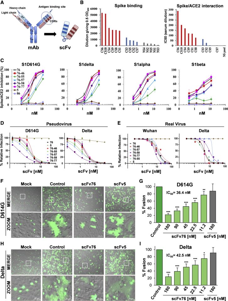
As of December 2021, coronavirus disease 2019 (COVID-19), caused by the severe acute respiratory syndrome coronavirus 2 (SARS-CoV-2), remains a global emergency, and novel therapeutics are urgently needed. Here we describe human single-chain variable fragment (scFv) antibodies (76clAbs) that block an epitope of the SARS-CoV-2 spike protein essential for ACE2-mediated entry into cells. 76clAbs neutralize the Delta variant and other variants being monitored (VBMs) and inhibit spike-mediated pulmonary cell-cell fusion, a critical feature of COVID-19 pathology. In two independent animal models, intranasal administration counteracted the infection. Because of their high efficiency, remarkable stability, resilience to nebulization, and low cost of production, 76clAbs may become a relevant tool for rapid, self-administrable early intervention in SARS-CoV-2-infected subjects independently of their immune status.

Keywords: COVID-19 pandemic; COVID-19 therapy; SARS-CoV-2 variant neutralization; aerosol therapy; anti-COVID-19 antibody; human single-chain antibody; inhalation; intranasal administration; phage display.


Conflict of interest statement

Declaration of interests O.M., E.M.P., and R.D.S. are employees of Alfasigma SpA and are named as inventors on a patent application in the name of the same company.

Figures

None
Graphical abstract
Figure 1
Figure 1
Selection and functional characterization of anti-SARS-CoV-2 human scFv antibodies (A) Schematic representation of scFv and mAb. (B) Analysis of COVID-19 convalescent (CS) and negative control (NS) sera for binding to SARS-CoV-2 spike (left) and inhibition of spike/hACE2 interaction (right) by ELISA. Data expressed as the dilution giving 0.5 OD and as IC50, respectively. (C) Inhibition of interaction of indicated spikes with hACE2 measured using ELISA. Data are the average ± SE of 2 or 3 experiments. (D) Neutralization of pseudotyped virus expressing indicated spikes assessed by luciferase assay in hACE2-expressing Caco-2 cells. Data are the average ± SD of two replicates from one representative experiment. (E) Microneutralization assay of SARS-CoV-2 Wuhan and delta strains in Vero E6 cells. (F–I) Inhibition of SARS-CoV-2 spike-mediated cell-cell fusion using HEK293T donor cells expressing GFP and D614G-mutated (F and G) or delta (H and I) spike, or GFP only (mock), incubated 1 h with saturating dose (180 nM) of scFv76 or scFv5 (F and H) or with scalar doses of scFv76 (G and I), and then overlaid on monolayers of hACE2-expressing A549 cells for 4 h. The overlay of bright-field and fluorescence images is shown (MERGE). Scale bar, 200 μm; zoom, 50 μm. Cell-cell fusion quantification expressed as percentage relative to control (average ± SD of 5 fields from two biological replicates). ∗p < 0.05, ∗∗p < 0.01, and ∗∗∗p < 0.001 (ANOVA).
Figure 2
Figure 2
Epitope mapping of anti-SARS-CoV-2 human scFv antibodies (A) Flow cytometry dot plot representation of antibody binding to alanine-mutated clones (left). For each point, background fluorescence was subtracted from the raw data, which were then normalized to reactivity with WT target protein. To identify preliminary primary critical clones (red circles), a threshold (dashed lines) of >65% WT binding to control Ab and <25% WT binding to test Abs was applied. Secondary clones (blue circles) are highlighted for clones that did not meet the set thresholds but whose decreased binding activity and proximity to critical residues suggested that the mutated residue may be part of the antibody epitope. Critical residues (red spheres) for scFv antibodies binding, and secondary residues (blue spheres) that may contribute to binding, are also visualized on crystal structure of the SARS-CoV-2 spike protein trimer (PDB: 6XCN) (center) and on SARS-CoV-2 spike protein receptor-binding domain (PDB: 6Z2M) (right). (B) Reactivity of scFv76 toward indicated spikes by ELISA.
Figure 3
Figure 3
Biochemical and structural characterization of 76clAbs (A) UV chromatogram at 280 nm from SEC-HPLC analysis of scFv76 in 50 mM phosphate buffer, 150 mM NaCl, 10% acetonitrile (pH 7.2) on TSKgel G3000 SWXL 30 cm × 7.8 mm column (Tosoh Bioscience). (B) Total ionic current (TIC) chromatogram from SEC-HPLC analysis of scFv76 in 20 mM ammonium formate (pH 6.8) on Mab Pac SEC-1 column (Thermo Fisher Scientific). Inset: mass spectrum as average of spectra recorded between 5 and 7 min. (C) Binding to SARS-CoV-2 RBD of thermally stressed (1 mg/mL concentration for 1 h at indicated temperatures) and not treated (NT) 76clAbs by ELISA. Data from one representative experiment. (D) Far-UV circular dichroism spectra of antibodies. Analysis was performed in PBS buffer at 20°C (solid lines) and 90°C (dashed lines). (E) UV chromatograms at 280 nm from SEC-HPLC analysis, as in (A), of scFv76 pre- and post-nebulization at 1 mg/mL. (F) Binding of scFv76, pre- and post-nebulization at 5 or 1 mg/mL, to SARS-CoV-2 RBD by ELISA. Data are from one representative experiment.
Figure 4
Figure 4
Intranasally administered scFv76 antibody inhibits viral infectivity in animal models (A) Representative bioluminescence images (BLI) at different time points after intranasal administration of scFv76 in K18-hACE2 mice infected with a luciferase-expressing SARS-CoV-2 spike (D614G) pseudotyped virus. Imaging by Xenogen IVIS200. (B and C) BLI photons/sec in nasal turbinates (B) and (C) total body. BLI data expressed as average ± SE of 5 animals. ∗∗p < 0.01 and ∗∗∗p < 0.001, Student’s t test versus infected (vehicle-treated) mice. (D and E) Body weight (D) and (E) nasal discharge of hamsters intranasally infected with SARS-CoV-2 (D614G) 105 pfu/animal on day 0 (D0) and treated with ScFv76 or vehicle 2 h before and once daily for 2 days post-infection. Body weight expressed as percentage versus D0. Nasal discharge scored as in the inset. Data are average ± SD of 5 animals/group. (F) Immunohistochemistry of pulmonary sections of mice, 1 h after PBS (panel 1) or 1 h after 4 min (panel 2), 8 min (panel 3), or 12 min (panel 4) scFv76 (5 mg/mL solution) aerosol exposure. (G) SARS-CoV-2 RBD binding by ELISA of lung proteins extracted from mice as in (F). Data expressed as average ± SD (n = 4). ∗p < 0.05, ∗∗p < 0.01, and ∗∗∗p < 0.001, Student’s t test versus 4-min-nebulized mice. §p < 0.05 and §§p < 0.01, Student’s t test versus 8-min-nebulized mice.

References

    1. Krause P.R., Fleming T.R., Longini I.M., Peto R., Briand S., Heymann D.L., Beral V., Snape M.D., Rees H., Ropero A.M., et al. SARS-CoV-2 variants and vaccines. N. Engl. J. Med. 2021;385:179–186. - PMC - PubMed
    1. Bernal J.L., Andrews N., Gower C., Gallagher E., Simmons R., Thelwall S., Stowe J., Tessier E., Groves N., Dabrera G., et al. Effectiveness of covid-19 vaccines against the B.1.617.2 (delta) variant. N. Engl. J. Med. 2021;385:585–594. - PMC - PubMed
    1. Wise J. Covid-19: delta variant doubles risk of hospital admission compared with alpha variant, study shows. Br. Med. J. 2021;374:n2152. - PubMed
    1. Papageorgiou A.C., Mohsin I. The SARS-CoV-2 spike glycoprotein as a drug and vaccine target: structural insights into its complexes with ACE2 and antibodies. Cells. 2020;9:2343. - PMC - PubMed
    1. Santopolo S., Riccio A., Santoro M.G. The biogenesis of SARS-CoV-2 spike glycoprotein: multiple targets for host-directed antiviral therapy. Biochem. Biophys. Res. Commun. 2021;538:80–87. - PMC - PubMed

Supplementary concepts