

TDP-43-associated atrophy in brains with and without frontotemporal lobar degeneration
- PMID: 35168140
- PMCID: PMC8850800
- DOI: 10.1016/j.nicl.2022.102954
TDP-43-associated atrophy in brains with and without frontotemporal lobar degeneration
Abstract
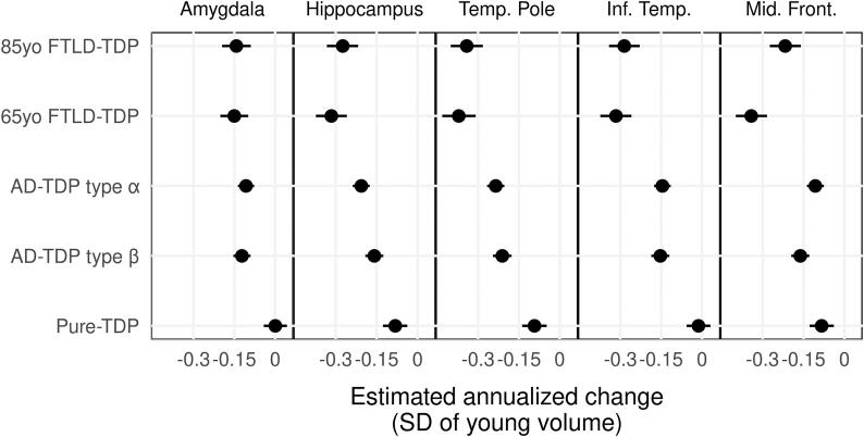
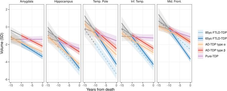
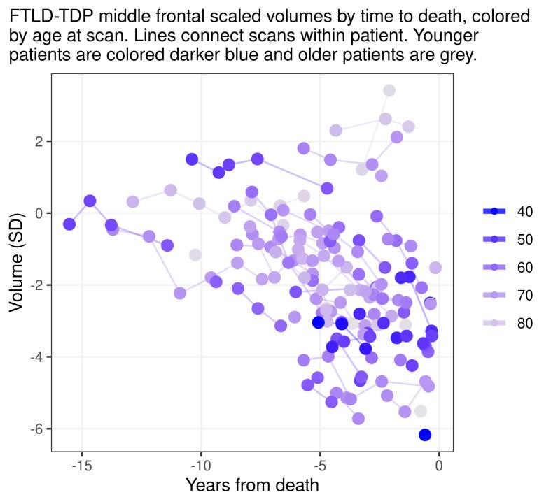
Transactive response DNA-binding protein of ∼43 kDa (TDP-43), a primary pathologic substrate in tau-negative frontotemporal lobar degeneration (FTLD), is also often found in the brains of elderly individuals without FTLD and is a key player in the process of neurodegeneration in brains with and without FTLD. It is unknown how rates and trajectories of TDP-43-associated brain atrophy compare between these two groups. Additionally, non-FTLD TDP-43 inclusions are not homogeneous and can be divided into two morphologic types: type-α and neurofibrillary tangle-associated type-β. Therefore, we explored whether neurodegeneration also varies due to the morphologic type. In this longitudinal retrospective study of 293 patients with 843 MRI scans spanning over ∼10 years, we used a Bayesian hierarchical linear model to quantify similarities and differences between the non-FTLD TDP-43 (type-α/type-β) and FTLD-TDP (n = 68) in both regional volume at various timepoints before death and annualized rate of atrophy. Since Alzheimer's disease (AD) is a frequent co-pathology in non-FTLD TDP-43, we further divided types α/β based on presence/absence of intermediate-high likelihood AD: AD-TDP type-β (n = 90), AD-TDP type-α (n = 104), and Pure-TDP (n = 31, all type-α). FTLD-TDP was associated with faster atrophy rates in the inferior temporal lobe and temporal pole compared to all non-FTLD TDP-43 groups. The atrophy rate in the frontal lobe was modulated by age with younger FTLD-TDP having the fastest rates. Older FTLD-TDP showed a limbic predominant pattern of neurodegeneration. AD-TDP type-α showed faster rates of hippocampal atrophy and smaller volumes of amygdala, temporal pole, and inferior temporal lobe compared to AD-TDP type-β. Pure-TDP was associated with slowest rates and less atrophy in all brain regions. The results suggest that there are differences and similarities in longitudinal brain volume loss between FTLD-TDP and non-FTLD TDP-43. Within FTLD-TDP age plays a role in which brain regions are the most affected. Additionally, brain atrophy regional rates also vary by non-FTLD TDP-43 type.
Keywords: Alzheimer’s disease; LATE; MRI; Old age FTLD; TDP-43.
Copyright © 2022 The Author(s). Published by Elsevier Inc. All rights reserved.
Conflict of interest statement
The authors report no competing interests. K.A.J., D.W.D., L.P., R.C.P., C.R.J., M.E.M. and J.L.W. receive research support from the National Institute of Health. L.P. has a U.S. patent #9,448,232 entitled ‘Methods and materials for detecting C9ORF72 hexanucleotide repeat expansion positive frontotemporal lobar degeneration or C9ORF72 hexanucleotide repeat expansion positive amyotrophic lateral sclerosis’. D.S.K. Knopman serves on a Data Safety Monitoring Board for the DIAN study. He serves on a Data Safety monitoring Board for Biogen but receives no personal compensation. He is an investigator in clinical trials sponsored by Biogen, Lilly Pharmaceuticals and the University of Southern California. He serves as a consultant for Roche, Samus Therapeutics, Third Rock and Alzeca Biosciences but receives no personal compensation; and receives research support from the NIH. R.C.P. serves as a consultant for Roche, Inc., Merck, Inc., Genentech, Inc., Biogen, Inc., Eli Lilly and Company and receives research support from the NIH. C.R.J. serves as a consultant for Janssen, Bristol Meyer-Squibb, General Electric, and Johnson & Johnson; is involved in clinical trials sponsored by Allon and Baxter, Inc.; and receives research support from Pfizer, Inc., the NIA, and the Alexander Family Alzheimer’s Disease Research Professorship of the Mayo Foundation.
Figures
References
-
- Arai T., Hasegawa M., Akiyama H., Ikeda K., Nonaka T., Mori H., Mann D., Tsuchiya K., Yoshida M., Hashizume Y. TDP-43 is a component of ubiquitin-positive tau-negative inclusions in frontotemporal lobar degeneration and amyotrophic lateral sclerosis. Biochem. Biophys. Res. Commun. 2006;351:602–611. - PubMed
-
- Arai T., Mackenzie I.R., Hasegawa M., Nonoka T., Niizato K., Tsuchiya K., Iritani S., Onaya M., Akiyama H. Phosphorylated TDP-43 in Alzheimer’s disease and dementia with Lewy bodies. Acta Neuropathol. 2009;117:125–136. - PubMed
-
- Baker M., Mackenzie I.R., Pickering-Brown S.M., Gass J., Rademakers R., Lindholm C., Snowden J., Adamson J., Sadovnick A.D., Rollinson S., Cannon A., Dwosh E., Neary D., Melquist S., Richardson A., Dickson D., Berger Z., Eriksen J., Robinson T., Zehr C., Dickey C.A., Crook R., McGowan E., Mann D., Boeve B., Feldman H., Hutton M. Mutations in progranulin cause tau-negative frontotemporal dementia linked to chromosome 17. Nature. 2006;442:916–919. - PubMed
-
- Barnes J., Whitwell J.L., Frost C., Josephs K.A., Rossor M., Fox N.C. Measurements of the amygdala and hippocampus in pathologically confirmed Alzheimer disease and frontotemporal lobar degeneration. Arch. Neurol. 2006;63:1434–1439. - PubMed
Publication types
MeSH terms
Substances
Grants and funding
LinkOut - more resources
Full Text Sources
Medical
