. 2022 Dec;17(1_suppl):87S-94S.
doi: 10.1177/15589447211068186. Epub 2022 Feb 15.

Extensor Tendon Injury After Volar Locking Plating for Distal Radius Fractures: A Systematic Review

Affiliations

Extensor Tendon Injury After Volar Locking Plating for Distal Radius Fractures: A Systematic Review

Jennifer A Kunes et al. Hand (N Y). 2022 Dec.

Abstract

Distal radius fractures are common orthopedic injuries. Treatment has varied historically, but volar locking plating currently predominates. Although flexor tendon injury is a well-studied complication of this operation, extensor tendon injury is less well studied. The purpose of this review is to search the literature and present the epidemiology, presentation, and treatment of this complication. The Cochrane, EMBASE, PubMed, and SCOPUS databases were searched for the terms "volar" + "radius" + ("plate" OR "plating") + "extensor." Ninety final studies were included for analysis in this review. The incidence of extensor tendon rupture varies from 0% to 12.5%; the extensor pollicis longus is most commonly ruptured. The presentation and management of extensor tendon injury after injury, intraoperatively, and postoperatively are summarized. Radiographic views are described to detect screw prominence and minimize intraoperative risk. Extensor tendon injury after volar locking plate for distal radius fractures is an uncommon injury with several risk factors including dorsal screw prominence and fracture fragments. Removal of hardware and tendon transfers or reconstruction may be necessary to prevent loss of extensor mechanism.

Keywords: anatomy; basic science; bone; diagnosis; distal radius; fracture/dislocation; research and health outcomes; specialty; surgery; tendon; treatment; wrist.


Conflict of interest statement

The author(s) declared no potential conflicts of interest with respect to the research, authorship, and/or publication of this article.

Figures
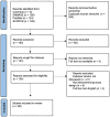
Figure 1.
Figure 1.
Preferred Reporting Items for Systematic Reviews and Meta-Analyses chart showing results of systematic review literature search.
Figure 2.
Figure 2.
Anatomy of the distal radius and volar plating. Note. (Right) Normal distal radius anatomy, marked landmarks for volar locking plate (VLP) placement. (1-6) Extensor tendons. (1) First compartment: extensor pollicis brevis, abductor pollicis longus. (2) Second compartment: extensor carpi radialis longus, extensor carpi radialis brevis. (3) Third compartment: extensor pollicis longus. (4) Fourth compartment: extensor indicis proprius, extensor digitorum communis. (5) Fifth compartment: extensor digiti minimi. (6) Sixth compartment: extensor carpi ulnaris. (A-E) Flexor tendons. (A) flexor carpi radialis, (B) flexor pollicis longus, (C) flexor digitorum profundus, (D) flexor digitorum superficialis, and (E) flexor carpi ulnaris. A wider margin between cortex and tendon exists on the volar side compared with the dorsal side. (Left) Distal radius with VLP. Lister’s tubercle may obstruct up to 1.4 to 6.6 mm of screw prominence (orange dotted line). Screws may protrude into extensor compartments at both distal screw holes and proximal screw holes.

References

    1. Nellans KW, Kowalski E, Chung KC. The epidemiology of distal radius fractures. Hand Clin. 2012;28(2):113-125. - PMC - PubMed
    1. Williksen JH, Husby T, Hellund JC, et al. External fixation and adjuvant pins versus volar locking plate fixation in unstable distal radius fractures: a randomized, controlled study with a 5-year follow-up. J Hand Surg Am. 2015;40(7):1333-1340. doi: 10.1016/j.jhsa.2015.03.008. - DOI - PubMed
    1. Yu YR, Makhni MC, Tabrizi S, et al. Complications of low-profile dorsal versus volar locking plates in the distal radius: a comparative study. J Hand Surg Am. 2011;36(7):1135-1141. doi: 10.1016/j.jhsa.2011.04.004. - DOI - PubMed
    1. Chappuis J, Bouté P, Putz P. Dorsally displaced extra-articular distal radius fractures fixation: dorsal IM nailing versus volar plating. A randomized controlled trial. Orthop Traumatol Surg Res. 2011;97(5):471-478. doi: 10.1016/j.otsr.2010.11.011. - DOI - PubMed
    1. Page MJ, McKenzie JE, Bossuyt PM, et al. The PRISMA 2020 statement: an updated guideline for reporting systematic reviews. BMJ. 2021;372:n71. doi: 10.1136/bmj.n71. - DOI - PMC - PubMed

Publication types