

Fatal Sarcocystis calchasi-associated meningoencephalitis in 2 captive vulturine guineafowl
- PMID: 35168421
- PMCID: PMC9254049
- DOI: 10.1177/10406387221078585
Fatal Sarcocystis calchasi-associated meningoencephalitis in 2 captive vulturine guineafowl
Abstract
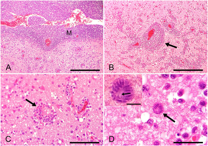
Two captive vulturine guineafowl (Acryllium vulturinum) were presented with lethargy, hyporexia, weight loss, and progressive neurologic signs. One of the guineafowl was seropositive for Sarcocystis falcatula (1:50 dilution). Both guineafowl died within 5 d of presentation. Histologic examination revealed nonsuppurative meningoencephalitis with gliosis, associated with occasional schizonts in the neuropil. Using fresh-frozen brain tissue, PCR was performed to amplify the ITS1 RNA region and portions of the 18S ribosomal RNA gene (18S gene) and the 28S ribosomal RNA gene (28S gene). Analysis of nucleic acid sequences from the resulting amplicons indicated that Sarcocystis calchasi was the likely cause of disease. To our knowledge, S. calchasi-associated disease has not been reported previously in the order Galliformes.
Keywords: Galliformes; Sarcocystis calchasi; aviary; guineafowl; meningoencephalitis.
Conflict of interest statement
Figures
References
-
- Clubb SL, Frenkel JK. Sarcocystis falcatula of opposums: transmission by cockroaches with fatal pulmonary disease in psittacine birds. J Parasitol 1992;78:116–124. - PubMed
-
- Dubey JP, et al.. Sarcocystosis of Animals and Humans. 2nd ed. CRC Press, 2016.
-
- Lindsay DS, et al.. Isolation, molecular characterization, and in vitro schizogonic development of Sarcocystis sp. ex Accipiter cooperii from a naturally infected Cooper’s hawk (Accipiter cooperii). Parasitol Int 2017;66:106–111. - PubMed
MeSH terms
Substances
LinkOut - more resources
Full Text Sources
